

FOXP3 inhibits angiogenesis by downregulating VEGF in breast cancer
- PMID: 29970908
- PMCID: PMC6030162
- DOI: 10.1038/s41419-018-0790-8
FOXP3 inhibits angiogenesis by downregulating VEGF in breast cancer
Abstract
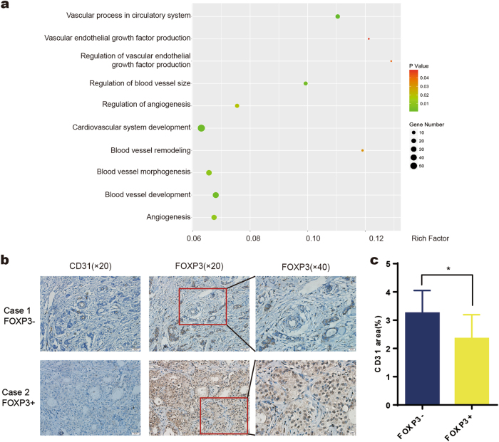
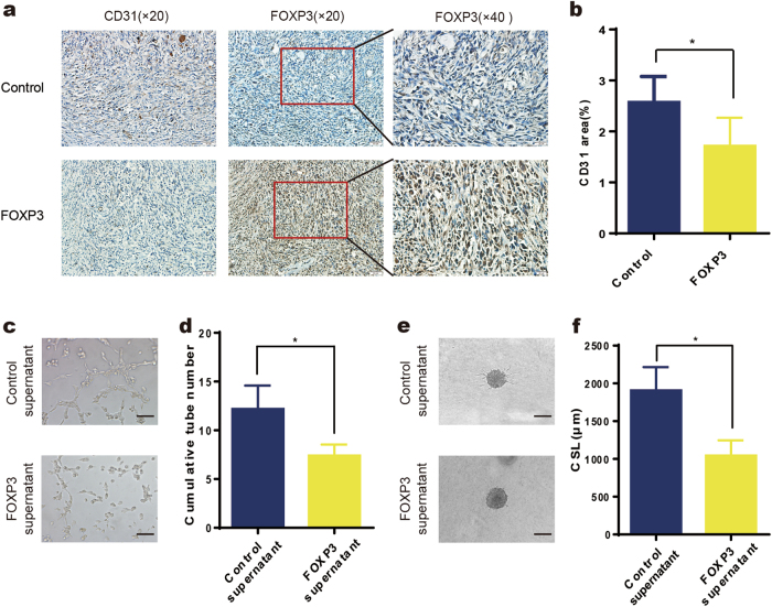
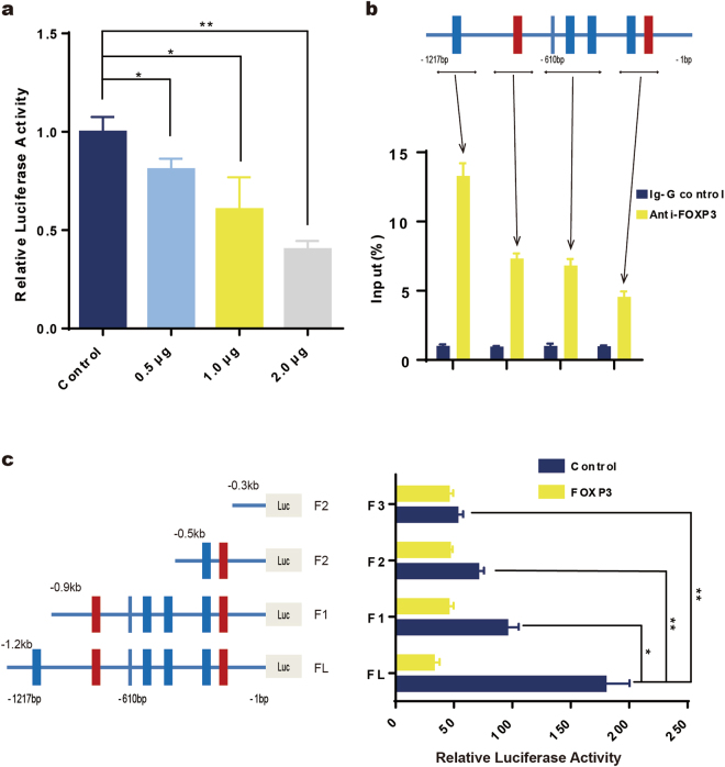
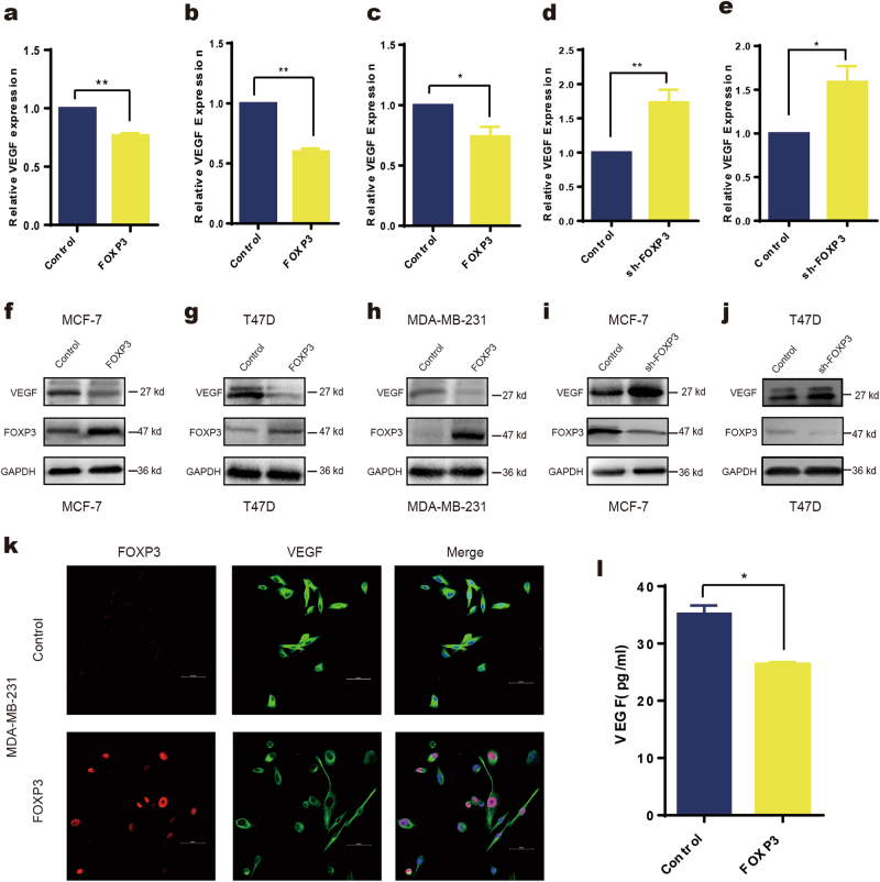
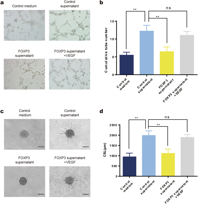
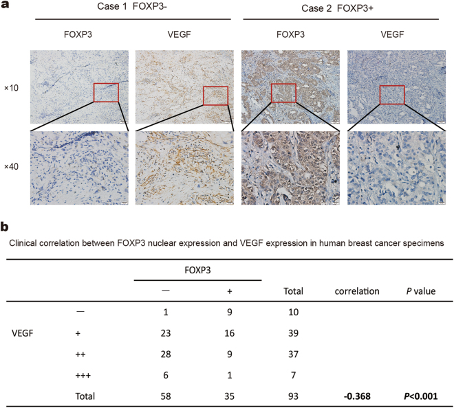
Forkhead box P3 (FOXP3), an X-linked tumor suppressor gene, plays an important role in breast cancer. However, the biological functions of FOXP3 in breast cancer angiogenesis remain unclear. Here we found that the clinical expression of nuclear FOXP3 was inversely correlated with breast cancer angiogenesis. Moreover, the animal study demonstrated that FOXP3 significantly reduced the microvascular density of MDA-MB-231 tumors transplanted in mice. The cytological experiments showed that the supernatant from FOXP3-overexpressing cells exhibited a diminished ability to stimulate tube formation and sprouting in HUVECs in vitro. In addition, expression of vascular endothelial growth factor (VEGF) was downregulated by FOXP3 in breast cancer cell lines. Luciferase reporter assays and chromatin immunoprecipitation assays demonstrated that FOXP3 can directly interact with the VEGF promoter via specific forkhead-binding motifs to suppress its transcription. Importantly, the inhibitory effects of FOXP3 in the supernatant on tube formation and sprouting in HUVECs could be reversed by adding VEGF in vitro. Nuclear FOXP3 expression was inversely correlated with VEGF expression in clinical breast cancer tissues, and FOXP3 downregulation and VEGF upregulation were both correlated with reduced survival in breast cancer data sets in the Kaplan-Meier plotter. Taken together, our data demonstrate that FOXP3 suppresses breast cancer angiogenesis by downregulating VEGF expression.
Conflict of interest statement
The authors declare that they have no conflict of interest.
Figures
Similar articles
-
FOXP3 suppresses breast cancer metastasis through downregulation of CD44.Int J Cancer. 2015 Sep 15;137(6):1279-90. doi: 10.1002/ijc.29482. Epub 2015 Feb 25. Int J Cancer. 2015. PMID: 25683728
-
Effects of MDM2 inhibitors on vascular endothelial growth factor-mediated tumor angiogenesis in human breast cancer.Angiogenesis. 2014 Jan;17(1):37-50. doi: 10.1007/s10456-013-9376-3. Epub 2013 Aug 2. Angiogenesis. 2014. PMID: 23907365
-
GABPα inhibits tumor progression and angiogenesis via a novel 18-bp indel within VEGF promoter in breast cancer.Cancer Biomark. 2024 Dec;41(3):CBM230541. doi: 10.3233/CBM-230541. Cancer Biomark. 2024. PMID: 39973818
-
Prognostic and predictive role of vascular endothelial growth factor polymorphisms in breast cancer.Pharmacogenomics. 2015 Jan;16(1):79-94. doi: 10.2217/pgs.14.148. Pharmacogenomics. 2015. PMID: 25560472 Review.
-
Signalling through FOXP3 as an X-linked tumor suppressor.Int J Biochem Cell Biol. 2010 Nov;42(11):1784-7. doi: 10.1016/j.biocel.2010.07.015. Epub 2010 Aug 1. Int J Biochem Cell Biol. 2010. PMID: 20678582 Free PMC article. Review.
Cited by
-
Targeting FOXP3 Tumor-Intrinsic Effects Using Adenoviral Vectors in Experimental Breast Cancer.Viruses. 2023 Aug 25;15(9):1813. doi: 10.3390/v15091813. Viruses. 2023. PMID: 37766222 Free PMC article.
-
Tumour infiltrating lymphocytes and immune-related genes as predictors of outcome in pancreatic adenocarcinoma.PLoS One. 2019 Aug 5;14(8):e0219566. doi: 10.1371/journal.pone.0219566. eCollection 2019. PLoS One. 2019. PMID: 31381571 Free PMC article.
-
Exogenous morphine inhibits the growth of human gastric tumor in vivo.Ann Transl Med. 2020 Mar;8(6):385. doi: 10.21037/atm.2020.03.116. Ann Transl Med. 2020. PMID: 32355829 Free PMC article.
-
Forkhead box P3 promotes breast cancer cell apoptosis by regulating programmed cell death 4 expression.Oncol Lett. 2020 Dec;20(6):292. doi: 10.3892/ol.2020.12155. Epub 2020 Sep 25. Oncol Lett. 2020. PMID: 33101486 Free PMC article.
-
The expression landscape of FOXP3 and its prognostic value in breast cancer.Ann Transl Med. 2022 Jul;10(14):801. doi: 10.21037/atm-22-3080. Ann Transl Med. 2022. PMID: 35965804 Free PMC article.
References
-
- Nico B, et al. Evaluation of microvascular density in tumors: pro and contra. Histol. Histopathol. 2008;23:601–607. - PubMed
Publication types
MeSH terms
Substances
Grants and funding
- 81472649/National Natural Science Foundation of China (National Science Foundation of China)/International
- 81702590/National Natural Science Foundation of China (National Science Foundation of China)/International
- 81402439/National Natural Science Foundation of China (National Science Foundation of China)/International
- 81472649/National Natural Science Foundation of China (National Science Foundation of China)/International
- 81672864/National Natural Science Foundation of China (National Science Foundation of China)/International
LinkOut - more resources
Full Text Sources
Other Literature Sources
Medical
Miscellaneous
