. 2018 Jun 19:12:45.
doi: 10.3389/fnana.2018.00045. eCollection 2018.

Identification of Functional Cell Groups in the Abducens Nucleus of Monkey and Human by Perineuronal Nets and Choline Acetyltransferase Immunolabeling

Affiliations

Identification of Functional Cell Groups in the Abducens Nucleus of Monkey and Human by Perineuronal Nets and Choline Acetyltransferase Immunolabeling

Anja K E Horn et al. Front Neuroanat. .

Abstract

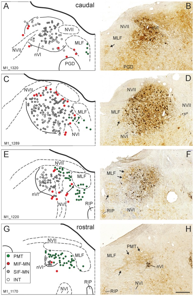
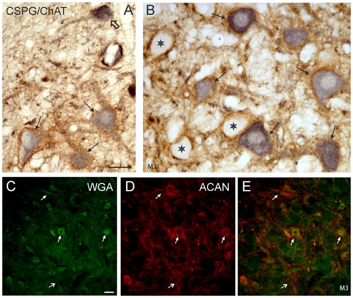
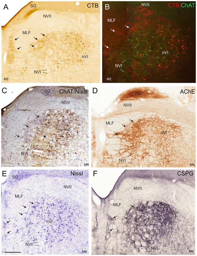
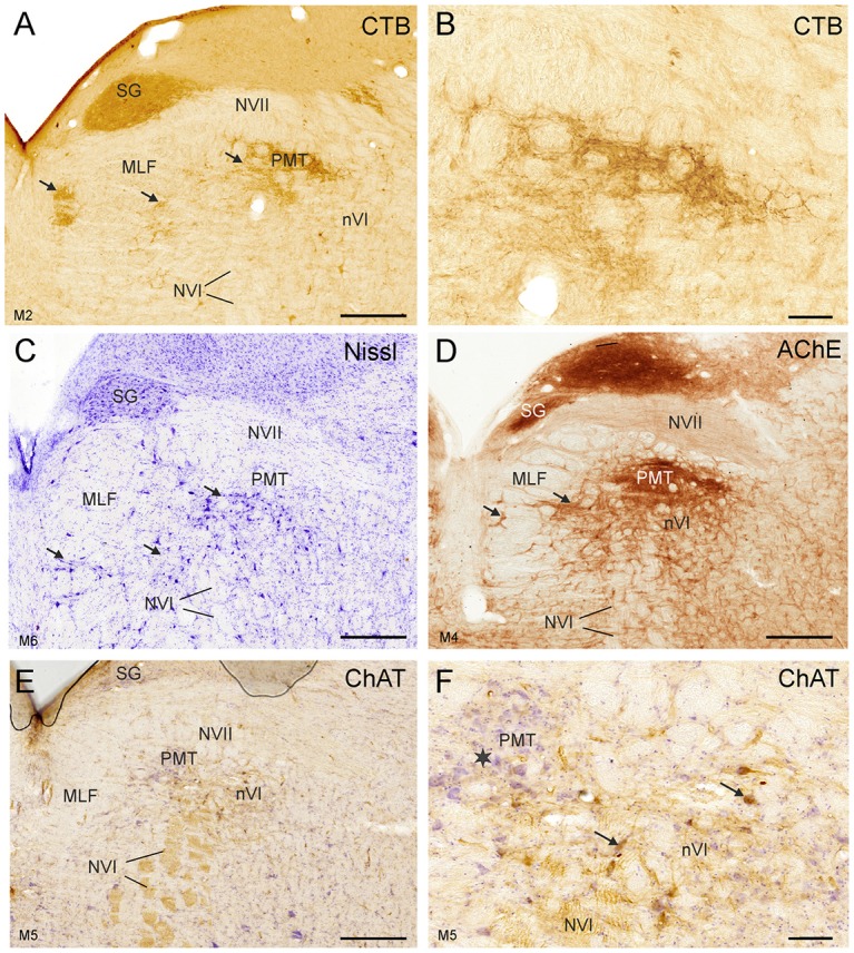
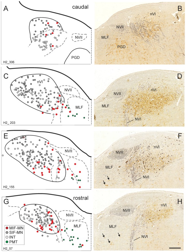
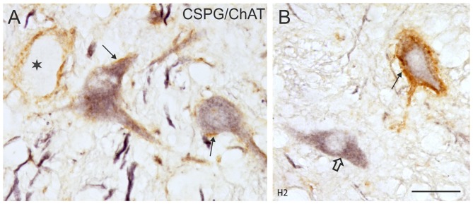
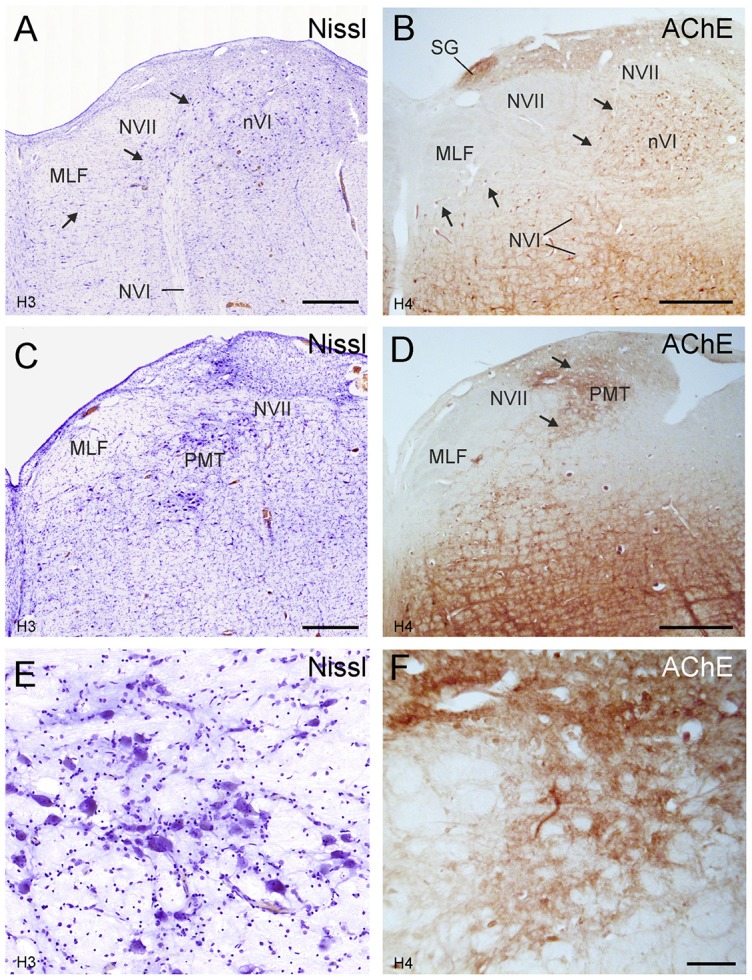
The abducens nucleus (nVI) contains several functional cell groups: motoneurons of the singly-innervated twitch muscle fibers (SIF) and those of the multiply-innervated muscle fibers (MIF) of the lateral rectus muscle (LR), internuclear neurons (INTs) projecting to the contralateral oculomotor nucleus (nIII) and paramedian tract-neurons (PMT) that receive input from premotor neurons of the oculomotor system and project to the floccular region. In monkey, these cell populations can be delineated by their chemical signature. For correlative clinico-pathological studies the identification of the homologous cell groups in the human nVI are required. In this study, we plotted the distribution of these populations in monkey nVI by combined tract-tracing and immunohistochemical staining facilitating the identification of homologous cell groups in man. Paraffin sections of two Rhesus monkeys fixed with 4% paraformaldhehyde and immunostained with antibodies directed against choline acetyltransferase (ChAT) as marker enzyme for cholinergic neurons and chondroitin sulfate proteoglycan (CSPG) to detect perineuronal nets (PNs) revealed four neuron populations in nVI with different chemical signatures: ChAT-positive and CSPG-positive SIF motoneurons, ChAT-positive, but CSPG-negative MIF motoneurons, and ChAT-negative neurons with prominent PNs that were considered as INTs. This was confirmed by combined immunofluorescence labeling of cholera toxin subunit B (CTB) or wheat germ agglutinin (WGA) and ChAT or CSPG in nVI sections from cases with tracer injections into nIII. In the rostral part of nVI and at its medial border, populations of ChAT-negative groups with weak CSPG-staining, but with strong acetylcholinesterase (AChE) activity, were identified as PMT cell groups by correlating them with the location of anterograde tracer labeling from INTs in nIII. Applying ChAT- and CSPG-immunostaining as well as AChE staining to human brainstem sections four neuron groups with the same chemical signature as those in monkey could be identified in and around the nVI in human. In conclusion, the distribution of nVI neuron populations was identified in human based on findings in monkey utilizing their markers for cholinergic neurons and their different ensheathment by PNs of the extracellular matrix.

Keywords: extracellular matrix; internuclear neurons; lateral rectus muscle; oculomotor; paramedian tract neurons; perineuronal nets.


Figures

Figure 1
Figure 1
Series of transverse brainstem sections through the monkey abducens nucleus (nVI) from caudal to rostral. Immunoreactivities of choline acetyltransferase (ChAT) in black and chondroitin sulfate proteoglycan (CSPG) in brown to reveal perineuronal nets (PNs). The left column (A,C,E,G) shows schematic drawings of the sections presented in the right column (B,D,F,H), where the functional cell groups, e.g., motoneurons of singly-innervated (SIF-MN, gray dots) and multiply-innervated muscle fibers (MIF-MN, red dots), internuclear neurons (INTs, open circles) and paramedian tract neurons (PMT; green dots) are plotted. Scale bar = 500 μm in (H; applies to A–H). MLF, medial longitudinal fascicle; NVII, facial nerve; PGD, nucleus paragigantocellularis dorsalis; RIP, nucleus raphe interpositus.
Figure 2
Figure 2
(A,B) Detailed microphotographs of sections from Figure 1 showing the monkey nVI stained for ChAT (black) and CSPG (brown). ChAT-positive neurons lacking PNs represent motoneurons of MIF (open arrow) (A), those enwrapped by PNs represent motoneurons of singly-innervated muscle fibers (thin arrows) (A,B), and the ChAT-negative ones INTs (B, star). (C,D,E) Image from double immunoflourescence in nVI of a monkey that had received a wheat germ agglutinin (WGA) injection into the oculomotor nucleus to backlabel INTs shown in (C) (green). Detection of aggrecan (ACAN) revealed that INTs (C, small arrows) are enwrapped by PNs (D, small arrows), more clearly seen in the overlay (E, arrows). The large arrow indicates a WGA-negative neuron enwrapped by a PN, which could represent a motoneuron. Scale bar = 30 μm in (A; applies to A,B); 30 μm in (C; applies to C–E).
Figure 3
Figure 3
Detailed views of corresponding planes of the right nVI in transverse monkey sections from different experiments. (A,B) Identification of INTs via retrograde labeling with choleratoxin subunit B (CTB) injection into the oculomotor nucleus (nIII; A,B, red) and their absence of ChAT-immunoreactivity (B, green). Anterograde CTB-labeling from nIII highlights (A) the paramedian tractneurons (PMT) in the supragenual nucleus (SG) and (B) the “intrafascicular nucleus of the preabducens area” forming bridges between the medial nVI and midline (A–F; arrows). The PMT neurons are not ChAT-positive (C, arrows), but are highlighted by acetylcholinesterase staining (AChE) in the SG and the “intrafascicular nucleus of the preabducens area” (D, arrows). The appearance of PMT neurons is further demonstrated in corresponding sections stained for Nissl (E, arrows) and CSPG (F, arrows). Scale bar = 500 μm in (E; applies to A–F). MLF, medial longitudinal fascicle; NVI, abducens nerve.
Figure 4
Figure 4
Detailed views of corresponding planes immediately rostral to the right nVI in transverse sections of monkey from different experiments. (A,B) Identification of PMT by anterograde choleratoxin subunit B (CTB)-labeling from the oculomotor nucleus. This includes the SG (A), the “intrafascicular nucleus of the preabducens area” and the’ rostral cap of the nVI’ (A, arrows) shown in detail in (B). The cytoarchitecture of these PMT groups is shown by Nissl staining in (C) (arrows). AChE staining highlights the PMT cells (D), but their cell bodies are devoid of ChAT-immunoreactivity (E,F), more clearly seen in the detailed view demonstrating the different morphology of ChAT-positive motoneurons (F, arrows) and ChAT-negative PMT neurons (star). Scale bar = 500 μm in (E; applies to A,C,D,E); 100 μm in (B; applies to B,F).
Figure 5
Figure 5
Series of transverse brainstem sections through the human nVI from caudal to rostral. Immunoreactivities for ChAT in black and CSPG in brown to reveal PNs. The left column (A,C,E,G) shows schematic drawings of the sections presented in the right column (B,D,F,H), where the functional cell groups, e.g., motoneurons of singly-innervated (SIF-MN, gray dots) and multiply-innervated muscle fibers (MIF-MN, red dots), INTs (open circles) and PMT (green dots in A,C,E,G and arrows in F,H) are plotted. Scale bar = 500 μm in (H; applies to A–H). MLF, medial longitudinal fascicle; NVI, abducens nerve; NVII, facial nerve; PGD, nucleus paragigantocellularis dorsalis.
Figure 6
Figure 6
(A,B) Detailed microphotographs of sections from Figure 5 showing the human nVI stained for ChAT (black) and CSPG (brown). ChAT-positive neurons lacking PNs represent motoneurons of multiply-innervated muscle fibers (MIF-MN; open arrow) (A), those enwrapped by PNs represent motoneurons of singly-innervated muscle fibers (A,B; thin arrows), and the ChAT-negative ones INTs (B, star). Scale bar = 30 μm in (B; applies to A,B).
Figure 7
Figure 7
Human transverse brainstem sections at the levels of the nVI (A,B) and immediately rostral to nVI (C,D) to demonstrate the PMT. These planes correspond to those in monkey shown in Figures 3, 4, respectively. The PMT cell groups, which include the SG, the “intrafasciular neurons of the preabducens area” and “rostral cap of nVI” are high-lighted in AChE staining (B,D arrows), whereas their cytoarchitecture is seen in Nissl staining (A,C, arrows). Detailed views of the “rostral cap of nVI” are shown by Nissl and AChE staining in (E,F), respectively. Scale bar = 500 μm (applies to A–D); 100 μm in (F; applies to E,F).

References

    1. Adamczyk C., Strupp M., Jahn K., Horn A. K. E. (2015). Calretinin as a marker for premotor neurons involved in upgaze in human brainstem. Front. Neuroanat. 9:153. 10.3389/fnana.2015.00153 - DOI - PMC - PubMed
    1. Ahlfeld J., Mustari M., Horn A. K. E. (2011). Sources of calretinin inputs to motoneurons of extraocular muscles involved in upgaze. Ann. N Y Acad. Sci. 1233, 91–99. 10.1111/j.1749-6632.2011.06168.x - DOI - PMC - PubMed
    1. Anagnostou E., Spengos K., Margeti S., Vassilopoulou S., Paraskevas G. P., Zis V. (2009). Vertical and horizontal integrator failure in a ponto-medullary infarction: a possible role for paramedian tract neurons. J. Neurol. Sci. 280, 118–119. 10.1016/j.jns.2009.02.356 - DOI - PubMed
    1. Armstrong D. M., Saper C. B., Levey A. I., Wainer B. H., Terry R. D. (1983). Distribution of cholinergic neurons in rat brain: demonstrated by immunocytochemical localization of choline acetyltransferase. J. Comp. Neurol. 216, 53–68. 10.1002/cne.902160106 - DOI - PubMed
    1. Baizer J. S., Baker J. F. (2005). Immunoreactivity for calcium-binding proteins defines subregions of the vestibular nuclear complex of the cat. Exp. Brain Res. 164, 78–91. 10.1007/s00221-004-2211-8 - DOI - PMC - PubMed

LinkOut - more resources