

Alterations of Both Dendrite Morphology and Weaker Electrical Responsiveness in the Cortex of Hip Area Occur Before Rearrangement of the Motor Map in Neonatal White Matter Injury Model
- PMID: 29971036
- PMCID: PMC6018077
- DOI: 10.3389/fneur.2018.00443
Alterations of Both Dendrite Morphology and Weaker Electrical Responsiveness in the Cortex of Hip Area Occur Before Rearrangement of the Motor Map in Neonatal White Matter Injury Model
Abstract
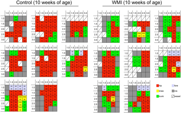
Hypoxia-ischemia (H-I) in rats at postnatal day 3 causes disorganization of oligodendrocyte development in layers II/III of the sensorimotor cortex without apparent neuronal loss, and shows mild hindlimb dysfunction with imbalanced motor coordination. However, the mechanisms by which mild motor dysfunction is induced without loss of cortical neurons are currently unclear. To reveal the mechanisms underlying mild motor dysfunction in neonatal H-I model, electrical responsiveness and dendrite morphology in the sensorimotor cortex were investigated at 10 weeks of age. Responses to intracortical microstimulation (ICMS) revealed that the cortical motor map was significantly changed in this model. The cortical area related to hip joint movement was reduced, and the area related to trunk movement was increased. Sholl analysis in Golgi staining revealed that layer I-III neurons on the H-I side had more dendrite branches compared with the contralateral side. To investigate whether changes in the motor map and morphology appeared at earlier stages, ICMS and Sholl analysis were also performed at 5 weeks of age. The minimal ICMS current to evoke twitches of the hip area was higher on the H-I side, while the motor map was unchanged. Golgi staining revealed more dendrite branches in layer I-III neurons on the H-I side. These results revealed that alterations of both dendrite morphology and ICMS threshold of the hip area occurred before the rearrangement of the motor map in the neonatal H-I model. They also suggest that altered dendritic morphology and altered ICMS responsiveness may be related to mild motor dysfunction in this model.
Keywords: cortical layer I-III; dendritic branches; golgi staining; hip area; hypoxia-ischemia in premature infants; intracortical microstimulation (ICMS); white matter injury.
Figures


Similar articles
-
Disorganization of Oligodendrocyte Development in the Layer II/III of the Sensorimotor Cortex Causes Motor Coordination Dysfunction in a Model of White Matter Injury in Neonatal Rats.Neurochem Res. 2018 Jan;43(1):136-146. doi: 10.1007/s11064-017-2352-3. Epub 2017 Jul 31. Neurochem Res. 2018. PMID: 28762105
-
Dysfunction in Motor Coordination in Neonatal White Matter Injury Model Without Apparent Neuron Loss.Cell Transplant. 2016;25(7):1381-93. doi: 10.3727/096368915X689893. Epub 2015 Nov 11. Cell Transplant. 2016. PMID: 26564423
-
Information processing within the motor cortex. I. Responses of morphologically identified motor cortical cells to stimulation of the somatosensory cortex.J Comp Neurol. 1994 Jul 8;345(2):161-71. doi: 10.1002/cne.903450202. J Comp Neurol. 1994. PMID: 7929897
-
Intracortical Microstimulation (ICMS) Activates Motor Cortex Layer 5 Pyramidal Neurons Mainly Transsynaptically.Brain Stimul. 2015 Jul-Aug;8(4):742-50. doi: 10.1016/j.brs.2015.03.003. Epub 2015 Mar 27. Brain Stimul. 2015. PMID: 25892002
-
Chapter 5--face sensorimotor cortex: its role and neuroplasticity in the control of orofacial movements.Prog Brain Res. 2011;188:71-82. doi: 10.1016/B978-0-444-53825-3.00010-3. Prog Brain Res. 2011. PMID: 21333803 Review.
References
LinkOut - more resources
Full Text Sources
Other Literature Sources