

Anatomic structure of the internal iliac artery and its educative dissection for peripartum and pelvic hemorrhage
- PMID: 29971190
- PMCID: PMC6022419
- DOI: 10.4274/tjod.23245
Anatomic structure of the internal iliac artery and its educative dissection for peripartum and pelvic hemorrhage
Abstract
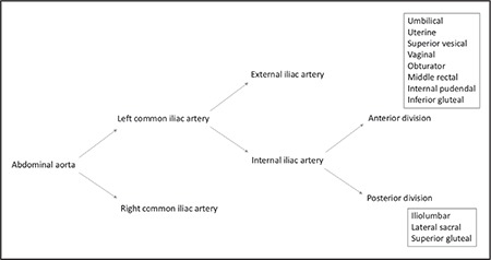
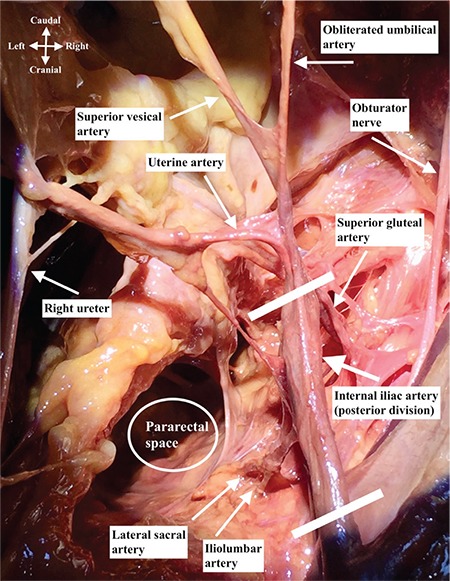
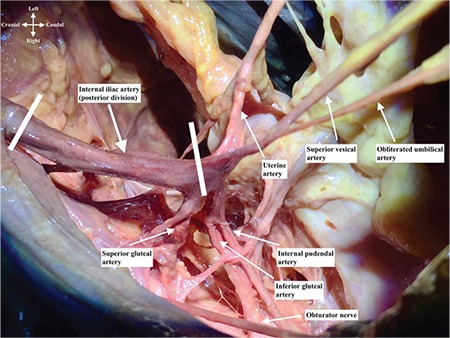
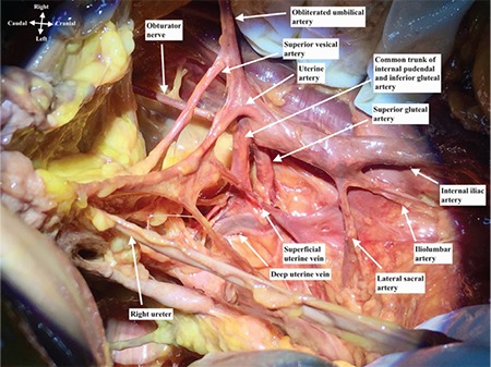
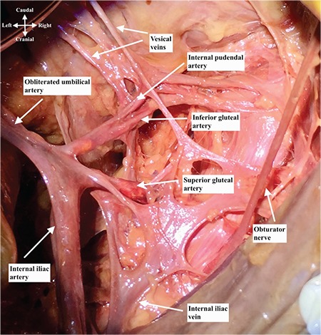
The abdominal aorta is divided into two parts (right and left) at the level of the fourth-fifth lumbar vertebra and called the common iliac artery. Anterior to the sacroiliac joint, common iliac arteries are divided into external and internal iliac arteries. The external iliac artery supplies the lower limb, and the internal iliac artery is the major vascular supply of the pelvis. Internal iliac artery is divided into anterior and posterior trunk. The anterior trunk supplies the pelvis, visceral organs, and the posterior trunk supplies pelvic parietal structures. The broad ligament envelopes the uterus anteriorly and posteriorly with its sheets and continues as the pelvic peritoneum at the lateral side of the pelvic wall. After cutting the pelvic peritoneum, the retroperitoneal area is visualized and the internal iliac artery with other great vessels of the abdomen can be noted.
Keywords: Internal iliac artery; dissection; hemorrhage; obstetrics; postpartum.
Conflict of interest statement
Conflict of Interest: No conflict of interest was declared by the authors.
Figures
Similar articles
-
Anatomical Pitfalls of Excision of Deep Endometriosis Nodules of the Sciatic Nerve: A three-dimensional Reconstruction and Surgical Educational Video.J Minim Invasive Gynecol. 2023 Apr;30(4):264-265. doi: 10.1016/j.jmig.2023.01.018. Epub 2023 Feb 4. J Minim Invasive Gynecol. 2023. PMID: 36740017
-
Laparoscopic Variants of Temporary Uterine Artery Ligation.J Minim Invasive Gynecol. 2020 May-Jun;27(4):811-812. doi: 10.1016/j.jmig.2019.08.026. Epub 2019 Sep 5. J Minim Invasive Gynecol. 2020. PMID: 31493570
-
Pelvic Ring Injuries.2024 Mar 2. In: StatPearls [Internet]. Treasure Island (FL): StatPearls Publishing; 2025 Jan–. 2024 Mar 2. In: StatPearls [Internet]. Treasure Island (FL): StatPearls Publishing; 2025 Jan–. PMID: 31335050 Free Books & Documents.
-
Anatomic variations of the Uterine Artery. Review of the literature and their clinical significance.Turk J Obstet Gynecol. 2020 Mar;17(1):58-62. doi: 10.4274/tjod.galenos.2020.33427. Epub 2020 Apr 6. Turk J Obstet Gynecol. 2020. PMID: 32341832 Free PMC article. Review.
-
Anatomy of the Internal Iliac Vein: Implications for Uterine Transplant.J Minim Invasive Gynecol. 2018 Feb;25(2):329. doi: 10.1016/j.jmig.2017.06.022. Epub 2017 Jun 29. J Minim Invasive Gynecol. 2018. PMID: 28669893 Review.
Cited by
-
Basic clinical retroperitoneal anatomy for pelvic surgeons.Turk J Obstet Gynecol. 2018 Dec;15(4):259-269. doi: 10.4274/tjod.88614. Epub 2019 Jan 9. Turk J Obstet Gynecol. 2018. PMID: 30693143 Free PMC article. Review.
-
Pelvic lymphadenectomy: Step-by-step surgical education video.J Turk Ger Gynecol Assoc. 2020 Mar 6;21(1):66-69. doi: 10.4274/jtgga.galenos.2019.2018.0167. Epub 2019 Mar 25. J Turk Ger Gynecol Assoc. 2020. PMID: 30905139 Free PMC article.
-
Anatomy of Internal Iliac Artery and Its Ligation to Control Pelvic Hemorrhage.JNMA J Nepal Med Assoc. 2020 Oct 15;58(230):826-830. doi: 10.31729/jnma.4958. JNMA J Nepal Med Assoc. 2020. PMID: 34504379 Free PMC article.
-
[Basic Arterial Anatomy and Interpretation of CT Angiography for Intra-Abdominal or Gastrointestinal Bleeding: Correlation with Conventional Angiographic Findings for Beginners].Taehan Yongsang Uihakhoe Chi. 2020 Jan;81(1):119-134. doi: 10.3348/jksr.2020.81.1.119. Epub 2020 Jan 31. Taehan Yongsang Uihakhoe Chi. 2020. PMID: 36238116 Free PMC article. Review. Korean.
-
Internal Iliac Artery Ligation in Obstetrics and Gynecology: Surgical Anatomy and Surgical Considerations.Clin Pract. 2023 Dec 27;14(1):32-51. doi: 10.3390/clinpract14010005. Clin Pract. 2023. PMID: 38248429 Free PMC article. Review.
References
-
- Standring S. Gray’s Anatomy. The Anatomical Basis of Clinical Practice, 40st Edition, Elsevier. 2016.
-
- Baggish M, Karram M. Atlas of Pelvic Anatomy and Gynecologic Surgery, 4th Edition. Elsevier. 2016.
-
- Agur AMR, Dalley AF. Grant’s Atlas of Anatomy. Wolters Kluwer Health. 2016.
-
- Hansen JT. Netter’s Clinical Anatomy. 3rd Edition. Saunders/Elsevier. 2014.
LinkOut - more resources
Full Text Sources
Other Literature Sources