. 2018 Aug;23(6):903-916.
doi: 10.1007/s00775-018-1589-x. Epub 2018 Jul 3.

Targeting the mitochondrial VDAC in hepatocellular carcinoma using a polyclonal antibody-conjugated to a nitrosyl ruthenium complex

Affiliations

Targeting the mitochondrial VDAC in hepatocellular carcinoma using a polyclonal antibody-conjugated to a nitrosyl ruthenium complex

Loyanne C B Ramos et al. J Biol Inorg Chem. 2018 Aug.

Abstract

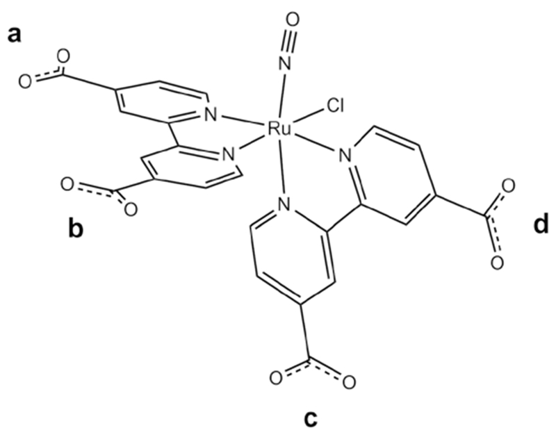
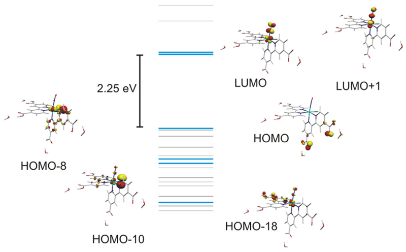
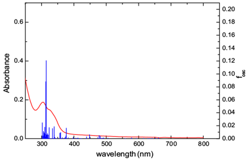
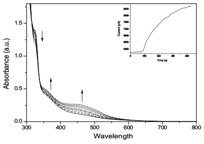
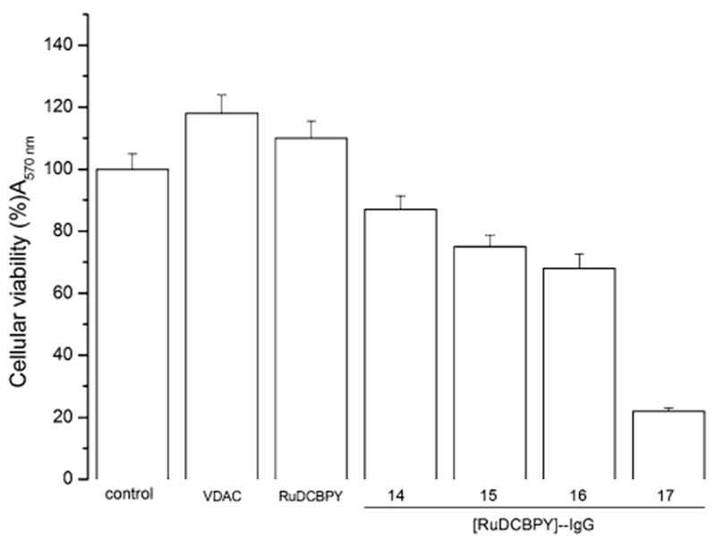
The rational design of anti-cancer agents includes a new approach based on ruthenium complexes that can act as nitric oxide (NO) donor agents against specific cellular targets. One of the most studied classes of those compounds is based on bis(bipyridine) ruthenium fragment and its derivative species. In this work, we present the chemical and cytotoxicity properties against the liver hepatocellular carcinoma cell line HepG2 of cis-[RuII(NO+)Cl(dcbpy)2]2- conjugated to a polyclonal antibody IgG (anti-VDAC) recognizing a cell surface marker. UV-visible bands of the ruthenium complex were assigned with the aid of density functional theory, which also allowed estimation of the structures that explain the biological effects of the ruthenium complex-IgG conjugate. The interaction of cis-[RuII(NO+)Cl(dcbpy)2]3- with mitochondria was evaluated due to the potential of these organelles as anti-cancer targets, and considering they interact with the anti-VDAC antibody. The cytotoxicity of cis-[RuII(NO+)Cl(dcbpy)2]3--anti-VDAC antibody was up to 80% greater in comparison to the free cis-[RuII(NO+)Cl(dcbpy)2]3- complex. We suggest that this effect is due to site-specific interaction of the complex followed by NO release.

Keywords: Conjugated ruthenium-antibody complex; Nitric oxide delivery agent; Nitrosyl ruthenium complexes.


Figures

Fig. 1
Fig. 1
Schematic representation of the molecular structure of cis-[Ru(NO)Cl(dcbpy)2]2−
Fig. 2
Fig. 2
Molecular orbital diagram for species number 6
Fig. 3
Fig. 3
Experimental and (TD)DFT-computed spectra for [Ru(NO) Cl(dcbpy-H)2]. The computed spectrum corresponds to species number 7
Fig. 4
Fig. 4
Spectroelectrochemistry measurements of (Ru-DCBPY) (7.4 × 10⁻⁵ M) in aqueous 0.01 M KCl solution, pH = 3.0. Inset: Chronoamperometry with NO sensor of aqueous (Ru-DCBPY) (7.4 × 10⁻⁵ M) containing ascorbic acid (0.015 M)
Fig. 5
Fig. 5
Release of NO from (Ru-DCBPY), evaluated by mitochondria-dependent oxy-reduction systems, detected directly through selective electrode. (Ru-DCBPY) (100 μM) was incubated with mitochondria in standard medium. The plot, representative of three independent experiments, shows NO release. Gray area shows time course after mitochondria addition
Fig. 6
Fig. 6
Time course of mitochondrial ROS generation in the presence of NO released from complex (Ru-DCBPY), as assessed with H2DCFDA. Mitochondria (0.5 mg protein mL−1) were incubated in the standard medium described in Fig. 1, at 30 °C, in the presence of 5 μM succinate + 2.5 μM rotenone. Plots are representative of at least three experiments with different mitochondrial preparations
Fig. 7
Fig. 7
Time course of mitochondrial swelling induced by NO released from complex (Ru-DCBPY). Mitochondria (0.5 mg protein mL−1) were incubated in the standard medium described in Fig. 1, at 30 °C, in the presence of 5 μM succinate + 2.5 μmol L−1 rotenone. Data are representative of at least three experiments with different mitochondrial preparations
Fig. 8
Fig. 8
Time course of membrane potential induced by NO released from complex (Ru-DCBPY) in succinate-energized mitochondria. Mitochondria (1 mg of protein mL−1) were incubated with 10–100 μM of (Ru-DCBPY) in the standard medium, in the presence of 5 μM succinate (plus 2.5 μmol L−1 rotenone), at 30 °C. Plots are representative of at least three experiments with different mitochondrial preparations
Fig. 9
Fig. 9
MTT cell viability assay. Effects on HepG2 cell viability of follow treatments: control, DMEM cell culture medium; VDAC (IgG anti-VDAC antibody solubilized in DMEM medium); complex (I), ruthenium nitrosyl complex Ru-DCBPY not conjugated with IgG anti-VDAC antibody, e.g., Ru-DCBPY; Fractions 14, 15, 16, and 17 were from the conjugate purification on a Sephadex column, as described in “Materials and methods”, (Ru-DCBPY)–IgG. The experiments were performed in triplicate, independently, and expressed as the mean and standard deviation
Scheme 1
Scheme 1
Effect of pH on the chemical composition of cis-[Ru(NO)Cl(dcbpy-H2)2]2+ complex
Scheme 2
Scheme 2
Control reduction potential electrolysis of (Ru-DCBPY) specie at 0.1 V vs. Ag/AgCl in pH 3.0
Scheme 3
Scheme 3
Chemical synthesis of the NHS-ester of (Ru-DCBPY) ligation, in basic medium, with IgG anti-VDAC antibody (protein)

Similar articles

Cited by

References

    1. Miller MR, Megson IL (2007) Recent developments in nitric oxide donor drugs. Br J Pharmacol 151(3):305–321 - PMC - PubMed
    1. Bonavida B (ed) (2010) Nitric oxide (NO) and cancer: prognosis, prevention, and therapy. Springer Science and Business Media, New York
    1. Serafim RA, Primi MC, Trossini GH, Ferreira EI (2012) Nitric oxide: state of the art in drug design. Curr Med Chem 19(3):386–405 - PubMed
    1. Tfouni E, Truzzi DR, Tavares A, Gomes AJ, Figueiredo LE, Franco DW (2012) Biological activity of ruthenium nitrosyl complexes. Nitric Oxide 26(1):38–53 - PubMed
    1. de Lima RG, Silva BR, da Silva RS, Bendhack LM (2014) Ruthenium complexes as NO donors for vascular relaxation induction. Molecules 19(7):9628–9654 - PMC - PubMed

Publication types

MeSH terms