. 2018 Jul 3;29(7):106.
doi: 10.1007/s10856-018-6106-9.

Biomechanical and morphological stability of acellular scaffolds for tissue-engineered heart valves depends on different storage conditions

Affiliations

Biomechanical and morphological stability of acellular scaffolds for tissue-engineered heart valves depends on different storage conditions

Piotr Wilczek et al. J Mater Sci Mater Med. .

Abstract

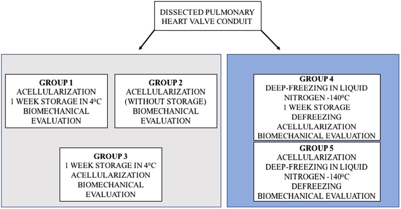
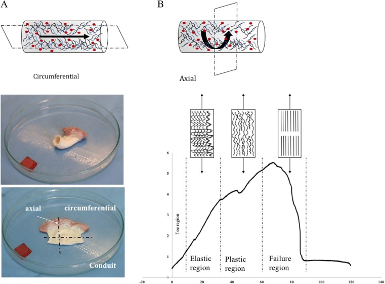
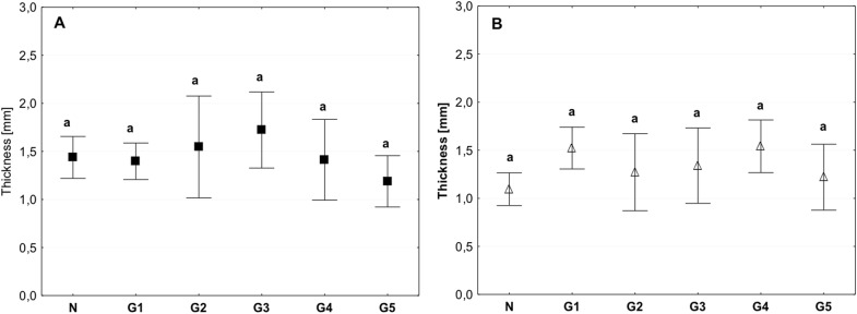
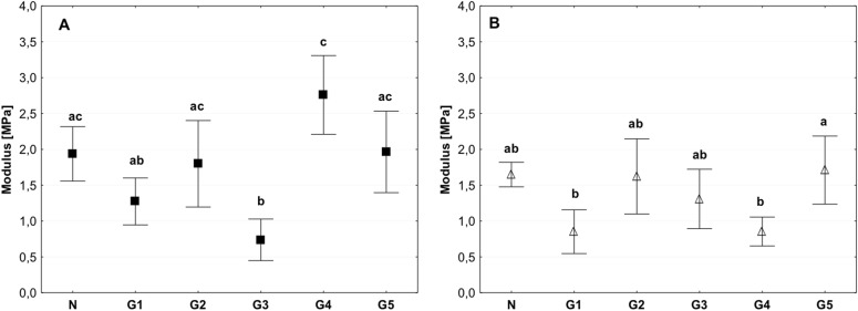
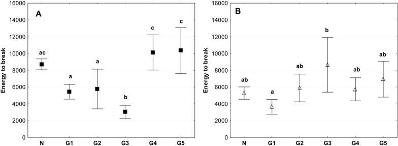
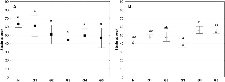
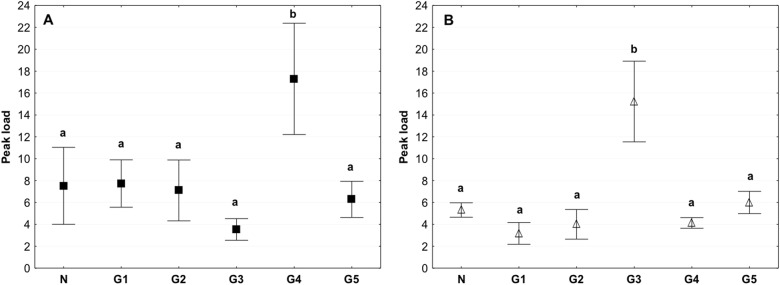
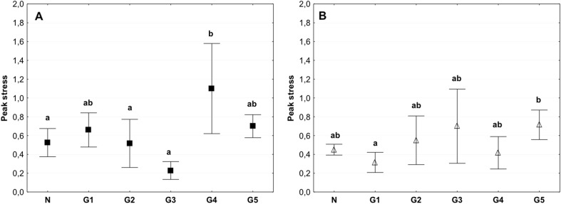
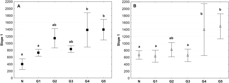
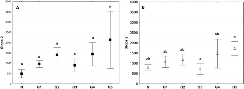
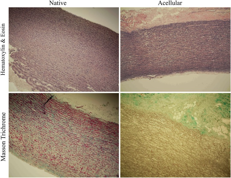
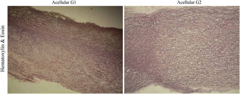
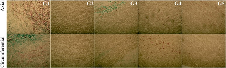
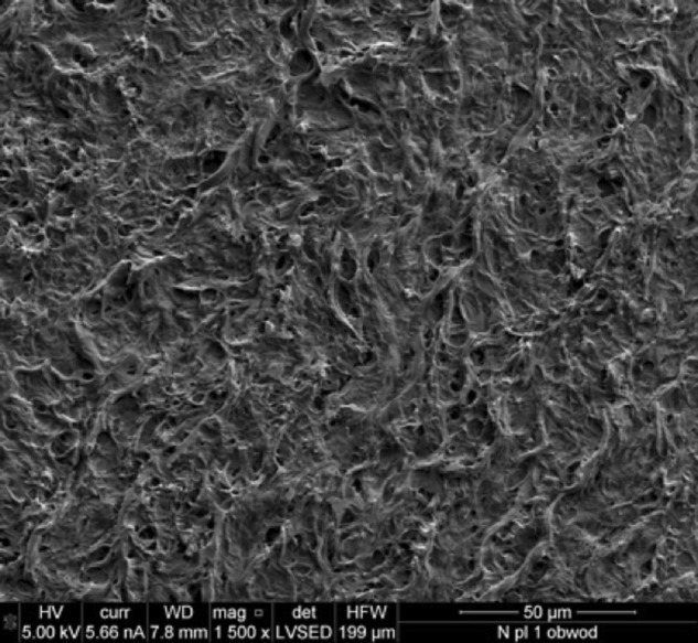
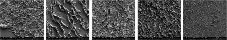
Currently available bioprosthetic heart valves have been successfully used clinically; however, they have several limitations. Alternatively, tissue-engineering techniques can be used. However, there are limited data concerning the impact of storage conditions of scaffolds on their biomechanics and morphology. The aim of this study was to determine the effect of different storage conditions on the biomechanics and morphology of pulmonary valve dedicated for the acellular scaffold preparation to achieve optimal conditions to obtain stable heart valve prostheses. Scaffold can then be used for the construction of tissue-engineered heart valve, for this reason evaluation of these parameters can determine the success of the clinical application this type of bioprosthesis. Pulmonary heart valves were collected from adult porcines. Materials were divided into five groups depending on the storage conditions. Biomechanical tests were performed, both the static tensile test, and examination of viscoelastic properties. Extracellular matrix morphology was evaluated using transmission electron microscopy and immunohistochemistry. Tissue stored at 4 °C exhibited a higher modulus of elasticity than the control (native) and fresh acellular, which indicated the stiffening of the tissue and changes of the viscoelastic properties. Such changes were not observed in the radial direction. Percent strain was not significantly different in the study groups. The storage conditions affected the acellularization efficiency and tissue morphology. To the best of our knowledge, this study is the first that attributes the mechanical properties of pulmonary valve tissue to the biomechanical changes in the collagen network due to different storage conditions. Storage conditions of scaffolds for tissue-engineered heart valves may have a significant impact on the haemodynamic and clinical effects of the used bioprostheses.


Conflict of interest statement

The authors certify that they have NO affiliations with or involvement in any organization or entity withany financial interest (such as honoraria; educational grants; participation in speakers’ bureaus;membership, employment, consultancies, stock ownership, or other equity interest; and expert testimony orpatent-licensing arrangements), or non-financial interest (such as personal or professional relationships,affiliations, knowledge or beliefs) in the subject matter or materials discussed in this manuscript.

Figures

Fig. 1
Fig. 1
Division of the evaluated study groups depending on the temperature and time of tissue storage
Fig. 2
Fig. 2
a, b Orientation of the samples for biomechanics. Valve fragments were dissected in two orientations, circumferential (sens of blood flow) and axial (perpendicular to blood flow). b Typical stress–strain curve for destructive tensile tensing of collagen tissue. Collagen fibril straightening and failure related to different regions of the stress–strain curve
Fig. 3
Fig. 3
Mean value of tissue thickness in the circumferential (a) and axial directions (b) (average ± SD) in the study groups. The same letters (a) indicate homogeneous groups within individual valves (Tukey’s test, P < 0.05; N = 8–10)
Fig. 4
Fig. 4
Mean value of the modulus of elasticity in the circumferential (a) and axial directions (b) (average ± SD) in the study groups. The same letters (a, b, c) indicate homogeneous groups within individual valves (Tukey’s test, P < 0.05; N = 8–10)
Fig. 5
Fig. 5
Mean value of energy to break in circumferential (a) and axial direction (b) (average ± SD) in study groups. The same letters (a, b, c) indicate homogeneous groups within individual valves (Tukey’s test, P < 0.05; N = 8–10)
Fig. 6
Fig. 6
Mean value of the peak strain in the circumferential (a) and axial directions (b) (average ± SD) in the study groups. The same letters (a, b) indicate homogeneous groups within individual valves (Tukey’s test, P < 0.05; N = 8–10)
Fig. 7
Fig. 7
Mean value of the peak load in the circumferential (a) and axial directions (b) (average ± SD) in the study groups. The same letters (a, b) indicate homogeneous groups within individual valves (Tukey’s test, P < 0.05; N = 8–10 per group)
Fig. 8
Fig. 8
Mean value of the peak stress in the circumferential (a) and axial directions (b) (average ± SD) in the study groups. The same letters (a, b) indicate homogeneous groups within individual valves (Tukey’s test, P < 0.05; N = 8–10)
Fig. 9
Fig. 9
Mean value of Slope 1 in the circumferential (a) and axial directions (b) (average ± SD) in the study groups. The same letters (a, b) indicate homogeneous groups within individual valves (Tukey’s test, P < 0.05; N = 8–10)
Fig. 10
Fig. 10
Mean value of Slope 2 in the circumferential (a) and axial (b) directions (average ± SD) in the study groups. The same letters (a, b) indicate homogeneous groups within individual valves (Tukey’s test, P < 0.05; N = 8–10)
Fig. 11
Fig. 11
Representative image of a control tissue sample and after the acellularization procedure—Haematoxylin and Eosin staining. Cross-section of the control and acellular tissue samples—Trichrome Masson staining. The red colour represents muscle fibres, and the green colour represents collagen. After acellularization, the muscle cells were removed from the tissue, and the reduction of green colouration (collagen) was observed in parallel (colour figure online)
Fig. 12
Fig. 12
Cross-section of the acellular tissue sample derived from the G1 group (a) and G2 group (b). The haematoxylin and eosin staining indicated that the tissue in Group 2 has a more relaxed structure, with visible interfibrillar spaces
Fig. 13
Fig. 13
Cross-section of the acellular tissue sample prepared in the axial and circumferential directions, derived from the investigated group. EVG staining in particular investigated groups
Fig. 14
Fig. 14
Cross-section of the acellular tissue sample prepared in the axial and circumferential directions, derived from the investigated group. Fibronectin staining in particular investigated groups
Fig. 15
Fig. 15
Cross-section of the acellular tissue sample prepared in the axial and circumferential directions, derived from the investigated group. Collagen IV staining in particular investigated groups
Fig. 16
Fig. 16
Cross-section of the acellular tissue sample prepared in the axial and circumferential directions, derived from the investigated group. Collagen I staining in particular investigated groups
Fig. 17
Fig. 17
Cross-section of the acellular tissue sample prepared in the axial and circumferential directions, derived from the investigated group. Trichrome Masson staining in particular investigated groups
Fig. 18
Fig. 18
Electron microscopy section of a control pulmonary valve conduit
Fig. 19
Fig. 19
Electron microscopy section of the pulmonary valve conduit after acellularization in the study group

Similar articles

Cited by

References

    1. Carapetis JR. Cumulative incidence of rheumatic fever in an endemic region: a guide to susceptibility of the population. Epidemiol Infect. 2000;124:239–44. doi: 10.1017/S0950268800003514. - DOI - PMC - PubMed
    1. World Population Prospects: The 2017 Revision, Key Findings and Advance Tables. ESA/P/WP/248. United Nations, Department of Economic and Social Affairs, Population Division. 2017.
    1. Tofield A. European cardiovascular disease statistics 4th edition 2012: EuroHeart II. Eur Heart J. 2013;34:3007–14. doi: 10.1093/eurheartj/eht379. - DOI
    1. d’Arcy JL, Prendergast BD, Chambers JB, Ray SG, Bridgewater B. Valvular heart disease: the next cardiac epidemic. Heart. 2011;97:91–3. doi: 10.1136/hrt.2010.205096. - DOI - PubMed
    1. Marijon E, Celermajer DS, Tafflet M, El-Haou S, Jani DN, Ferreira B, et al. Rheumatic heart disease screening by echocardiography the inadequacy of world health organization criteria for optimizing the diagnosis of subclinical disease. Circulation. 2009;120:663–8. doi: 10.1161/CIRCULATIONAHA.109.849190. - DOI - PubMed

LinkOut - more resources