. 2018 Sep;144(9):1717-1730.
doi: 10.1007/s00432-018-2699-0. Epub 2018 Jul 3.

Cell growth potential drives ferroptosis susceptibility in rhabdomyosarcoma and myoblast cell lines

Affiliations

Cell growth potential drives ferroptosis susceptibility in rhabdomyosarcoma and myoblast cell lines

Silvia Codenotti et al. J Cancer Res Clin Oncol. 2018 Sep.

Abstract

Purpose: Ferroptosis is a programmed form of iron-dependent cell death caused by lipid hydroperoxide accumulation, which can be prevented by glutathione peroxidase 4 (GPx4) activity. Here we investigated the effects of ferroptosis inducers called erastin and RSL3, which act by glutathione depletion and GPx4 inactivation, respectively, on muscle-derived cell lines of embryonal and alveolar rhabdomyosarcoma (RMS), and mouse normal skeletal C2C12 myoblasts.

Methods: Myogenic lines were exposed to stepwise increasing concentrations of ferroptosis inducers either alone or in combination with iron supplementation, iron chelating agents (bathophenanthrolinedisulfonic acid, BPS), antioxidant molecules (glutathione, N-acetylcysteine), lipid peroxidation inhibitors (ferrostatin-1), and chemotherapeutic agents (doxorubicin and actinomycin D). Drug susceptibility was quantified by measuring cell viability, proliferation and differentiation via neutral red assay, crystal violet assay and Giemsa staining, respectively. The detection of lipid hydroperoxide and protein levels was performed by immunofluorescence and Western blot analysis, respectively.

Results: Erastin and RSL3 increased lipid hydroperoxide levels preferentially in the embryonal U57810 and myoblast C2C12 lines, leading to ferroptosis that was accentuated by iron supplementation or prevented by co-treatment with BPS, glutathione, N-acetylcysteine and ferrostatin-1. The inhibition of extracellular regulated kinases (ERK) pathway prevented ferroptosis in U57810 and C2C12 cells, whereas its increased activation in the embryonal RD cells mediated by caveolin-1 (Cav-1) overexpression led to augmented ferroptosis susceptibility. Finally, we observed the combination of erastin or RSL3 with chemotherapeutic doxorubicin and actinomycin D agents to be effective in increasing cell death in all RMS lines.

Conclusions: Erastin and RSL3 trigger ferroptosis in highly proliferating myogenic lines through a ERK pathway-dependent fashion.

Keywords: Erastin; Ferroptosis; GPx4; Iron; RSL3; Rhabdomyosarcoma.


Conflict of interest statement

The authors declare that they have no conflict of interest.

Figures

Fig. 1
Fig. 1
Ferroptosis inducers affect the cell viability of myogenic lines. a, b Neutral red assay performed on the different lines after exposure to increasing doses of erastin, RSL3 or DMSO for 24 and 48 h. Results are representative of four independent experiments. *p values < 0.05; **p values < 0.001; ***p values < 0.0001; one-way Anova test. c, d Light microscopy pictures representative of U57810 and RH30 lines treated with 0.5 or 5 µM erastin and RSL3 or DMSO up to 24 or 48 h
Fig. 2
Fig. 2
Dose response curves were generated to calculate the IC50 doses of ferroptosis inducers. The different lines were treated with erastin and RSL3 doses ranging from 0.1 to 5 µM, or DMSO for 24 and 48 h. The black dotted line corresponds to the concentration required for 50% inhibition in vitro
Fig. 3
Fig. 3
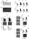
Erastin and RSL3 trigger ferroptosis by lipid ROS formation in U57810 and C2C12 lines. a Cells treated with IC50 doses of erastin and RSL3 or DMSO were probed with the lipid peroxidation sensor BODIPY 581/591 C11. At the indicated time points the staining was visualized by fluorescence microscopy and the intensity signal was quantified using the Image Pro Plus software. Results are representative of two independent experiments. *p values < 0.05; **p values < 0.001; ***p values < 0.0001; one-way Anova test. b Neutral red assay performed on cells treated with IC50 doses of erastin and RSL3 either alone or in presence of 100 μM FAC, 100 μM BPS, 10 μM ferrostatin-1 (Fer-1), 5 mM glutathione (GSH) and 5 mM N-acetylcysteine (NAC) up to 24 h. Results are representative of three independent experiments. *p values < 0.05; **p values < 0.001; ***p values < 0.0001 vs vehicle-treated cells. #p values < 0.05; ##p values < 0.001; ###p values < 0.0001 vs cells treated with erastin, RSL3 or FAC alone; unpaired Student’s t test
Fig. 4
Fig. 4
Ferroptosis susceptibility is blunted by ERK pathway inhibition in U57810 and C2C12 lines. a The growth rate of the different myogenic lines was calculated over a 72 h time course (top graph). The expression of phosphorylated and total ERK1/2 proteins was assessed by immunoblotting analysis. Quantification of protein bands after normalization with respect to beta-tubulin is shown at the top of each lane. Results are representative of three independent experiments. b Crystal violet assay performed on cell lines incubated with 10 μM PD098059, 10 μM U0126 or DMSO for 48 h (top graph). The amount of phosphorylated and total ERK1/2 proteins was detected by immunoblotting. Quantification of protein bands was obtained after normalization with respect to beta-tubulin. Results are representative of three independent experiments. c Neutral red assay performed on cell lines pre-treated with 10 μM PD098059, 10 μM U0126 or DMSO for 48 h and then administered with IC50 doses of erastin or RSL3 for 24 h. Results are representative of four independent experiments. d Neutral red assay performed on cell lines differentiated for 72 h before treatment with IC50 doses of erastin and RSL3 or DMSO up to 48 h. Cells morphology was evidenced by Giemsa staining. The expression of myogenin and MHC was assessed by immunoblotting analysis. Beta-tubulin was used as a loading control. Results are representative of three independent experiments. **p values < 0.001; ***p values < 0.0001; unpaired Student’s t test
Fig. 5
Fig. 5
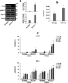
Ferroptosis susceptibility is augmented in RD cells by increased ERK pathway activation due to Cav-1 overexpression. a The expression of Cav-1 and phosphorylated vs total ERK1/2 proteins was assessed by immunoblotting analysis. Quantification of protein bands was obtained after normalization with respect to beta-tubulin. Results are representative of three independent experiments. b The growth rate of RD/empty and RD/Cav-1 cells was calculated over a 72 h time course. Results are representative of four independent experiments. c Neutral red assay performed on RD/empty and RD/Cav-1 cell lines after exposure to increasing doses of erastin and RSL3 or DMSO for 24 and 48 h. Results are representative of two independent experiments. *p values < 0.05; **p values < 0.001; ***p values < 0.0001; unpaired Student’s t test
Fig. 6
Fig. 6
Combination of erastin or RSL3 with chemotherapeutic agents increase RMS cell death. a Neutral red assay performed on RMS lines treated with increasing doses of doxorubicin (doxo) or actinomycin D (actD) for 24 h. Results are representative of two independent experiments. Neutral red assay performed on RMS lines co-treated with IC50 doses of erastin and RSL3 plus 0.5 μM doxo (b) or 0.5 nM actD (c) up to 24 h. Results are representative of two independent experiments. *p values < 0.05; **p values < 0.001; ***p values < 0.0001 vs vehicle-treated cells. #p values < 0.05; ##p values < 0.001 vs cells treated with doxo, actD, erastin or RSL3 alone; unpaired Student’s t test

Similar articles

Cited by

References

    1. Adelman R, Saul RL, Ames BN (1988) Oxidative damage to DNA: relation to species metabolic rate and life span. Proc Natl Acad Sci USA 85:2706–2708 - PMC - PubMed
    1. Borinstein SC, Steppan D, Hayashi M, Loeb DM, Isakoff MS, Binitie O, Brohl AS, Bridge JA, Stavas M, Shinohara ET et al (2018) Consensus and controversies regarding the treatment of rhabdomyosarcoma. Pediatr Blood Cancer 65:e26809 - PubMed
    1. Breuer W, Shvartsman M, Cabantchik ZI (2008) Intracellular labile iron. Int J Biochem Cell Biol 40:350–354 - PubMed
    1. Brigelius-Flohé R, Maiorino M (2013) Glutathione peroxidases. Biochim Biophys Acta 1830:3289–3303 - PubMed
    1. Chardin P, Yeramian P, Madaule P, Tavitian A (1985) N-ras gene activation in the RD human rhabdomyosarcoma cell line. Int J Cancer 35:647–652 - PubMed

MeSH terms

LinkOut - more resources