. 2018 Jul 4;10(1):13.
doi: 10.1186/s13089-018-0096-1.

The impact of heart, lung and diaphragmatic ultrasound on prediction of failed extubation from mechanical ventilation in critically ill patients: a prospective observational pilot study

Affiliations

The impact of heart, lung and diaphragmatic ultrasound on prediction of failed extubation from mechanical ventilation in critically ill patients: a prospective observational pilot study

Kavi Haji et al. Crit Ultrasound J. .

Abstract

Background: Failed extubation from mechanical ventilation in critically ill patients is multifactorial, complex and not well understood. We aimed to identify whether combined transthoracic echocardiography, lung and diaphragmatic ultrasound can predict extubation failure in critically ill patients.

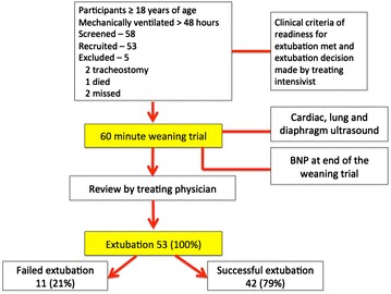
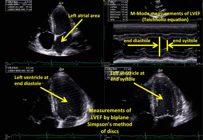
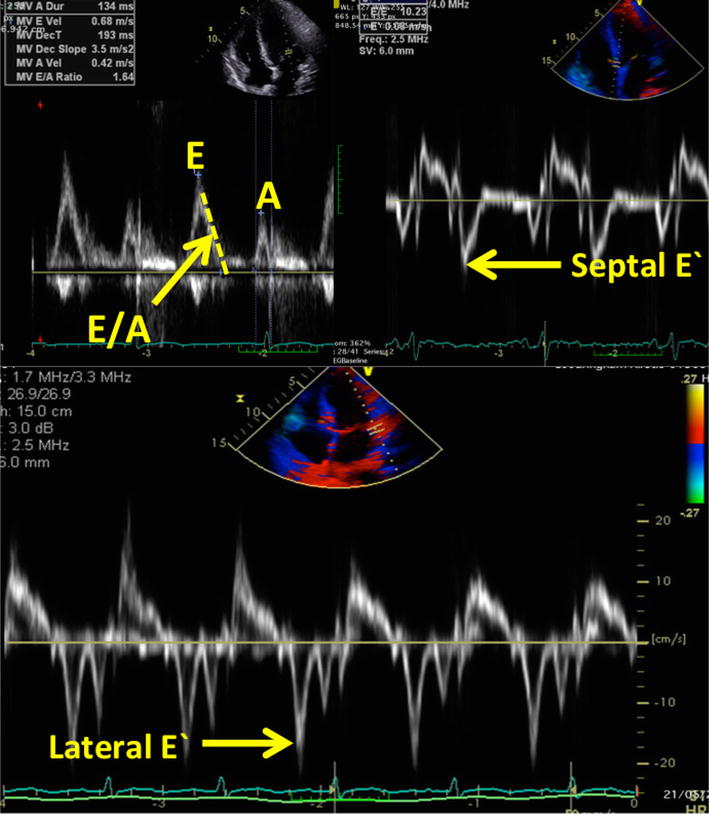
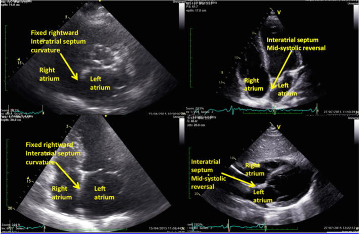
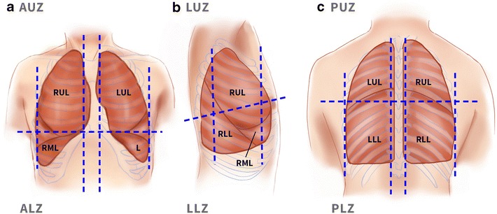
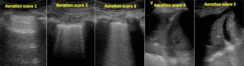
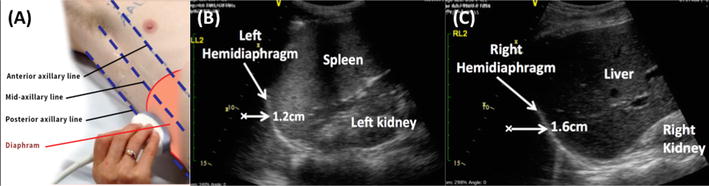
Results: Fifty-three participants who were intubated > 48 h and deemed by the treating intensivist ready for extubation underwent a 60-min pre-extubation weaning trial (pressure support ≤ 10 cmH2O and positive end expiratory pressure 5 cmH2O). Prior to extubation, data collected included ultrasound assessment of left ventricular ejection fraction, left atrial area, early diastolic trans-mitral flow velocity wave (E), early diastolic trans-mitral flow velocity wave/late diastolic trans-mitral flow velocity wave (E/A), early diastolic trans-mitral flow velocity wave/early diastolic mitral annulus velocity (E/E'), interatrial septal motion, lung loss of aeration score and diaphragm movement. At the end of the weaning trial, the rapid shallow breathing index and serum B-type natriuretic peptide concentration were measured. Success and failure of weaning was assessed by defined criteria. Decision to extubate was at the discretion of the treating intensivist. Failure of extubation was defined as re-intubation, non-invasive ventilation or death within 48 h after extubation. Of 53 extubated participants, 11 failed extubation. Failed extubation was associated with diabetes, ischaemic heart disease, higher E/E' (OR 1.27, 95% CI 1.05-1.54), left atrial area (OR 1.14, CI 1.02-1.28), fixed rightward curvature of the interatrial septum (OR 12.95, CI 2.73-61.41), and higher loss of aeration score of anterior and lateral regions of the lungs (OR 1.41, CI 1.01-1.82).

Conclusions: Failed extubation in mechanically ventilated patients is more prevalent if markers of left ventricular diastolic dysfunction and loss of lung aeration are present.

Keywords: Diaphragm; Echocardiography; Lung; RSBI; Ultrasound; Weaning.


Figures

Fig. 1
Fig. 1
The participants’ flow diagram
Fig. 2
Fig. 2
Echocardiographic assessment of left ventricular systolic function (Simpson’s method and Techoltz equation) as defined by Lang et al. [22]
Fig. 3
Fig. 3
Examples of Doppler assessments used for assessment of left ventricular diastolic function (trans-mitral inflow and tissue Doppler assessment of the septal and lateral mitral valve annulus). A, late diastolic trans-mitral flow velocity wave, E early diastolic trans-mitral flow velocity wave, E′, early diastolic mitral annulus velocity
Fig. 4
Fig. 4
Interatrial septal movements during the cardiac cycle. Fixed rightward interatrial septum curvature is bowing of the interatrial septal to right throughout the cardiac cycle, and mid-systolic reversal is reversal of the septum from right to left during mid-systole
Fig. 5
Fig. 5
Surface anatomy of the regions of the lung. Ultrasound surface anatomy: the parasternal, anterior axillary and posterior axillary lines divide the chest wall into anterior, lateral and posterior regions. a The anterior region, delineated superiorly by the clavicle, inferiorly by the liver or the spleen, medially by parasternal line and laterally by the anterior axillary line. The anterior region is further divided by an arbitrary line to anterior upper zone and anterior lower zone. b The lateral region, delineated superiorly by the axilla, inferiorly by the liver or the spleen, anteriorly by the anterior axillary line and posteriorly by the posterior axillary line. An arbitrary line further divides the lateral region to lateral upper zone and lateral lower zone. c The posterior region, delineated by the posterior axillary line laterally and paravertebral line medially. A horizontal arbitrary line at the tip of the scapula further divides the posterior region to upper and lower zone. ALZ anterior lower zone, AUZ anterior upper zone, L lingula, LLL left lower lobe, LLZ left lower zone, LUL left upper lobe, PLZ posterior lower zone, PUZ posterior upper zone, RLL right lower lobe, RML right middle lobe, RUL right upper lobe
Fig. 6
Fig. 6
Lung ultrasound aeration scoring. The pattern and the extent of aeration defect is quantified as follows: A normally aerated or fewer than 3 B-lines scored 1; multiple discrete B-lines scored 2; multiple and fused B-lines scored 3; unaerated consolidated lung scored 4; unaerated consolidation with pleural effusion scored 5. Hence, the maximum score for each half was 5 and the maximum score for the posterior zone was 5
Fig. 7
Fig. 7
Ultrasound technique and images of the diaphragm. A The location of probe placement on the chest wall for assessment of diaphragm movement. The probe is placed laterally and perpendicularly on the lateral chest wall on the lower intercostal spaces between mid- and posterior axillary line. B, C The ultrasound images of the left and the right hemidiaphragm and the measurement of the diaphragmatic excursion from end of end inspiration to end expiration

References

    1. Boles JM, Bion J, Connors A, Herridge M, Marsh B, Melot C, et al. Weaning from mechanical ventilation. Eur Respir J. 2007;29(5):1033–1056. doi: 10.1183/09031936.00010206. - DOI - PubMed
    1. Esteban A, Ferguson ND, Meade MO, Frutos-Vivar F, Apezteguia C, Brochard L, et al. Evolution of mechanical ventilation in response to clinical research. Am J Respir Crit Care Med. 2008;177(2):170–177. doi: 10.1164/rccm.200706-893OC. - DOI - PubMed
    1. Esteban A, Alia I, Tobin MJ, Gil A, Gordo F, Vallverdu I, et al. Effect of spontaneous breathing trial duration on outcome of attempts to discontinue mechanical ventilation. Spanish Lung Failure Collaborative Group. Am J Respir Crit Care Med. 1999;159(2):512–518. doi: 10.1164/ajrccm.159.2.9803106. - DOI - PubMed
    1. Hurford WE, Favorito F. Association of myocardial ischemia with failure to wean from mechanical ventilation. Crit Care Med. 1995;23(9):1475–1480. doi: 10.1097/00003246-199509000-00006. - DOI - PubMed
    1. Jubran A, Lawm G, Duffner LA, Collins EG, Lanuza DM, Hoffman LA, et al. Post-traumatic stress disorder after weaning from prolonged mechanical ventilation. Intensive Care Med. 2010;36(12):2030–2037. doi: 10.1007/s00134-010-1972-8. - DOI - PMC - PubMed

LinkOut - more resources