. 2018 Jun;13(12):1417-1432.
doi: 10.2217/nnm-2018-0011. Epub 2018 Jul 4.

1α,25-dihydroxyvitamin D3-eluting nanofibrous dressings induce endogenous antimicrobial peptide expression

Affiliations

1α,25-dihydroxyvitamin D3-eluting nanofibrous dressings induce endogenous antimicrobial peptide expression

Jiang Jiang et al. Nanomedicine (Lond). 2018 Jun.

Abstract

Aim: The aim of this study was to develop a nanofiber-based dressing capable of local sustained delivery of 1α,25-dihydroxyvitamin D3 (1,25(OH)2D3) and augmenting human CAMP induction.

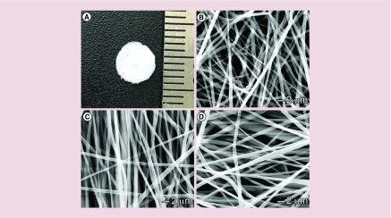
Materials & methods: Nanofibrous wound dressings containing 1,25(OH)2D3 were successfully prepared by electrospinning, which were examined in vitro, in vivo and ex vivo.

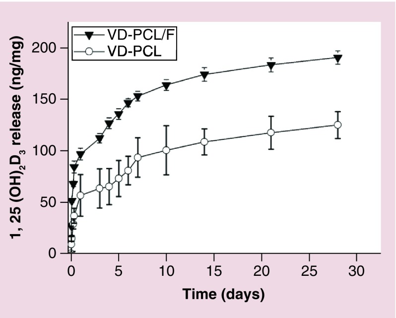
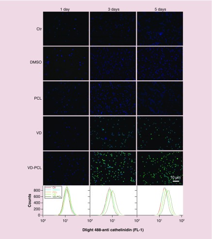
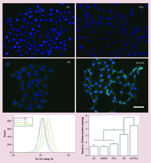
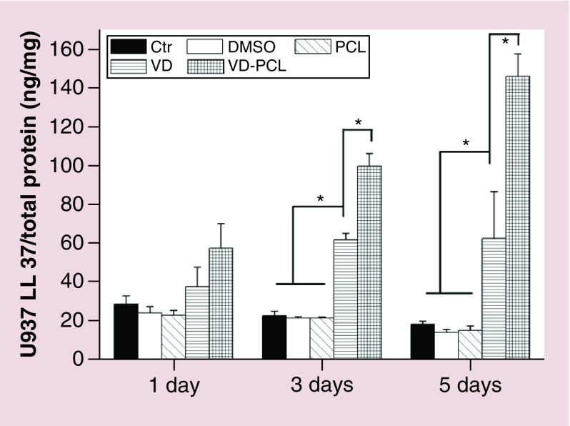
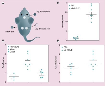
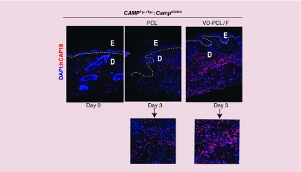
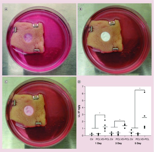
Results: 1,25(OH)2D3 was successfully loaded into nanofibers with encapsulation efficiency larger than 90%. 1,25(OH)2D3 showed a sustained release from nanofibers over 4 weeks. Treatment of U937 and HaCaT cells with 1,25(OH)2D3-loaded poly(ϵ-caprolactone) nanofibers significantly induced hCAP18/LL37 expression in monocytes and keratinocytes, skin wounds of humanized transgenic mice and artificial wounds of human skin explants.

Conclusion: 1,25(OH)2D3 containing nanofibrous dressings could enhance innate immunity by inducing antimicrobial peptide production.

Keywords: 1α,25-dihydroxyvitamin D3; antimicrobial peptides LL-37; electrospun nanofibers; endogenous production; innate immunity; sustained delivery.


Conflict of interest statement

Financial & competing interests disclosure

This work was supported by grants from the National Institute of General Medical Science (NIGMS) at the NIH (2P20 GM103480-06 and 1R01GM123081 to J Xie), the Otis Glebe Medical Research Foundation and startup funds from the University of Nebraska Medical Center. The content is solely the responsibility of the authors and does not necessarily represent the official views of the National Institutes of Health. The authors have no other relevant affiliations orfinancial involvement with any organization or entity with a financial interest in or financial conflict with the subject matter or materials discussed in the manuscript apart from those disclosed.

No writing assistance was utilized in the production of this manuscript.

Figures

<b>Figure 1.</b>
Figure 1.. Morphology characterization.
(A) Photograph shows a 1,25(OH)2D3-loaded PCL fiber membrane with a diameter of 5 mm. (B & C) SEM images of (B) PCL fibers, (C) 1,25(OH)2D3-loaded PCL fibers and (D) 1,25(OH)2D3-loaded PCL/pluronic F127 fibers. PCL: Poly(ϵ-caprolactone); SEM: Scanning electron microscope.
<b>Figure 2.</b>
Figure 2.. In vitro release kinetics of 1,25(OH)2D3.
Release profiles of VD from VD-PCL membranes and VD-PCL/pluronic F127 nanofiber (VD-PCL/F) membranes. Each data point represents the arithmetic mean ± standard deviation values from three samples. F: Plutonic F127; PCL: Poly(ϵ-caprolactone); VD-PCL: 1,25(OH)2D3-loaded PCL nanofiber.
<b>Figure 3.</b>
Figure 3.. 1,25(OH)2D3-loaded poly(ϵ-caprolactone) nanofibers induce hCAP18/LL37 protein expression.
Immunofluorescent staining with anti-hCAP18 antibody shows the expression of cathelicidin in U937 cells that were incubated in the absence (Ctr: without any treatment) and presence of pristine 1 mg/ml PCL fibers, 0.52% DMSO, 2.0 × 10-7 M VD or VD-PCL fibers for 1, 3 and 5 days. Expression levels were determined by flow cytometry (lower panels). Ctr: Control; DMSO: Dimethyl sulfoxide; PCL: Poly(ϵ-caprolactone); VD: 1,25(OH)2D3.
<b>Figure 4.</b>
Figure 4.. hCAP18/LL37 expression is induced in HaCaT cells after treatment with VD-PCL fibers.
Fluorescence microscopy images show hCAP18/LL37 expression in HaCaT cells that were incubated in the presence of 0.52% DMSO (Ctr: treatment with 0.52% DMSO), pristine 1 mg/ml PCL fibers, 2.0 × 10-7 M VD and 1 mg/ml VD-PCL fibers for 5 days. Ctr: Control; DMSO: Dimethyl sulfoxide; PCL: Poly(ϵ-caprolactone); VD: 1,25(OH)2D3.
<b>Figure 5.</b>
Figure 5.. Quantification of hCAP18/LL-37 expressed in U937 cells after treatment with different formulations.
Cells were incubated in the absence (Ctr: without any treatment) and presence of 1 mg/ml pristine PCL fibers, 0.52% DMSO, 2.0 × 10-7 M VD or 1 mg/ml VD-PCL fibers for 1, 3 and 5 days. The levels of hCAP18/LL-37 were measured by ELISA. Each data point represents the arithmetic mean ± standard deviation values from three samples. Statistical significance was evaluated by one-way analysis of variance (*p < 0.05). Ctr: Control; DMSO: Dimethyl sulfoxide; PCL: Poly(ϵ-caprolactone); VD: 1,25(OH)2D3.
<b>Figure 6.</b>
Figure 6.. 1,25(OH)2D3-loaded poly(ϵ-caprolactone)/pluronic F127 nanofiber (VD-PCL/F) dressings induce CAMP gene expression in full-thickness excisional wounds in human CAMPTg+/Tg+; murine CampKO/KO mice 3 days postwounding.
(A) Schematic showing location of nanofiber dressings in the wounds on each flank, the day 0 skin taken from the wound and the skin distal from the wound collected on day 3. (B) Cyp24a1 mRNA expression in the kidneys of mice treated with PCL or VD-PCL/F dressings collected on day 3. (C) CAMP mRNA expression in the day 0 skin, wound and skin distal to the wounds in mice treated with VD-PCL/F dressings. (D) CAMP mRNA expression in wounds containing PCL nanofibers (PCL) versus expression in wounds containing VD-PCL/F. Data are means ± SEM of two independent experiments (n = 6) and are normalized to mouse Ywhaz and expressed as a ratio. Panels (B) and (D), p < 0.05 by paired t-test. Panel (C), p < 0.01 by one-way analysis of variance and Tukey's multiple comparisons test. Statistical significance indicated by *p < 0.05. F: Pluronic F127; PCL: Poly(ϵ-caprolactone); SEM: Scanning electron microscopy; VD: 1,25(OH)2D3.
<b>Figure 7.</b>
Figure 7.. Induction of hCAP18/LL-37 protein in the dermis postwounding of the human CAMP transgenic mouse.
Immunofluorescence staining of hCAP18/LL-37 protein (in red) on day 3 samples in the presence of PCL fibers with or without 1,25(OH)2D3. Nuclei were counterstained with DAPI (in blue). The yellow-dotted lines indicate the separation of the epidermis (E) from the dermis (D). Insets indicate higher magnification (40×) images of hCAP18/LL-37 protein expression in the absence (left) or presence (right) of 1,25(OH)2D3. Original magnifications: 10×. DAPI: 4′,6-diamidino-2-phenylindole; PCL: Poly(ϵ-caprolactone).
<b>Figure 8.</b>
Figure 8.. 1,25(OH)2D3-loaded poly(ϵ-caprolactone) nanofibers induce hCAP18/LL37 production in human skin explants.
(A) An artificial wound (epidermal and partial dermal layer 1-mm thick) with diameter of 8 mm was created in each skin explant (Ctr: without any treatment). (B) Nanofibrous dressings containing either VD-PCL or vehicle (PCL) were placed in the wound. (C) Appearance of the skin tissue and nanofiber dressing after 5 days of culture. (D) Quantification of hCAP18/LL-37 production by ELISA after treatment for 1, 3 and 5 days. *p < 0.05. Ctr: Control; PCL: Poly(ϵ-caprolactone); VD: 1,25(OH)2D3.

Similar articles

Cited by

References

    1. Mangram AJ, Horan TC, Pearson ML, et al. Guideline for prevention of surgical site infection. Centers for Disease Control and Prevention (CDC) Hospital Infection Control Practices Advisory Committee. Am. J. Infect. Control. 1999;27(2):97–132. - PubMed
    1. Magill SS, Edwards JR, Bamberg W, et al. Multistate point-prevalence survey of health care-associated infections. N. Engl. J. Med. 2014;370(13):1198–1208. - PMC - PubMed
    1. Tsai DM, Caterson EJ. Current preventive measures for health-care associated surgical site infections: a review. Patient Saf. Surg. 2014;8(1):42. - PMC - PubMed
    1. Anderson DJ. Prevention of surgical site infection: beyond SCIP. AORN J. 2014;99(2):315–319. - PMC - PubMed
    1. Yosef I, Manor M, Qimron U. Counteracting selection for antibiotic-resistant bacteria. Bacteriophage. 2016;6(4):e1096996. - PMC - PubMed

Publication types

MeSH terms