. 2018 Jul 3;115(1):59-71.
doi: 10.1016/j.bpj.2018.05.029.

The Structural Properties in Solution of the Intrinsically Mixed Folded Protein Ataxin-3

Affiliations

The Structural Properties in Solution of the Intrinsically Mixed Folded Protein Ataxin-3

Alessandro Sicorello et al. Biophys J. .

Abstract

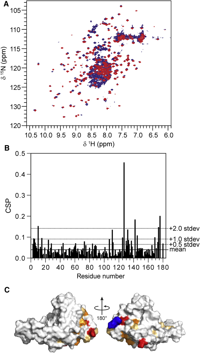
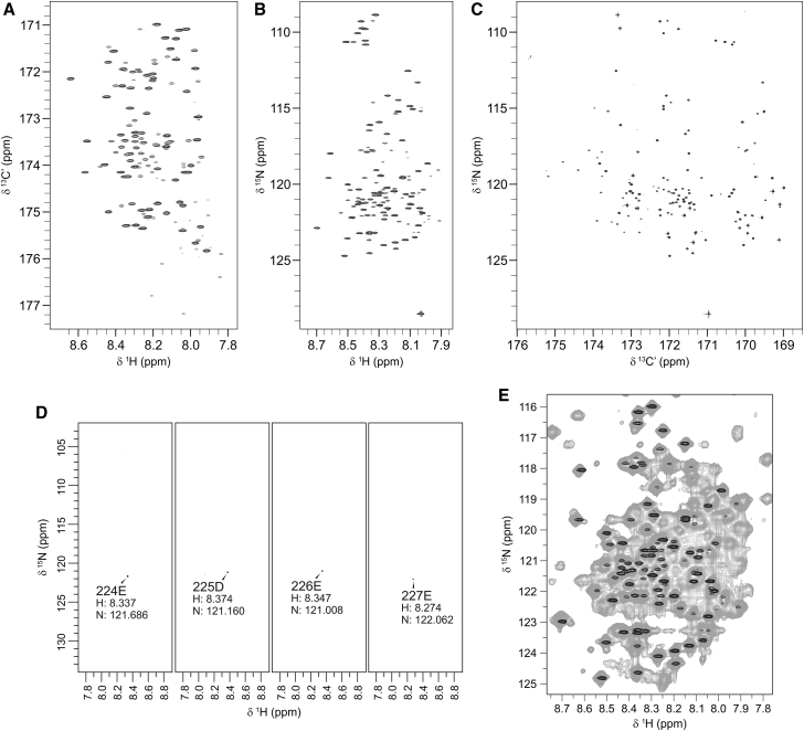
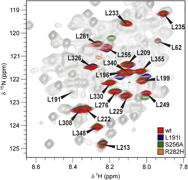
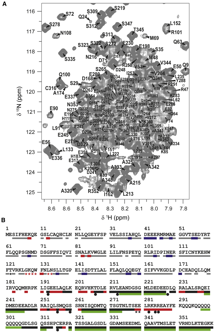
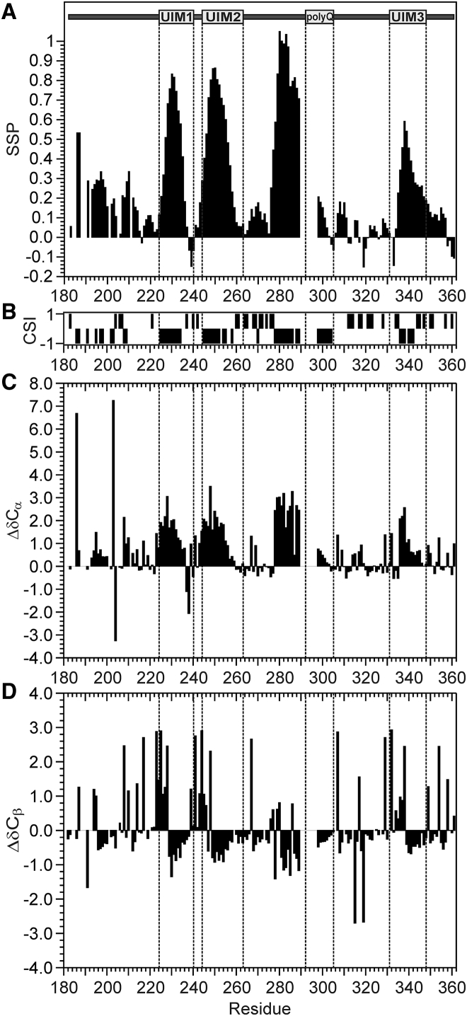
It has increasingly become clear over the last two decades that proteins can contain both globular domains and intrinsically unfolded regions that can both contribute to function. Although equally interesting, the disordered regions are difficult to study, because they usually do not crystallize unless bound to partners and are not easily amenable to cryo-electron microscopy studies. NMR spectroscopy remains the best technique to capture the structural features of intrinsically mixed folded proteins and describe their dynamics. These studies rely on the successful assignment of the spectrum, a task not easy per se given the limited spread of the resonances of the disordered residues. Here, we describe the structural properties of ataxin-3, the protein responsible for the neurodegenerative Machado-Joseph disease. Ataxin-3 is a 42-kDa protein containing a globular N-terminal Josephin domain and a C-terminal tail that comprises 13 polyglutamine repeats within a low complexity region. We developed a strategy that allowed us to achieve 87% assignment of the NMR spectrum using a mixed protocol based on high-dimensionality, high-resolution experiments and different labeling schemes. Thanks to the almost complete spectral assignment, we proved that the C-terminal tail is flexible, with extended helical regions, and interacts only marginally with the rest of the protein. We could also, for the first time to our knowledge, observe the structural propensity of the polyglutamine repeats within the context of the full-length protein and show that its structure is stabilized by the preceding region.


Figures

Figure 1
Figure 1
Assignment of the NH backbone resonances of the Josephin domain within the ataxin-3(Q13) construct. (A) Superimposition of the 15N-1H HSQC spectrum of the isolated Josephin domain (red) and ataxin-3(Q13) (blue). The spectra were recorded at 800 MHz and 25°C. (B) Chemical shift perturbation of Josephin NH backbone resonances in ataxin-3(Q13) with respect to the isolated domain. (C) Mapping of the chemical shift perturbations on the structure of the Josephin domain. Light orange, chemical shift perturbation > average + 0.5 SD; orange, chemical shift perturbation > average + 1 SD; red: chemical shift perturbation > average + 2 SD. The C-terminal arginine is colored in blue. The chemical shift perturbation (ΔδH-N) was calculated according to the formula ΔδHN=δH2+αδN2/2, where α = 0.14 for glycine and α = 0.20 for any other residue. To see this figure in color, go online.
Figure 2
Figure 2
Assisting assignment with 5D HNcaCONH experiments. (A) 13C-1H, (B) 15N-1H, and (C) 15N-13C projections of the HNCO experiment. (D15N-1H planes of consecutive residues associated to HNCO peaks in the 5D HNcaCONH experiment. (E) Superimposition of the HSQC spectrum of ataxin-3(Q13) (gray) with the 15N-1H projection of the HNCO spectrum used as reference for the 5D HNcaCONH (black).
Figure 3
Figure 3
15N-1H HSQC spectra of 15N-Leu selectively labeled wild-type and mutated ataxin-3(Q13). The spectrum of the wild-type protein (red) is superimposed to the spectra of three mutants (L191I: blue, S256A: green, R282H: orange). The counter levels were adjusted to show only the sharper resonances related mainly to residues in the C-terminal tail. The resonances of at least eight more leucines were also detectable in the experiment but with lower intensities. They all correspond to Josephin leucines. Minor scrambling was observed at the noise level. To see this figure in color, go online.
Figure 4
Figure 4
Summary of the assignment. (A) Highly degenerate areas of the 1H-15NHSQC spectrum of ataxin-3(Q13) annotated with the assignment. (B) Final achieved assignment reported on thesequence of ataxin-3(Q13). The bar colors are associated to the technique used for assignment. The backbone NH pairs of the Josephin domain (residues 1–182) were assigned by comparison with the spectrum of the isolated domain without (gray bars) or with the aid of 15N-amino acid selective labeling (red bars) or BEST-TROSY HSQC (red stars). Residues assigned by comparison of the NOESY-HSQC of ataxin-3(Q13) with that of the isolated Josephin domain are indicated by blue bars. The C-terminal tail was assigned via HNCACB and CBCAcoNH (black bars), 5D HNcaCONH and HabCabCONH (green bars), selective 15N-amino acid labeling (red bars), BEST-TROSY-HNCACB (black stars), and single-point mutations (black circles). To see this figure in color, go online
Figure 5
Figure 5
Secondary structure of the C-terminus of ataxin-3(Q13). (A) SSP algorithm. Values around zero indicate random coil regions; positive and negative values indicate α-helical or β-sheet conformations, respectively. (B) CSI values (−1 indicates α-helix, 0 random coil, and +1 β-strand). (C) Secondary Cα chemical shifts. (D) Secondary Cβ chemical shifts.
Figure 6
Figure 6
Description of the dynamics of ataxin-3(Q13). (A) Longitudinal and (B) transverse relaxation of the spectrum of the isolated Josephin domain (continuous line) and ataxin-3(Q13) (dashed line) in the range 7.7–10.5 ppm. (C) Correlation times (τC) of well-dispersed resonances of Josephin within the spectrum of ataxin-3(Q13). The dashed line indicates the average τC.

Similar articles

Cited by

References

    1. Paulson H.L., Perez M.K., Pittman R.N. Intranuclear inclusions of expanded polyglutamine protein in spinocerebellar ataxia type 3. Neuron. 1997;19:333–344. - PubMed
    1. Ellisdon A.M., Thomas B., Bottomley S.P. The two-stage pathway of ataxin-3 fibrillogenesis involves a polyglutamine-independent step. J. Biol. Chem. 2006;281:16888–16896. - PubMed
    1. Saunders H.M., Gilis D., Bottomley S.P. Flanking domain stability modulates the aggregation kinetics of a polyglutamine disease protein. Protein Sci. 2011;20:1675–1681. - PMC - PubMed
    1. Doss-Pepe E.W., Stenroos E.S., Madura K. Ataxin-3 interactions with rad23 and valosin-containing protein and its associations with ubiquitin chains and the proteasome are consistent with a role in ubiquitin-mediated proteolysis. Mol. Cell. Biol. 2003;23:6469–6483. - PMC - PubMed
    1. Scaglione K.M., Zavodszky E., Paulson H.L. Ube2w and ataxin-3 coordinately regulate the ubiquitin ligase CHIP. Mol. Cell. 2011;43:599–612. - PMC - PubMed

Publication types

LinkOut - more resources