. 2018 Jul 4;18(1):105.
doi: 10.1186/s12876-018-0833-8.

Secreted protein acidic and rich in cysteine-like 1 suppresses metastasis in gastric stromal tumors

Affiliations

Secreted protein acidic and rich in cysteine-like 1 suppresses metastasis in gastric stromal tumors

Chaoyong Shen et al. BMC Gastroenterol. .

Abstract

Background: Malignant growth and metastasis of gastrointestinal stromal tumors (GIST) occur in some patients even during the course of treatment, but their mechanisms remains poorly understand at the molecular level so far.

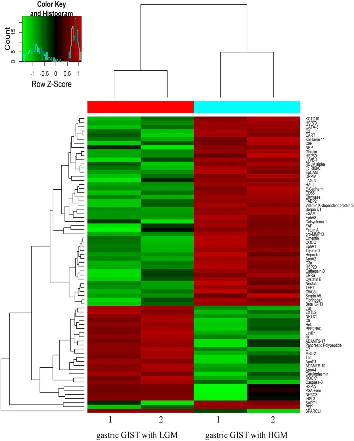
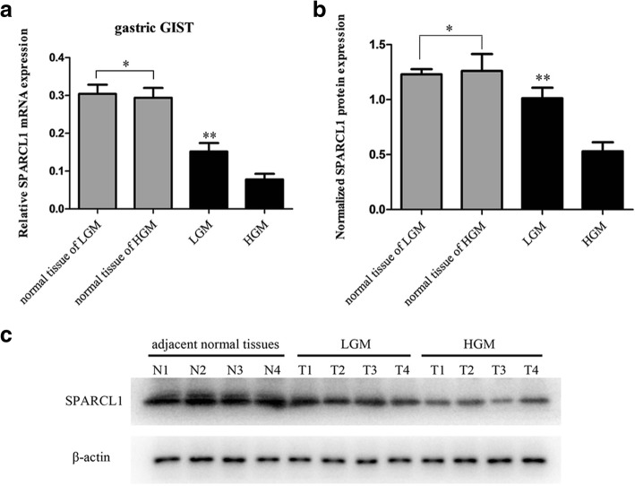
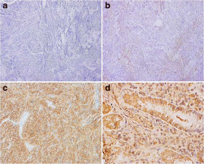
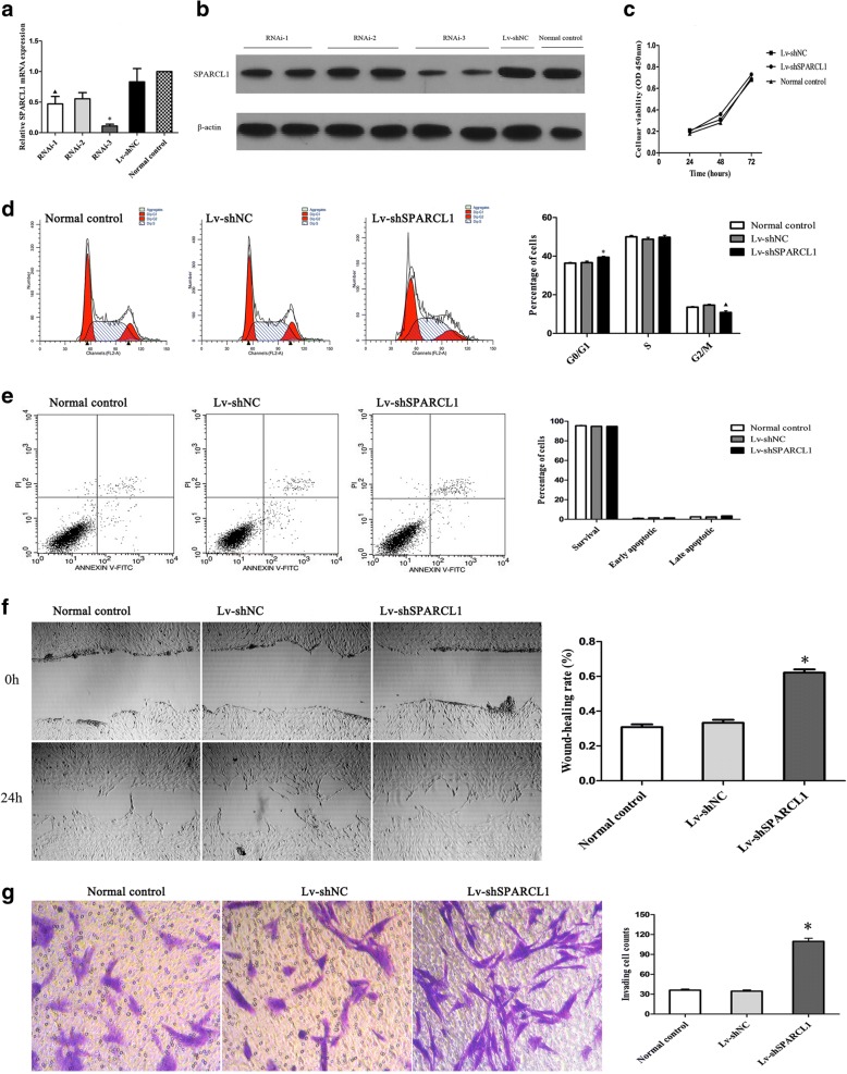
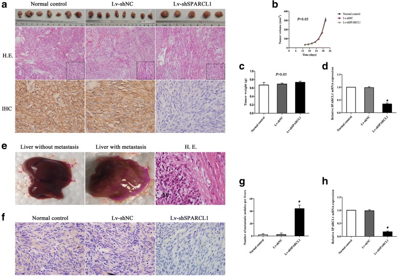
Methods: Profiles of protein expression in gastric GIST tissues were explored using protein microarray analysis, down-regulation of SPARCL1 (secreted protein acidic and rich in cysteine-like protein 1) was validated by RT-qPCR, western blot and immunohistochemistry. The effect of specific shRNA-induced SPARCL1 downregulation on the biological traits of GIST 882 cell was investigated. We then employed a mouse xenograft model to investigate whether the low-expression of SPARCL1 impact the metastasis ability of GIST cells in vivo.

Results: SPARCL1 was significantly downregulated in the gastric GIST with high-grade malignance as compared with low-grade malignance, its expression was closely correlated with tumor size, mitotic index, distant metastasis at the time of initial diagnosis and tumor progression of GIST (P < 0.05). Moreover, results of the Cox analysis showed that expression of SPARCL1 is an independent prognostic predictors for gastric GIST (P = 0.008; HR 0.157, 95% CI 0.040~ 0.612). Downregulation of SPARCL1 promoted cell migration and invasion, but did not affect proliferation, cell cycle and apoptosis of GIST 882 cells. In mouse xenograft model, GIST cells with the decreased expression of SPARCL1 presented an enhanced ability of liver metastasis (P < 0.05).

Conclusions: Taken together, our present study demonstrated that SPARCL1 have a certain degree of malignancy-suppressing potential through inhibiting the metastasis of gastric GIST.

Keywords: Gastrointestinal stromal tumors; Malignization; Metastasis; Microarray; SPARCL1.


Conflict of interest statement

Ethics approval

Informed consents were provided by each patient before surgery, and the protocol of this study was approved by the Research Ethics Board of West China Hospital, Sichuan University. All animal experiments were conducted under an approved protocol from Sichuan University Institutional Animal Care and Use Committee.

Consent for publication

Not applicable.

Competing interests

The authors declare that they have no competing interests.

Publisher’s Note

Springer Nature remains neutral with regard to jurisdictional claims in published maps and institutional affiliations.

Figures

Fig. 1
Fig. 1
Heatmap of the hierarchical cluster analysis of the differentially expressed proteins between LGM and HGM in gastric GIST. Each column represents a single tissue specimen and each row represents a protein. Pseudocolors indicate differential expression, with red indicating high expression, green for low expression and black indicating for the mean expression levels
Fig. 2
Fig. 2
SPARCL1 is downregulated in human gastric GIST. The protein expression of SPARCL1 was determined by RT-qPCR and western blot with β-actin as a loading control. a Relative SPARCL1 mRNA level was significantly decreased in gastric GSIT with HGM as compared with those with LGM (**P < 0.05), while no statistical significance was noted between matched normal tissues from HGM and LGM (*P > 0.05); b, c A significant reduced expression of SPARCL1 in gastric GIST with HGM compared to those with LGM was determined by western blot analysis (**P < 0.05). N1~N2 for adjacent normal tissue of LGM, while N3~N4 for adjacent tissue of HGM. The bars represent mean ± SD (n = 4)
Fig. 3
Fig. 3
Representative immunohistochemical staining of SPARCL1 in gastric GIST and normal gastric tissues. a, b Negative/low expression of SPARCL1 in gastric GIST; (c, d) Gastric GIST and normal gastric tissues showed high expression of SPARCL1, separately. Original magnification: × 200 for a, b, c and × 600 for d
Fig. 4
Fig. 4
Kaplan-Meier survival curves of progression-free survival in patients with primary gastric GIST (n = 98). a Comparison of progression-free survival between tumors with ≤6 cm and > 6 cm (P < 0.001); b The tumors with mitotic count ≤5/50HPF showed significant better PFS compared with those of 6~ 10/50HPF and > 10/50HPF (P < 0.001); c Patients with R0 resection showed better PFS than that who did not achieve R0 resection (P < 0.001); d A worse prognosis was noted in patients with SPARCL-low expression in comparison to those with SPARCL1-high expression (P < 0.001)
Fig. 5
Fig. 5
The roles of SPARCL1 knockdown in GIST 882 cell proliferation, cell cycle, apoptosis, migration and invasion. a, b Downregulation of SPARCL1 in GIST cells was confirmed by RT-qPCR and western blot. ▲compared with RNAi-3, P < 0.05; * compared with RNAi-2, P < 0.05. c: A CCK-8 assay was performed to evaluate the influence of SPARCL1 knockdown on GIST 882 cell proliferation. d: SPARCL1 downregulation could induce the arrest of cell cycles in G0/G1 phase. ▲ * compared with the other two groups, both P < 0.05. e SPARCL1 downregulation did not affect apoptosis of GIST 882 cell. f Wound-healing assay showed that SPARCL1 knockdown promoted migration of GIST 882 cells after cultured for 24 h. * compared with the other two groups, both P < 0.05. g The invasive GIST cells and quantitation of invasive GIST cell counts. * compared with the other two groups, both P < 0.05. Data represent the mean of three independent experiments, and the error bars refer to SD from the mean
Fig. 6
Fig. 6
Downregulation of SPARCL1 in GIST 882 cells promoted liver metastasis in xenograft mouse models. a Representative images of hematoxylin and eosin staining (HE × 100) and IHC staining (× 600) of SPARCL1 in xenografted tumors; b, c Quantification of tumor volume and tumor weight for tumorigenesis among three groups in nude mice (P > 0.05). The error bars refer to SD (n = 7, 8, and 5 for normal control, Lv-shNC and Lv-shSPARCL1, respectively); d Expression level of SPARCL1 mRNA was significantly decreased in Lv-shSPARCL1 group as compared to other two groups (*P < 0.05). The error bars present SD (n = 7, 8, and 5 for normal control, Lv-shNC and Lv-shSPARCL1, respectively); e Representative of gross view of liver without and with metastasis, and HE staining of liver metastatic tumor (M); f Low expression of SPARCL1 in liver metastatic nodules was observed in Lv-shSPARCL1 group as compared to other two groups; g The number of liver metastatic nodules was increased in Lv-shSPARCL1 group when compared to other two groups (*P < 0.05); h The relative SPARCL1 mRNA level of liver metastatic nodules of nude mice (*P < 0.05). The error bars refer to SD from the mean

Similar articles

Cited by

References

    1. Wang X, Mori I, Tang W, Utsunomiya H, Nakamura M, Nakamura Y, Zhou G, Kakudo K. Gastrointestinal stromal tumors: are they of cajal cell origin? Exp Mol Pathol. 2002;72:172–177. doi: 10.1006/exmp.2001.2419. - DOI - PubMed
    1. Miettinen M, Lasota J. Gastrointestinal stromal tumors--definition, clinical, histological, immunohistochemical, and molecular genetic features and differential diagnosis. Virchows Arch. 2001;438:1–12. doi: 10.1007/s004280000338. - DOI - PubMed
    1. Lin JX, Chen QF, Zheng CH, Li P, Xie JW, Wang JB, Lu J, Chen QY, Cao LL, et al. Is 3-years duration of adjuvant imatinib mesylate treatment sufficient for patients with high-risk gastrointestinal stromal tumor? A study based on long-term follow-up. J Cancer Res Clin Oncol. 2017;143:727–734. doi: 10.1007/s00432-016-2334-x. - DOI - PMC - PubMed
    1. Joensuu H, Wardelmann E, Sihto H, Eriksson M, Sundby Hall K, Reichardt A, Hartmann JT, Pink D, Cameron S, et al. Effect of KIT and PDGFRA mutations on survival in patients with gastrointestinal stromal tumors treated with adjuvant Imatinib: an exploratory analysis of a randomized clinical trial. JAMA Oncol. 2017;3:602–609. doi: 10.1001/jamaoncol.2016.5751. - DOI - PMC - PubMed
    1. Baskin Y, Kocal GC, Kucukzeybek BB, Akbarpour M, Kayacik N, Sagol O, Ellidokuz H, Oztop I. PDGFRA and KIT mutation status and its association with Clinicopathological properties, including DOG1. Oncol Res. 2016;24:41–53. doi: 10.3727/096504016X14576297492418. - DOI - PMC - PubMed

MeSH terms