. 2018 Jul 4;18(1):205.
doi: 10.1186/s12906-018-2265-y.

Oleanolic acid attenuates TGF-β1-induced epithelial-mesenchymal transition in NRK-52E cells

Affiliations

Oleanolic acid attenuates TGF-β1-induced epithelial-mesenchymal transition in NRK-52E cells

Wei-Ming He et al. BMC Complement Altern Med. .

Abstract

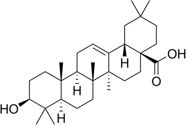
Background: Epithelial-to-mesenchymal transition (EMT) plays an important role in the progression of renal interstitial fibrosis, which finally leads to renal failure. Oleanolic acid (OA), an activator of NF-E2-related factor 2 (Nrf2), is reported to attenuate renal fibrosis in mice with unilateral ureteral obstruction. However, the role of OA in the regulation of EMT and the underlying mechanisms remain to be investigated. This study aimed to evaluate the effects of OA on EMT of renal proximal tubular epithelial cell line (NRK-52E) induced by TGF-β1, and to elucidate its underlying mechanism.

Methods: Cells were incubated with TGF-β1 in the presence or absence of OA. The epithelial marker E-cadherin, the mesenchymal markers, α-smooth muscle actin (α-SMA), fibronectin, Nrf2, klotho, the signal transducer (p-Smad2/3), EMT initiator (Snail), and ILK were assayed by western blotting.

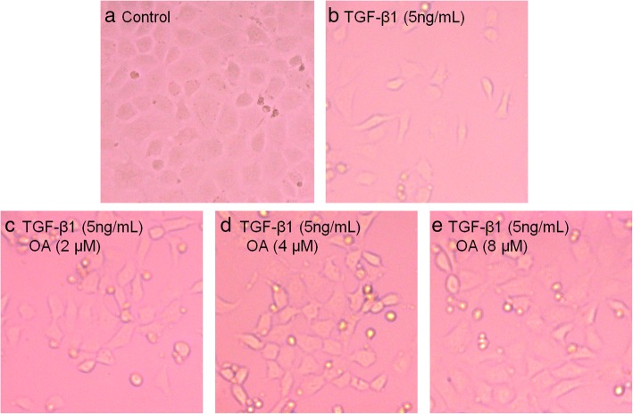
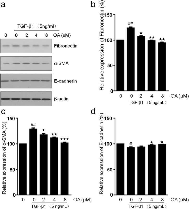
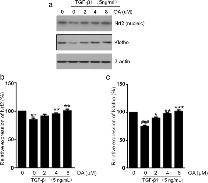
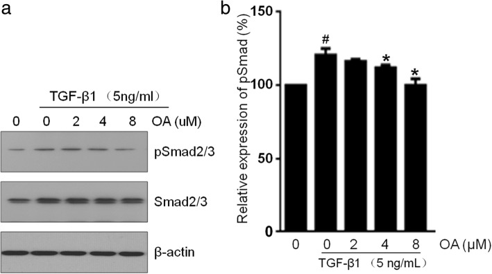
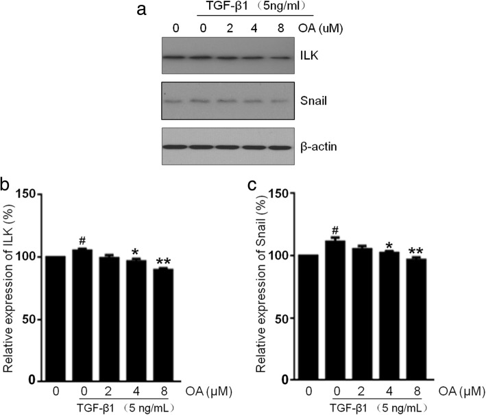
Results: Our results showed that the NRK-52E cells incubated with TGF-β1 induced EMT with transition to the spindle-like morphology, down-regulated the expression of E-cadherin but up-regulated the expression of α-SMA and fibronectin. However, the treatment with OA reversed all EMT markers in a dose-dependent manner. OA also restored the expression of Nrf2 and klotho, decreased the phosphorylation of Smad2/3, ILK, and Snail in cells which was initiated by TGF-β1.

Conclusion: OA can attenuate TGF-β1 mediate EMT in renal tubular epithelial cells and may be a promising therapeutic agent in the treatment of renal fibrosis.

Keywords: EMT; Klotho; Nrf2; Oleanolic acid; TGF-β1.


Conflict of interest statement

Ethics approval and consent to participate

Not applicable.

Consent for publication

Not applicable.

Competing interests

The authors declare that they have no competing interests.

Publisher’s Note

Springer Nature remains neutral with regard to jurisdictional claims in published maps and institutional affiliations.

Figures

Fig. 1
Fig. 1
Chemical structure of Oleanolic acid
Fig. 2
Fig. 2
Effects of OA on TGF-β1-induced Morphological changes in NRK-52E cells. The cells were incubated with 5 ng/mL of TGF-β1 for 48 h with different concentrations of OA (0, 2, 4, 8 μM). a Untreated NRK-52E cells showed a pebble-like shape is clearly observed. b TGF-β1-treated cells showed a more elongated morphological shape. c, d, e showed OA reversed TGF-β1-induced morphological changes (Magnification of 200×)
Fig. 3
Fig. 3
Effects of OA on TGF-β1-induced EMT in NRK-52E cells. The cells were incubated with 5 ng/mL of TGF-β1 for 48 h with different concentrations of OA (0, 2, 4, 8 μM). a The expression of Fibronectin, α-SMA and E-cadherin was determined by Western blotting. b, c, d The expression level was quantitatively analyzed with Image J software. The data showing mean ± SD. # P < 0.05, ## P < 0.01, and ### P < 0.005 vs. 0 ng/mL TGF-β1. * P < 0.05, ** P < 0.01, and *** P < 0.005 vs. 0 μM OA in the presence of 5 ng/mL TGF-β1
Fig. 4
Fig. 4
Effects of OA on the Nrf2 and klotho expression in NRK-52E cells. The cells were incubated with 5 ng/mL of TGF-β1 for 48 h with different concentrations of OA (0, 2, 4, 8 μM). a The expression level of Nrf2 and klotho was determined by Western blotting. b, c The expression level was quantitatively analyzed with Image J software. The data showing mean ± SD. # P < 0.05, ## P < 0.01, and ### P < 0.005 vs. 0 ng/mL TGF-β1. * P < 0.05, ** P < 0.01, and *** P < 0.005 vs. 0 μM OA in the presence of 5 ng/mL TGF-β1
Fig. 5
Fig. 5
Effects of OA on the expression of pSmad2/3 in NRK-52E cells. The cells were incubated with 5 ng/mL of TGF-β1 for 48 h with different concentrations of OA (0, 2, 4, 8 μM). a The expression level of Smad2/3 and pSmad2/3 was determined by Western blotting. b The pSmad2/3 was quantitatively analyzed with Image J software. The data showing mean ± SD. # P < 0.05 vs. 0 ng/mL TGF-β1. * P < 0.05 vs. 0 μM OA in the presence of 5 ng/mL TGF-β1
Fig. 6
Fig. 6
Effects of OA on the ILK and Snail expression in NRK-52E cells. The cells were incubated with 5 ng/mL of TGF-β1 for 48 h with different concentrations of OA (0, 2, 4, 8 μM). a The expression level of ILK and Snail was determined by Western blotting. b, c The expression level was quantitatively analyzed with Image J software. The data showing mean ± SD. # P < 0.05 vs. 0 ng/mL TGF-β1. * P < 0.05, and ** P < 0.01 vs. 0 μM OA in the presence of 5 ng/mL TGF-β1

Similar articles

Cited by

References

    1. Kalluri R, Neilson EG. Epithelial-mesenchymal transition and its implications for fibrosis. J Clin Invest. 2003;112:1776–1784. doi: 10.1172/JCI200320530. - DOI - PMC - PubMed
    1. Lee JM, Dedhar S, Kalluri R, Thompson EW. The epithelial-mesenchymal transition: new insights in signaling, development, and disease. J Cell Biol. 2006;172:973–981. doi: 10.1083/jcb.200601018. - DOI - PMC - PubMed
    1. Kalluri R, Weinberg RA. The basics of epithelial-mesenchymal transition. J Clin Invest. 2009;119:1420–1428. doi: 10.1172/JCI39104. - DOI - PMC - PubMed
    1. Liu Y. Epithelial to mesenchymal transition in renal fibrogenesis: pathologic significance, molecular mechanism, and therapeutic intervention. J Am Soc Nephrol. 2004;15:1–12. doi: 10.1097/01.ASN.0000106015.29070.E7. - DOI - PubMed
    1. Hewitson TD. Renal tubulointerstitial fibrosis: common but never simple. Am J Physiol Renal Physiol. 2009;296:1239–1244. doi: 10.1152/ajprenal.90521.2008. - DOI - PubMed

MeSH terms

Substances

LinkOut - more resources