

Wounding promotes ovarian cancer progression and decreases efficacy of cisplatin in a syngeneic mouse model
- PMID: 29973223
- PMCID: PMC6032528
- DOI: 10.1186/s13048-018-0428-6
Wounding promotes ovarian cancer progression and decreases efficacy of cisplatin in a syngeneic mouse model
Abstract
Background: Primary cytoreductive surgery followed by adjuvant chemotherapy is the standard treatment for advanced epithelial ovarian cancer. The average interval between surgery and chemotherapy initiation is approximately 4-weeks at most centers; however, since surgery may accelerate residual tumor growth, a shorter interval may be more beneficial.
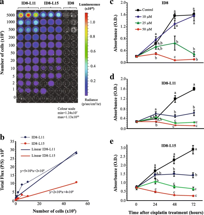
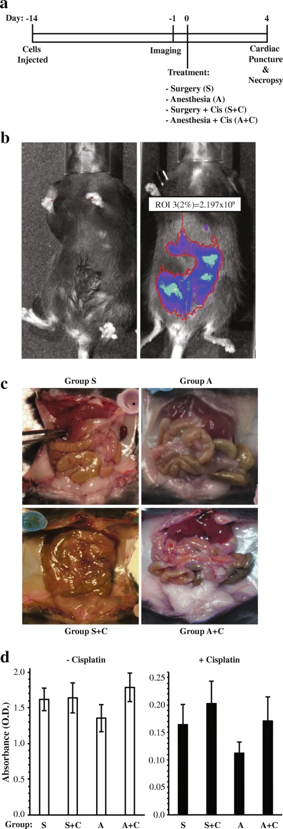
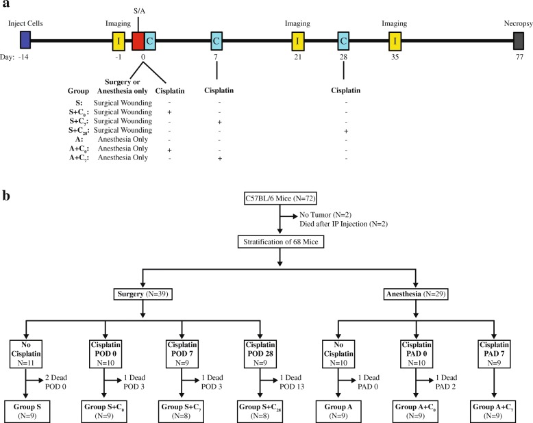
Methods: The murine ID8 cell model of ovarian cancer was used to examine the efficacy of cisplatin treatment administered perioperatively or 7 days after surgical wounding. Luciferase-expressing cells ID8 cells were injected intraperitoneally (i.p.) into female C57/Bl6 mice. Fourteen days post-injection, animals received an abdominal incision or anesthesia alone and received i.p. cisplatin either on the surgical day or 7 days later, or received no chemotherapy. Additional animals received cisplatin 28 days after wounding for comparison.
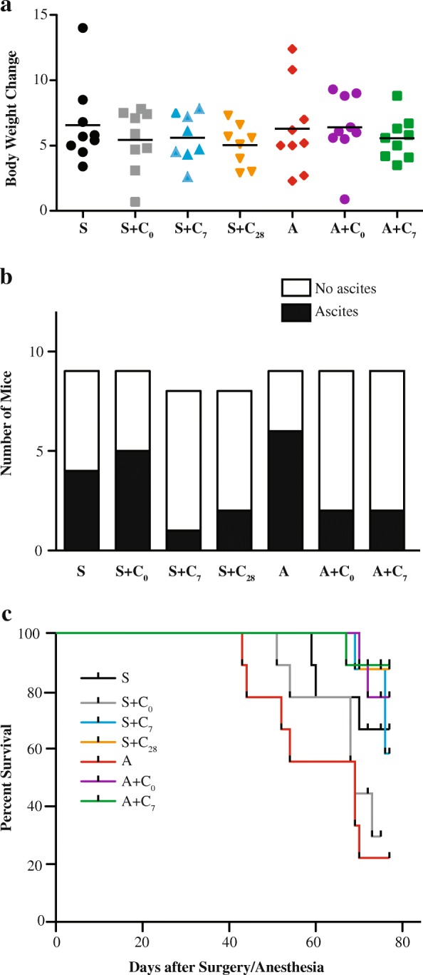
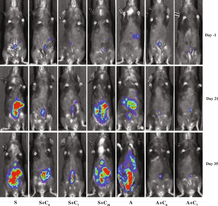
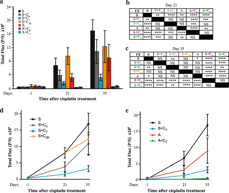
Results: Abdominal tumor mass increased 2.5-fold in wounded vs. unwounded animals as determined by bioluminescent in vivo tumor imaging. Cisplatin administered on the day of wounding decreased tumor burden by 50%, as compared to 90% in unwounded animals. Cisplatin on day 7 or day 28 decreased tumor burden by 80 and 37% respectively.
Conclusions: Surgical wounding increases ovarian tumor mass and decreases perioperative cisplatin efficacy in this animal model. Administration of cisplatin 1 week after surgery was more effective than cisplatin administered perioperatively or 4 weeks after surgery.
Keywords: Chemoresistance; Cisplatin; ID8 cells; Mouse; Ovarian cancer; Wound healing.
Conflict of interest statement
Ethics approval
All animal procedures were approved by the University of Toronto Animal Care and Use Committee.
Consent for publication
Not applicable.
Competing interests
The authors declare that they have no competing interests.
Publisher’s Note
Springer Nature remains neutral with regard to jurisdictional claims in published maps and institutional affiliations.
Figures
Similar articles
-
A mouse model of neoadjuvant chemotherapy followed by interval cytoreductive surgery indicates impaired efficacy of perioperative cisplatin.J Ovarian Res. 2021 Nov 16;14(1):157. doi: 10.1186/s13048-021-00895-w. J Ovarian Res. 2021. PMID: 34784944 Free PMC article.
-
Thymoquinone enhances cisplatin-response through direct tumor effects in a syngeneic mouse model of ovarian cancer.J Ovarian Res. 2015 Jul 28;8:46. doi: 10.1186/s13048-015-0177-8. J Ovarian Res. 2015. PMID: 26215403 Free PMC article.
-
A comparison of the toxicity and tolerability of two intraperitoneal chemotherapy regimens for advanced-stage epithelial ovarian cancer.Gynecol Oncol. 2016 Jan;140(1):36-41. doi: 10.1016/j.ygyno.2015.11.005. Epub 2015 Nov 4. Gynecol Oncol. 2016. PMID: 26546964
-
Intraperitoneal chemotherapy in advanced epithelial ovarian cancer: a survey.Arch Gynecol Obstet. 2014 Sep;290(3):425-34. doi: 10.1007/s00404-014-3252-2. Epub 2014 May 21. Arch Gynecol Obstet. 2014. PMID: 24845970 Review.
-
Intraperitoneal chemotherapy requires expertise and should be the standard of care for optimally surgically resected epithelial ovarian cancer patients.Ann Oncol. 2013 Dec;24 Suppl 10:x41-45. doi: 10.1093/annonc/mdt469. Ann Oncol. 2013. PMID: 24265403 Review.
Cited by
-
Clinical factors associated with prognosis in low-grade serous ovarian carcinoma: experiences at two large academic institutions in Korea and Taiwan.Sci Rep. 2020 Nov 17;10(1):20012. doi: 10.1038/s41598-020-77075-1. Sci Rep. 2020. PMID: 33203969 Free PMC article.
-
The impact of interval between primary cytoreductive surgery with bowel resection and initiation of adjuvant chemotherapy on survival of women with advanced ovarian cancer: a multicenter cohort study.J Gynecol Oncol. 2022 Nov;33(6):e76. doi: 10.3802/jgo.2022.33.e76. Epub 2022 Aug 4. J Gynecol Oncol. 2022. PMID: 36047378 Free PMC article.
-
Surgical trauma-induced immunosuppression in cancer: Recent advances and the potential therapies.Clin Transl Med. 2020 Jan;10(1):199-223. doi: 10.1002/ctm2.24. Clin Transl Med. 2020. PMID: 32508035 Free PMC article. Review.
-
Preoperative stimulation of resolution and inflammation blockade eradicates micrometastases.J Clin Invest. 2019 Jun 17;129(7):2964-2979. doi: 10.1172/JCI127282. eCollection 2019 Jun 17. J Clin Invest. 2019. PMID: 31205032 Free PMC article.
-
A mouse model of neoadjuvant chemotherapy followed by interval cytoreductive surgery indicates impaired efficacy of perioperative cisplatin.J Ovarian Res. 2021 Nov 16;14(1):157. doi: 10.1186/s13048-021-00895-w. J Ovarian Res. 2021. PMID: 34784944 Free PMC article.
References
-
- Chi DS, Eisenhauer EL, Lang J, Huh J, Haddad L, Abu-Rustum NR, Sonoda Y, Levine DA, Hensley M, Barakat RR. What is the optimal goal of primary cytoreductive surgery for bulky stage IIIC epithelial ovarian carcinoma (EOC)? Gynecol Oncol. 2006;103(2):559–564. doi: 10.1016/j.ygyno.2006.03.051. - DOI - PubMed
-
- Tewari KS, Java JJ, Eskander RN, Monk BJ, Burger RA. Early initiation of chemotherapy following complete resection of advanced ovarian cancer associated with improved survival: NRG oncology/gynecologic oncology group study. Ann Oncol. 2016;27(1):114–121. doi: 10.1093/annonc/mdv500. - DOI - PMC - PubMed
MeSH terms
Substances
Grants and funding
LinkOut - more resources
Full Text Sources
Other Literature Sources
Medical
