. 2018 Jul 4;12(1):35.
doi: 10.1186/s40246-018-0165-3.

A novel LRAT mutation affecting splicing in a family with early onset retinitis pigmentosa

Affiliations

A novel LRAT mutation affecting splicing in a family with early onset retinitis pigmentosa

Yabin Chen et al. Hum Genomics. .

Abstract

Background and purpose: Retinitis pigmentosa is an important cause of severe visual dysfunction. This study reports a novel splicing mutation in the lecithin retinol acyltransferase (LRAT) gene associated with early onset retinitis pigmentosa and characterizes the effects of this mutation on mRNA splicing and structure.

Methods: Genome-wide linkage analysis followed by dideoxy sequencing of the linked candidate gene LRAT was performed in a consanguineous Pakistani family with autosomal recessive retinitis pigmentosa. In silico prediction and minigene assays were used to investigate the effects of the presumptive splicing mutation.

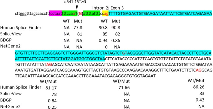
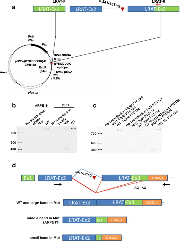
Results: ARRP in this family was linked to chromosome 4q31.21-q32.1 with a maximum LOD score of 5.40. A novel homozygous intronic mutation (NM_004744.4: c.541-15T>G) was detected in LRAT. In silico tools predicted that the AG-creating mutation would activate an intronic cryptic acceptor site, but cloning fragments of wild-type and mutant sequences of LRAT into Exontrap Cloning Vector pET01 and Expression Cloning Vector pCMV-(DYKD4K)-C showed that the primary effect of the sequence change was to weaken the nearby authentic acceptor site and cause exon skipping, with only a small fraction of transcripts utilizing the acceptor site producing the reference transcript.

Conclusions: The c.541-15T>G mutation in LRAT results in aberrant splicing and is therefore predicted to be causal for the early onset retinitis pigmentosa in this family. In addition, this work suggests that minigenes adapted to the specific gene and exon may need to be designed for variants in the first and last exon and intron to mimic the authentic splicing mechanism in vivo.

Keywords: Cryptic splice site; Exon splicing; LRAT; Linkage; Minigene assay; Retinitis pigmentosa; Splicing mutation.


Conflict of interest statement

Ethics approval and consent to participate

This study was approved by the Institutional Review Boards (IRB) of the National Centre of Excellence in Molecular Biology, Lahore, Pakistan, and the CNS IRB at the National Institutes of Health. Participating individuals or their guardians gave written informed consent to participate and to publish consistent with the tenets of the Declaration of Helsinki before the study.

Consent for publication

Not applicable.

Competing interests

The authors declare that they have no competing interests.

Publisher’s Note

Springer Nature remains neutral with regard to jurisdictional claims in published maps and institutional affiliations.

Figures

Fig. 1
Fig. 1
Pedigree, LRAT Region, and sequence. a Haplotypes of the LRAT region of family 61254 showing the LRAT c.541-15T>G mutation and surrounding microsatellite markers included in Table 1. The risk haplotype is marked in black. b Electropherograms of the LRAT homozygous mutation c.541-15T>G (up, individual 7), carrier sequence (middle, individual 22), and wildtype alleles (down, individual 41)
Fig. 2
Fig. 2
Fundus photographs of family 61254. a, b Oculus dexter (OD) and oculus sinister (OS) of an affected 35-year-old individual (08), which show waxy pale optic discs, attenuation of retinal arteries, bone spicule-like pigment deposits in the midperiphery of the retina, and macular changes. c, d OD and OS of an affected 10-year-old individual (32), which show attenuation of retinal arteries. e, f OD and OS of an unaffected 60-year-old individual (05), which show no signs of retinal dystrophy but does show haziness secondary to age-related cataracts
Fig. 3
Fig. 3
Electroretinography recordings of family members of 61254. ad ERG responses of individual 8 (affected, 35-year-old). eh ERG responses of individual 32 (affected, 10-year-old). il ERG response of individual 5 (unaffected, 60-year-old). a, e, and i OD combined rod and cone response; b, f, and j OD cone response; c, g, and k OS combined rod and cone response; d, h, and l OS cone response
Fig. 4
Fig. 4
Results of in silico splice prediction tools. Lowercase nucleotides indicate intron 2 sequences, upper case nucleotides indicate exon 3 sequences, and the fragment highlighted in blue is the coding region. Nucleotides marked in red indicate potential splice donor or acceptor sites. The YAG sequence is shown in yellow, the polypyrimidine tract (PPT) is shown in green, and the branch point sequence (BPS) is shown in purple. The scores calculated for wildtype sequence (WT) and mutated sequence (Mut) using Human Splice Finder, SpliceView, BDGP, and NetGene2 are displayed below each splice site. A higher score implies greater potential for a splice site. NA means no splice site predictions above threshold of the in silico tool
Fig. 5
Fig. 5
Splicing analysis using Exontrap pET01. a Diagram of LRAT genomic DNA, the location of the c.541-15T>G mutation and the fragment inserted into pET01 construct. In the pET01 construct, the multiple cloning site (MCS) is in an intron flanked by pre-proinsulin 5′ and 3′ exons. The primers used for cDNA amplification are within the pre-proinsulin 5′ and 3′ exons and indicated by black arrows. b Diagrams of transcript splicing of WT- and Mut- LRAT-pET01. Splicing events in the WT construct are shown by a blue line, and transcript splicing of c.541-15T>G Mut-LRAT-pET01 construct are shown by a red line. Results derived from in silico prediction (top), results obtained from minigene assay using pET01 vector (middle), and results obtained from minigene assay using pET01 vector with the splice acceptor site of 3′ exon abolished (bottom). c Electrophoresis results of cDNA amplification obtained from 293T cells transfected with empty construct pET01, wildtype construct (WT), c.541-15T>G mutant construct (Mut), modified empty construct (pET01-2), modified wildtype construct (WT-2), and modified c.541-15T>G mutant construct (Mut-2). The major PCR products of cDNA amplification from each construct as verified by sequencing are indicated in d
Fig. 6
Fig. 6
a Diagram of the LRAT gene showing the location of the c.541T>G variation and the fragment inserted into the pCMV-(DYKD4K)-C construct. b Electrophoresis results of cDNA amplification obtained from 293T cells and ARPE-19 cells transfected with the wildtype construct WT, mutated construct Mut, and empty construct pCMV. c Non-transfected and transfected ARPE19 cells were treated with 0, 5 μM, and 10 μM PTC124 separately for 24 h. There were no extra transcripts under PTC124 treatment. d Diagram of transcript in which splicing of WT-LRAT-pCMV is shown by a blue line; transcript splicing of Mut-LRAT-pCMV is shown by a red line. The forward primer used in cDNA amplification is within the exon 2 of LRAT and the reverse primer binds to the construct sequence of DYKD4K epitope, which are indicated by arrows (the AG shown in the 3′ UTR of LRAT refers to the last two AG in Fig. 4). A diagram of the PCR product of cDNA amplification from each construct is also shown in d

Similar articles

Cited by

References

    1. Hartong DT, Berson EL, Dryja TP. Retinitis pigmentosa. Lancet. 2006;368:1795–1809. doi: 10.1016/S0140-6736(06)69740-7. - DOI - PubMed
    1. Bird AC. Retinal photoreceptor dystrophies LI. Edward Jackson memorial lecture. AmJOphthalmol. 1995;119:543–562. - PubMed
    1. Bunker CH, Berson EL, Bromley WC, Hayes RP, Roderick TH. Prevalence of retinitis pigmentosa in Maine. AmJOphthalmol. 1984;97:357–365. - PubMed
    1. Rivolta C, Sharon D, DeAngelis MM, Dryja TP. Retinitis pigmentosa and allied diseases: numerous diseases, genes, and inheritance patterns. HumMolGenet. 2002;11:1219–1227. - PubMed
    1. Sears AE, Palczewski K. Lecithin:retinol acyltransferase: a key enzyme involved in the retinoid (visual) cycle. Biochemistry. 2016;55:3082–3091. doi: 10.1021/acs.biochem.6b00319. - DOI - PMC - PubMed

Publication types

Substances

LinkOut - more resources