. 2018 Jul 4;6(1):55.
doi: 10.1186/s40478-018-0555-8.

Dipeptide repeat proteins activate a heat shock response found in C9ORF72-ALS/FTLD patients

Affiliations

Dipeptide repeat proteins activate a heat shock response found in C9ORF72-ALS/FTLD patients

Daniel A Mordes et al. Acta Neuropathol Commun. .

Abstract

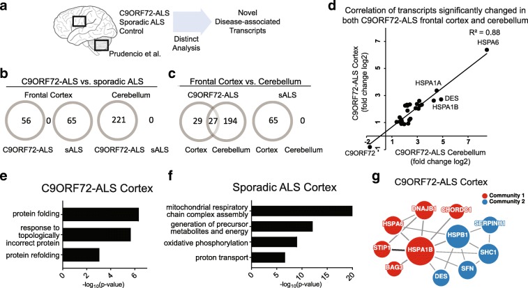
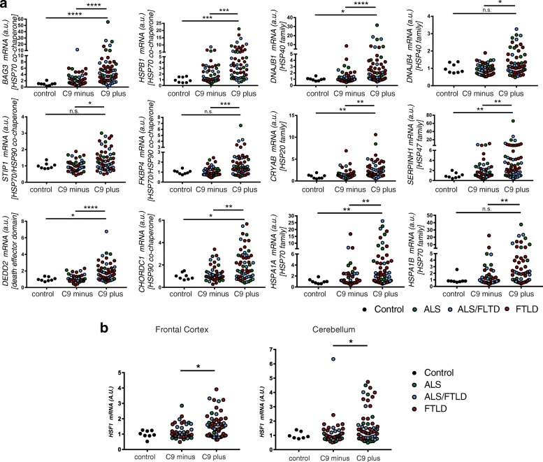
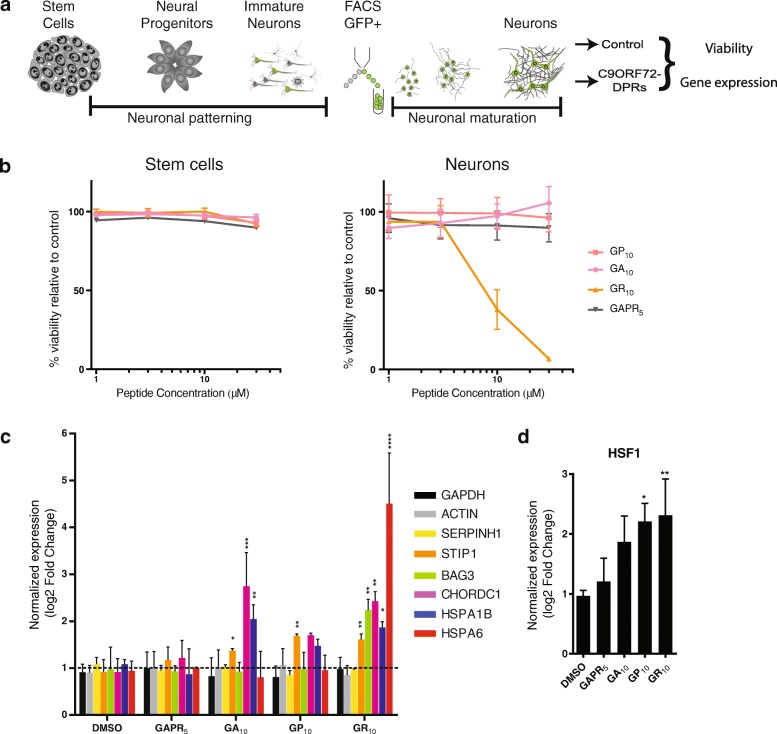
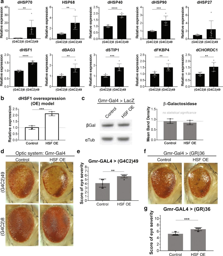
A hexanucleotide (GGGGCC) repeat expansion in C9ORF72 is the most common genetic contributor to amyotrophic lateral sclerosis (ALS) and frontotemporal lobar degeneration (FTLD). Reduced expression of the C9ORF72 gene product has been proposed as a potential contributor to disease pathogenesis. Additionally, repetitive RNAs and dipeptide repeat proteins (DPRs), such as poly-GR, can be produced by this hexanucleotide expansion that disrupt a number of cellular processes, potentially contributing to neural degeneration. To better discern which of these mechanisms leads to disease-associated changes in patient brains, we analyzed gene expression data generated from the cortex and cerebellum. We found that transcripts encoding heat shock proteins (HSPs) regulated by the HSF1 transcription factor were significantly induced in C9ORF72-ALS/FTLD patients relative to both sporadic ALS/FTLD cases and controls. Treatment of human neurons with chemically synthesized DPRs was sufficient to activate a similar transcriptional response. Expression of GGGGCC repeats and also poly-GR in the brains of Drosophila lead to the upregulation of HSF1 and the same highly-conserved HSPs. Additionally, HSF1 was a modifier of poly-GR toxicity in Drosophila. Our results suggest that the expression of DPRs are associated with upregulation of HSF1 and activation of a heat shock response in C9ORF72-ALS/FTLD.

Keywords: Amyotrophic lateral sclerosis; C9ORF72 repeat expansion; Dipeptide repeat proteins; Drosophila; Frontotemporal dementia; Frontotemporal lobar degeneration; HSF1; Heat shock response.


Conflict of interest statement

Ethics approval and consent to participate

Written informed consent was given by all participants or authorized family members in accordance with institutional review board guidelines of Mayo Clinic.

Competing interests

The authors declare that they have no competing interests.

Publisher’s Note

Springer Nature remains neutral with regard to jurisdictional claims in published maps and institutional affiliations.

Figures

Fig. 1
Fig. 1
Identification of specific cellular pathways perturbed in sporadic ALS and C9ORF72-ALS a Diagram of RNA-seq datasets obtained from the frontal cortex and cerebellum by Prudencio et al. b Comparison of the significant (FDR < 0.05) differentially expressed transcripts in C9ORF72-ALS (C9-ALS) and sporadic ALS (sALS). Note, there were no common transcripts between C9ORF72-ALS and sporadic ALS in either brain region. c Comparison of the differentially expressed transcripts by brain region. d Correlation of the fold change (log2) of changed transcripts in C9ORF72-ALS that were common to both brain regions (Spearman’s R2) e, f Gene ontology (GO) analysis revealed cellular processes affected in C9ORF72-ALS and sALS. g Protein-protein interaction analysis of proteins encoded by the transcripts changed in C9ORF72-ALS revealed a protein chaperone network
Fig. 2
Fig. 2
Activation of HSF1 in C9ORF72-ALS, FTLD, and combined ALS/FTLD patients. a Quantitative real-time PCR (qRT-PCR) for HSF1 target genes in the frontal cortex of sporadic and C9ORF72-associated disease (n = 56 C9ORF72-ALS/FTLD, n = 46 sporadic ALS/FTLD, n = 9 controls) (one-way ANOVA with Bonferonni post-hoc test for multiple comparisons, *p < 0.05, **p < 0.01, ***p < 0.001). No significant changes were detected between the sporadic cases and controls (mean values for each gene are provided in Additional file: 3 Table S3). b qRT-PCR for HSF1 in the frontal cortex and cerebellum of these same cases
Fig. 3
Fig. 3
DPRs induce expression of C9ORF72 signature transcripts in human neurons. a Diagram of generation of human neurons from stem cells. b Viability dose response curve of human stem cells and stem-cell derived neurons exposed to various DPRs (n = 6). c, d qRT-PCR of C9ORF72-chaperome transcripts (c) and HSF1 (d) in human stem cell-derived motor neurons following treatment with DPRs (poly-GA, poly-GP, poly-GR) or a scrambled poly-GAPR (5 uM for 24 h) normalized to control (DMSO-treated) neurons (mean ± SD, n = 3, one-way ANOVA with Dunnett’s post-hoc test for upregulated genes in DPR-treated neurons compared to control, * p < 0.05, ** p < 0.01, *** p < 0.001, **** p < 0.0001)
Fig. 4
Fig. 4
a Control UAS-(G4C2)8 and expanded UAS-(G4C2)49 transgenes were expressed in the adult fly nervous system using the drug-inducible Gal4 driver, elavGS, for 16d. Quantitative PCR (qPCR) analysis of endogenous dHSF1 and HSF1-regulated genes revealed significant upregulation with (G4C2)49 expression compared to (G4C2)8 controls. Differences in expression are likely underestimated as the analyses include neuronal and non-neuronal tissue while (G4C2)n was expressed only in neurons. b qPCR analysis of a dHSF1 overexpression mutant fly line shows endogenous HSF is upregulated approximately 2-fold in mutant flies compared to control. c Western immunoblot analysis of expression of a control UAS-LacZ transgene confirmed that the HSF OE mutant did not affect the Gal4/UAS expression system. d (G4C2)49 was expressed in the optic system of control animals or HSF OE animals using Gmr-Gal4. (G4C2)49 causes toxicity seen by pigment loss, reduced eye size, and disruptions in the normal ommatidial organization. In HSF OE animals, toxicity of (G4C2)49 is enhanced – animals have increased pigment loss, increased disruption of ommatidial organization, and further reduced eye size. Expression of control (G4C2)8 in the fly optic system (Gmr-Gal4) of control and HSF OE animals does not affect the external eye. e Quantification of the external eye degenerative phenotype caused by (G4C2)49 expression shows enhancement in HSF OE animals versus control animals to be consistent and statistically significant (n = 6). Animals received a score between 0 (WT eye) and 8 (lethality caused by extreme degeneration in the optic system). (G4C2)49 expression causes an average score of 4 in controls. f Gmr-GAL4 driven expression of (GR)36 shows toxicity in control scenarios like (G4C2)49. HSF OE in these animals also causes enhanced toxicity (increased pigment loss, increased disruption of ommatidial organization, and reduced eye size) g Quantification of the external eye degenerative phenotype caused by (GR)36 expression shows enhancement in HSF OE animals versus control animals to be consistent and statistically significant (n = 7). Animals received a score between 0 (WT eye) and 11 (lethality caused by extreme degeneration in the optic system) while (GR)36 causes an average score of 5 in controls. (All plots: mean +/− SD, unpaired, student’s t-test, *p < 0.05, **p < 0.01, ***p < 0.001, ****p < 0.0001)

References

    1. Agosta F, Ferraro PM, Riva N, Spinelli EG, Domi T, Carrera P, Copetti M, Falzone Y, Ferrari M, Lunetta C, Comi G, Falini A, Quattrini A, Filippi M (2017) Structural and functional brain signatures of C9orf72 in motor neuron disease. Neurobiol Aging. 10.1016/j.neurobiolaging.2017.05.024 - PubMed
    1. Al-Sarraj S, King A, Troakes C, Smith B, Maekawa S, Bodi I, Rogelj B, Al-Chalabi A, Hortobagyi T, Shaw CE. p62 positive, TDP-43 negative, neuronal cytoplasmic and intranuclear inclusions in the cerebellum and hippocampus define the pathology of C9orf72-linked FTLD and MND/ALS. Acta Neuropathol. 2011;122:691–702. doi: 10.1007/s00401-011-0911-2. - DOI - PubMed
    1. Anders S, Huber W. Differential expression analysis for sequence count data. Genome Biol. 2010;11:R106. doi: 10.1186/gb-2010-11-10-r106. - DOI - PMC - PubMed
    1. Benatar M, Wuu J, Andersen PM, Atassi N, David W, Cudkowicz M, Schoenfeld D. Randomized, double-blind, placebo-controlled trial of arimoclomol in rapidly progressive SOD1 ALS. Neurology. 2018;90:e565–e574. doi: 10.1212/WNL.0000000000004960. - DOI - PMC - PubMed
    1. Berson A, Sartoris A, Nativio R, Van Deerlin V, Toledo JB, Porta S, Liu S, Chung CY, Garcia BA, Lee VM, Trojanowski JQ, Johnson FB, Berger SL, Bonini NM (2017) TDP-43 promotes neurodegeneration by impairing chromatin remodeling. Curr Biol 27:3579–3590 e3576. doi:10.1016/j.cub.2017.10.024 - PMC - PubMed

Publication types

MeSH terms

LinkOut - more resources