. 2018 Jul 4;9(1):2613.
doi: 10.1038/s41467-018-05053-3.

microRNA-122 amplifies hepatitis C virus translation by shaping the structure of the internal ribosomal entry site

Affiliations

microRNA-122 amplifies hepatitis C virus translation by shaping the structure of the internal ribosomal entry site

Philipp Schult et al. Nat Commun. .

Abstract

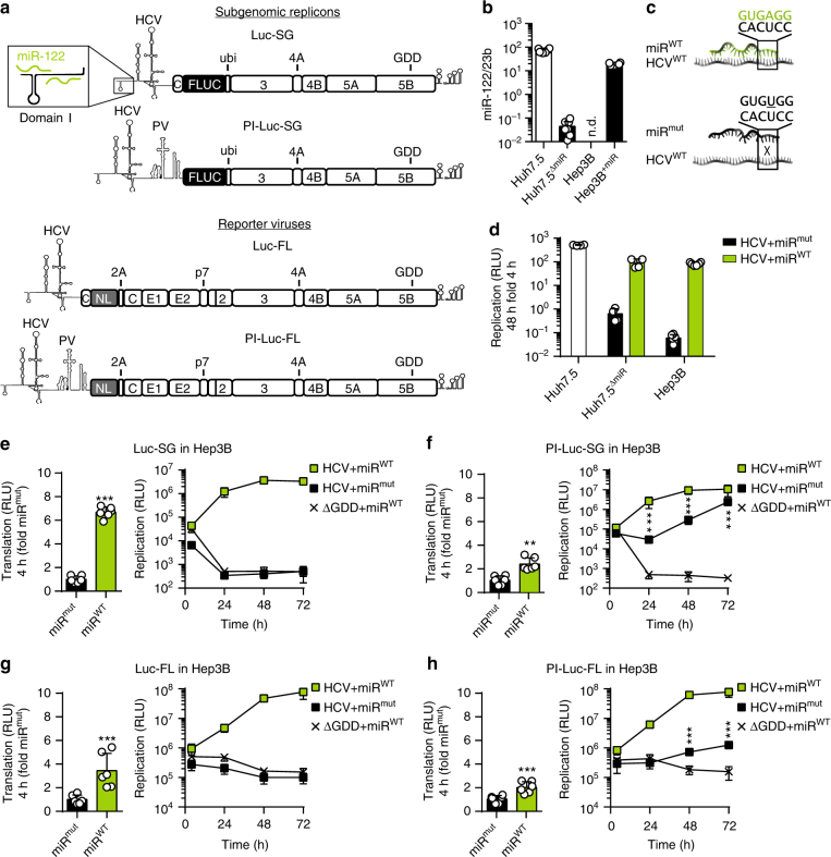
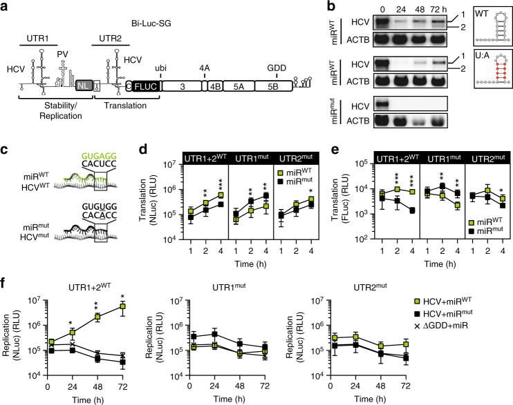
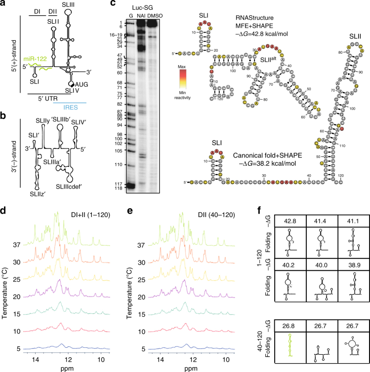
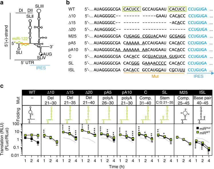
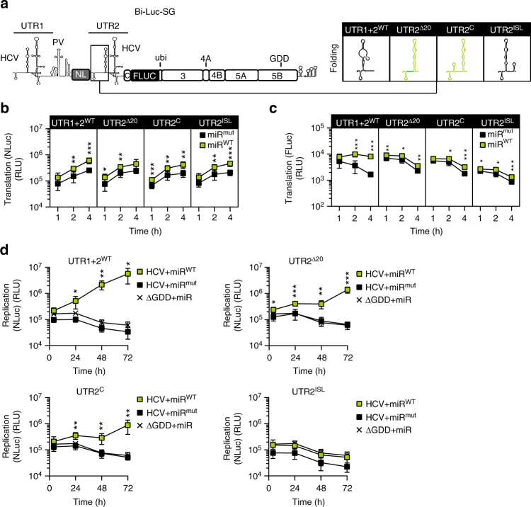
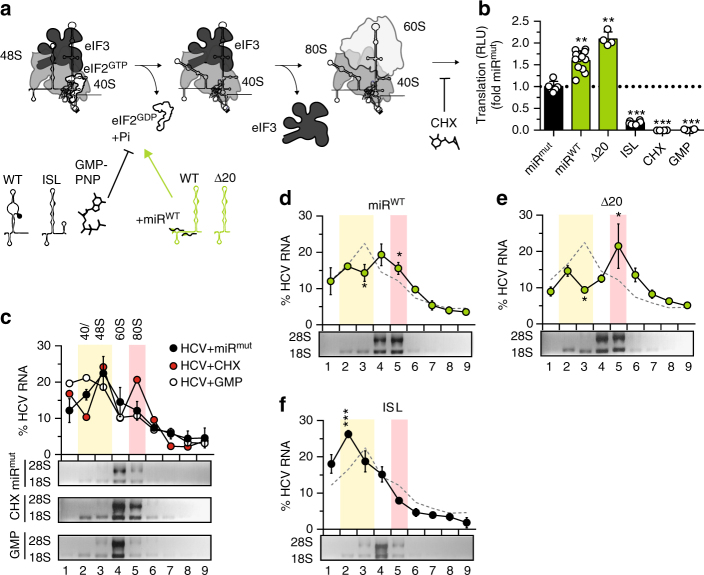
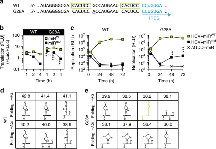
The liver-specific microRNA-122 (miR-122) recognizes two conserved sites at the 5' end of the hepatitis C virus (HCV) genome and contributes to stability, translation, and replication of the viral RNA. We show that stimulation of the HCV internal ribosome entry site (IRES) by miR-122 is essential for efficient viral replication. The mechanism relies on a dual function of the 5' terminal sequence in the complementary positive (translation) and negative strand (replication), requiring different secondary structures. Predictions and experimental evidence argue for several alternative folds involving the miR-binding region (MBR) adjacent to the IRES and interfering with its function. Mutations in the MBR, designed to suppress these dysfunctional structures indeed stimulate translation independently of miR-122. Conversely, MBR mutants favoring alternative folds show impaired IRES activity. Our results therefore suggest that miR-122 binding assists the folding of a functional IRES in an RNA chaperone-like manner by suppressing energetically favorable alternative secondary structures.


Conflict of interest statement

The authors declare no competing interests.

Figures

Fig. 1
Fig. 1
Initial translation and its importance for the HCV life cycle. a Representation of replicons and full-length viruses used in this study (FLUC firefly luciferase, NL nano luciferase). 5′UTR/IRES elements are indicated. The box shows an enlarged schematic of the miR-122-binding sites in the 5′ UTR. b Quantification of mature miR-122 by stem-loop qRT-PCR in naïve Huh7.5 cells, a Huh7.5 miR-122 knock-out cell clone (Huh7.5ΔmiR), Hep3B cells before (Hep3B), and after (Hep3B+miR) electroporation of miR-122 mimic. All values were normalized to miR-23b levels, which is homogenously expressed in hepatic cells. c Binding of wild type miR-122 (miRWT) to a target RNA (HCVWT) and influence of seed mutations (miRmut). d Comparison of HCV replication in different cell lines by luciferase assay after co-electroporation of reporter replicons (Luc-SG) and miRWT or miRmut into Hep3B cells. Reporter activity was measured at 4 and 48 h. e Luciferase assay of initial translation (left panel) and replication (right panel) of subgenomic HCV. f Luciferase assay to determine the effects of PV IRES-driven polyprotein translation on the viral life cycle (PI-Luc-SG). g, h Confirmation of results from (e) and (f), using full length reporter viruses (Luc-FL, PI-Luc-FL). n.d. not detectable. Mean values (±SD), n = 3, in technical duplicates. RLU relative light units, ΔGDD replication deficient mutant. For translation, statistical significance was determined for miRWT against miRmut; for replication HCV + miRmut was tested against ΔGDD. *P<0.05, **P<0.01, *P<0.001
Fig. 2
Fig. 2
Dissection of stability and replication using a dual-luciferase replicon.a Illustration of the Bi-Luc-SG construct. miR-122 binding to UTR1 acts on RNA stability and replication (as measured by Nano luciferase activity), translation of non-structural proteins is driven by UTR2 (as measured by firefly luciferase activity). b Northern blot analysis of HCV RNA stability over 72 h. Stability of different Bi-Luc construct RNAs was analyzed in the presence of miRmut or miRWT. Band #1: full-length RNA, #2: truncated product. U:A mutations in SLI of UTR2 (red dots) prevented the formation of truncated RNA. c Illustration of miRWT (green) and miRmut (black) binding upon introduction of a matching point mutation in the target (underlined). d, e Translation assay of point mutants cloned into the MBR of UTR1 or 2. The activity of Nano (d) and firefly (e) luciferase was measured in the presence of miRmut or miRWT. f Assessment of the full intracellular replication cycle of the Bi-Luc-SG constructs after electroporation with miRmut or miRWT. Replication was monitored with the nano luciferase signal. Mean values (±SD), n = 3, duplicates. RLU relative light units, ΔGDD replication deficient mutant. For translation, statistical significance was determined for miRWT against miRmut, for replication miRWT and miRmut conditions were tested against ΔGDD. *P<0.05, **P<0.01, ***P<0.001
Fig. 3
Fig. 3
Structural assessment of the HCV 5′ UTR.a Schematic overview of the HCV 5′ UTR and IRES. The miR-122-binding sites are indicated in green. Domain I and II are indicated above (DI and DII). b Schematic overview of the 3′ UTR of the negative strand RNA, which is complementary to the 5′UTR of the positive strand. Note the divergence in secondary structures, which is due to the different function in replication (3′(-)-strand) and translation (5′(+)-strand). c Representative autoradiography image showing the in vitro SHAPE reactivity of nucleotides 1–120 of a HCV reporter replicon. The SHAPE data were used to predict the secondary structure (minimum free energy, MFE) of DI and II with the RNA structure web tool and were visualized with R2R. The nucleotides of SLIIalt are colored according to their respective normalized reactivity. Yellow and red colors indicate unpaired nucleotides. The SHAPE data are also shown in context of the canonical fold of DI + II. d, e RNA conformation of DI + II (nt 1–120) or DII alone (nt 40–120), monitored by 1D 1H NMR spectra of imino protons in a temperature gradient. f Top conformations calculated by RNA structure and their respective free energy values (−ΔG [kcal/mol]) for DI + II or DII alone
Fig. 4
Fig. 4
DI mutants and their influence on translation using the Luc-SG replicon.a Illustration of the HCV 5′ UTR and IRES and location of the inserted mutations (orange). b Sequence alterations of DI mutants compared to WT. The miR-122 seed binding sites are marked by green boxes. Note that all mutants lack wild type miR-122 binding, due to mutations in the seed sequence. c Translation assays in Hep3B cells, using firefly luciferase (FLuc) reporter replicons and a capped Renilla (RLuc) control. miRmut or miRWT were co-electroporated, and luciferase activity was measured after 1, 2 and 4 h. All constructs were replication deficient ΔGDD mutants, to exclude potential effects of early replication. Mutations are described in the “mut” row, and a pictogram of the predicted minimum free energy structure in the “folding” row. Mutants enhancing SLII formation are depicted in green, SLIIalt stabilizing in red. Mean values (±SD), n = 3, duplicates. RLU relative light units. Statistical significance of the difference between WT and DI mutants in the presence of miRmut is indicated. The reference graph used to calculate is given in each subpanel in light gray. *P < 0.05, **P < 0.01, ***P < 0.001
Fig. 5
Fig. 5
Translation stimulation by DI mutations in the Bi-Luc context.a Schematic representation of the dual-luciferase construct (Bi-Luc-SG) and insertion region of the mutations (black box). The in silico predicted MFE structures of the inserted mutants are given in the table on the right. Mutants enhancing SLII formation are depicted in green. b, c Translation assay of selected mutants cloned into UTR2. The activity of Nano and firefly luciferase was measured in presence of miRmut or miRWT. d Assessment of the full intracellular replication cycle of the dual luciferase constructs after electroporation with miRmut or miRWT. Replication was monitored via the Nano luciferase signal. Mean values (±SD), n = 3, in technical duplicates. RLU relative light units, ΔGDD replication-deficient mutant. *P < 0.05, **P < 0.01, ***P < 0.001
Fig. 6
Fig. 6
Ribosome profiling.a Schematic representation of translation initiation and assembly of the 80S ribosome. The 48S preinitiation complex is constituted of the 40S ribosomal subunit, eIF3 and the (eIF2)–GTP-tRNAPMetP. Hydrolysis of GTP leads to release of eIF2. The L-shape of SLII is crucial for this process. Hence, the ISL mutant, misfolded WT and GMP-PNP, inhibiting GTPase activity, stall the reaction at this stage (bar-headed line). In contrast, miR-bound WT and Δ20 should activate eIF2-release, since SLII is effectively formed (green arrow). Subsequently, eIF3 is released and the 60S subunit is recruited to form the 80S complex. The following transition from initiation to elongation can be inhibited by CHX (bar-headed line). b Monitoring translation by luciferase assay in HeLa cell extracts, using Luc-SG in presence of miRmut or miRWT, DI mutants, GMP-PNP or CHX. c Analysis of HCV RNA distribution in ribosome profiles of HeLa cell extracts. Total lysates incubated with HCV in vitro transcripts were recorded; Relative abundance of HCV RNA from sucrose fractions was analyzed by qRT-PCR. Represented are percentages relative to the total amount in the gradient. The assay was performed with either no inhibitor, GMP-PNP or CHX in the reaction mix. The HCV RNA profile for the control reactions is shown in the top panel. Total RNA was analyzed by denaturing agarose gel electrophoresis (lower panel) to confirm the presence of 18S and 28S ribosomal RNA and distinguish precisely 40/48S and 80S ribosomes. Note that in fraction 4 and 9 the CHX data points are obscured by the GMP data. d Comparison of translation initiation efficiency of Luc-SG in presence of miRWT. The control with miRmut is shown as dashed line for comparison. e, f As in (d), comparing the WT reporter replicon and DI mutants. Mean values (±SD), n = 3, in technical duplicates. RLU relative light units. Significance was determined compared to the miRmut control. The reference graph used to calculate is given in light gray. *P<0.05, **P<0.01, ***P<0.001. 40/48S and 80S containing fractions are highlighted by yellow or orange boxes, respectively. Note that results shown in panels (df) were obtained in absence of CHX
Fig. 7
Fig. 7
Impact of natural variants on IRES-mediated translation.a Sequence of the G28A mutant compared to wild type. b Translation assay of WT versus G28A in the presence of miRmut or miRWT. c Replication assay of WT versus G28A in presence of miRmut or miRWT. d Representation of the predicted structural ensemble for the first 120 nt of the WT, as predicted by RNA structure and their respective free energy values (−ΔG [kcal/mol]). e As in (d) for the G28A mutant. Mean values (±SD), n = 3, in technical duplicates. RLU relative light units, ΔGDD replication deficient mutant. For the translation assay, statistical significance of the difference between WT and G28A mutants in presence of miRmut is indicated. The reference graph used to calculate is given in light gray. *P < 0.05, **P < 0.01, ***P < 0.001

References

    1. Lohmann V. Hepatitis C virus RNA replication. Curr. Top. Microbiol. Immunol. 2013;369:167–198. - PMC - PubMed
    1. Reynolds JE, et al. Unique features of internal initiation of Hepatitis C virus RNA translation. EMBO J. 1995;14:6010–6020. - PMC - PubMed
    1. Friebe P, Bartenschlager R. Role of RNA structures in genome terminal sequences of the Hepatitis C virus for replication and assembly. J. Virol. 2009;83:11989–11995. doi: 10.1128/JVI.01508-09. - DOI - PMC - PubMed
    1. Smith RM, Walton CM, Wu CH, Wu GY. Secondary structure and hybridization accessibility of Hepatitis C virus 3’-terminal sequences. J. Virol. 2002;76:9563–9574. doi: 10.1128/JVI.76.19.9563-9574.2002. - DOI - PMC - PubMed
    1. Khawaja A, Vopalensky V, Pospisek M. Understanding the potential of hepatitis C virus internal ribosome entry site domains to modulate translation initiation via their structure and function. Wiley Interdiscip. Rev. RNA. 2015;6:211–224. doi: 10.1002/wrna.1268. - DOI - PMC - PubMed

Publication types

MeSH terms