. 2018 Jul;559(7713):274-278.
doi: 10.1038/s41586-018-0281-1. Epub 2018 Jul 4.

Mechanism for remodelling of the cell cycle checkpoint protein MAD2 by the ATPase TRIP13

Affiliations

Mechanism for remodelling of the cell cycle checkpoint protein MAD2 by the ATPase TRIP13

Claudio Alfieri et al. Nature. 2018 Jul.

Abstract

The maintenance of genome stability during mitosis is coordinated by the spindle assembly checkpoint (SAC) through its effector the mitotic checkpoint complex (MCC), an inhibitor of the anaphase-promoting complex (APC/C, also known as the cyclosome)1,2. Unattached kinetochores control MCC assembly by catalysing a change in the topology of the β-sheet of MAD2 (an MCC subunit), thereby generating the active closed MAD2 (C-MAD2) conformer3-5. Disassembly of free MCC, which is required for SAC inactivation and chromosome segregation, is an ATP-dependent process driven by the AAA+ ATPase TRIP13. In combination with p31comet, an SAC antagonist6, TRIP13 remodels C-MAD2 into inactive open MAD2 (O-MAD2)7-10. Here, we present a mechanism that explains how TRIP13-p31comet disassembles the MCC. Cryo-electron microscopy structures of the TRIP13-p31comet-C-MAD2-CDC20 complex reveal that p31comet recruits C-MAD2 to a defined site on the TRIP13 hexameric ring, positioning the N terminus of C-MAD2 (MAD2NT) to insert into the axial pore of TRIP13 and distorting the TRIP13 ring to initiate remodelling. Molecular modelling suggests that by gripping MAD2NT within its axial pore, TRIP13 couples sequential ATP-driven translocation of its hexameric ring along MAD2NT to push upwards on, and simultaneously rotate, the globular domains of the p31comet-C-MAD2 complex. This unwinds a region of the αA helix of C-MAD2 that is required to stabilize the C-MAD2 β-sheet, thus destabilizing C-MAD2 in favour of O-MAD2 and dissociating MAD2 from p31comet. Our study provides insights into how specific substrates are recruited to AAA+ ATPases through adaptor proteins and suggests a model of how translocation through the axial pore of AAA+ ATPases is coupled to protein remodelling.


Conflict of interest statement

Author information. The authors declare no competing interests.

Figures

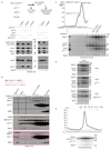
Extended Data Figure 1
Extended Data Figure 1. Biochemical characterization of Mad2-containing complexes with TRIP13.
a, TRIP13 and p31comet can extract O-Mad2 from MCC and APC/CMCC regardless of Apc15. Western blot showing the disassembly reactions together with the respective input material (i) of APC/C-bound MCC (APC/CMCC), in either the absence (lanes 1-2) or presence of Apc15 (lanes 3-6) and MCC alone (lanes 7-10) (experimental design is shown on top). Negative control reactions (lanes 3-4 and 9-10) were performed with the TRIP13E253Q mutant. Mad2 levels were detected with an anti-Mad2 antibody. Loading controls of Apc4-Strep, Apc15, Cdc20, TRIP13, Bub3 were detected with antibodies specific for Strep, Apc15, Cdc20, TRIP13 and Bub3 respectively. BubR1 (lanes 7-10) was detected with an anti-Strep antibody. b, Western blots showing eluted size exclusion (Superdex 200 10/300 column) fractions of the MCC and Mad2 remodelling reactions catalysed by TRIP13:p31comet in the context of free MCC and APC/CMCC. The fractions corresponding to (i) APC/CMCC, (ii) MCC, (iii) p31comet:C-Mad2 and (iv) monomeric C-Mad2 are shown. A reference gel for size exclusion column elution fractions corresponding to p31comet:C-Mad2:Cdc20-MBP, p31comet:C-Mad2 and monomeric C-Mad2 is shown in Extended Data Fig. 2a. c, Analysis of TRIP13-p31comet complexes with the MCC using size exclusion chromatography in the presence of ATP. Coomassie-stained gel showing the gel filtration fractions (chromatogram above) of p31comet:TRIP13 complexes in complex with MCC (fraction 8 is the p31comet:TRIP13 complex with C-Mad2-Cdc20 and fraction 9 is the BubR1:Bub3 complex). Input material (i) is shown on the left. d, MCC binds p31comet and not TRIP13 alone. Coomassie-stained gel showing the gel filtration fractions of the MCC (top gel) and in the presence of p31comet (middle gel) and TRIP13E253Q (lower gel). e, Chromatogram (top) and SDS-PAGE (bottom) of the gel filtration performed with the TRIP13E253Q:p31comet:C-Mad2:Cdc20 complex in the presence of ATPγS. Experiments in a–e were performed in triplicate with similar results. See Supplementary Fig. 1 for gel source data.
Extended Data Figure 2
Extended Data Figure 2. Biochemical assay for TRIP13:p31comet-catalysed O-Mad2 generation.
Shown are size exclusion (Superdex 200 10/300 column) chromatograms and corresponding Coomassie-stained gels for the Mad2 remodelling reaction catalysed by TRIP13:p31comet. a, Reference chromatograms and Coomassie-stained gels for (i) p31comet:C-Mad2:MBP-Cdc20 (brown trace), (ii) p31comet:C-Mad2 (blue trace) and (iii) monomeric O-Mad2 (red trace). Chromatograms and gels are colour-coded. Monomeric O-Mad2 elutes in fractions 22-24, whereas C-Mad2 in complex with p31comet (p31comet:C-Mad2) elutes in fractions 17-19. b, Chromatograms and Coomassie-stained gels for the products of the reaction of TRIP13 with (i) wild type p31comet and Mad2 (orange trace) and (ii) WT p31comet and mutant C-Mad2 (C-Mad2Δ7 (7 N-terminal residues deleted)). c, Chromatograms and Coomassie-stained gels for the products of the reaction with (i) mutant C-Mad2LEE, wild type TRIP13 and p31comet (yellow trace), (ii) mutant TRIP13E269R,D272R and wild type p31comet and C-Mad2 (green trace) and (iii) mutant p31comet-α3-4 and wild type TRIP13 and C-Mad2 (black trace). Experiments in a–c were performed in triplicate with similar results. See Supplementary Fig. 1 for gel source data.
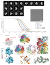
Extended Data Figure 3
Extended Data Figure 3. Cryo-EM analysis and resolution of TRIP13 complexes in this study.
a, Left: Gallery of two-dimensional class averages of TRIP13-p31-substrate showing different views representative of 50 two-dimensional classes. Right: A typical cryo-EM micrograph of TRIP13-p31-substrate representative of 3,630 micrographs. b, Fourier shell correlation (FSC) curves are shown for all the cryo-EM reconstructions determined in this study. c, local resolution maps calculated with RESMAP of the TRIP13p31-substrate complex. d, Cryo-EM density of the TRIP13-p31-substrate reconstruction shown as in Fig. 1b. e, g, Representative density quality for the ATPγS (e) and β-strand (g). In (e) critical residues for the TRIP13 catalytic site are indicated: WA (Walker A), WB (Walker B), S1 (sensor 1), S2 (sensor 2) and RF (Arg finger). f, Close up of the TRIP13 pore loops interacting with C-Mad2NT from the cryo-EM density of the TRIP13-p31-substrate. EM density for C-Mad2 shown in black mesh, TRIP13 in transparent coloured surface.
Extended Data Figure 4
Extended Data Figure 4. Three-dimensional classification of TRIP13-p31-substrate full dataset.
a, Local refinement (see Methods) by applying a mask covering TRIP13A/B/C/D/E monomers. b, c, 3D class averages obtained by classification (see Methods) of the TRIP13-p31-substrate full dataset (b) and TRIP13-p31-substrate (c) are shown. The percentages relative to the total number of TRIP13-p31-substrate particles are shown.
Extended Data Figure 5
Extended Data Figure 5. Comparative analysis of TRIP13 structures.
a, Comparison of TRIP13 cryo-EM structure (right, this study) and previous TRIP13 crystal structures (left, C. elegans PCH2 (ref. 17), middle, human TRIP13 (ref. 17)). b, Comparison of TRIP13 monomers within the TRIP13 cryo-EM structure, RMSDs between TRIP13A and other TRIP13 monomers are indicated in the insert table. A superimposition of all six TRIP13 monomers is shown, colour-coded according to Fig. 1. TRIP13F differs from all the other monomers in the relative orientation of the small and large AAA+ domains. Its conformation relative to TRIP13A is shown in the lower right. The open conformation prevents nucleotide binding. (The sensor 2 residue (S2) is positioned too far from the ATP-binding site). Lower left panel shows a superimposition of TRIP13F onto an open subunit C (grey) of the PCH2 structure . c, Conformational differences in pore loop-1 between the PCH2:ADP complex (grey) and the cryo-EM TRIP13:ATPγS complex (orange) (this study).
Extended Data Figure 6
Extended Data Figure 6. Structures of Mad2.
a, C-Mad2 (ref. 23); b, in the TRIP13:p31comet:C-Mad2 complex (this work); c, O-Mad2 (ref. 23); d, the S. pombe mitotic checkpoint complex ; e, p31comet:C-Mad2 complex . In all figures the regions of Mad2 that reposition on the O-Mad2 to C-Mad2 transition are coloured blue and red for N-terminal (residues 1-16) and C-terminal (158 to 204) regions respectively. In O-Mad2 these are the N-terminus (Mad2NT) and β1 strand (blue) and C-terminal β7-β8 hairpin (red). In C-Mad2 these are Mad2NT including the αN helix, and first turn of αA (blue), and C-terminal β8’-β8” hairpin, safety belt and C-terminus (Mad2CT) (red). On conversion of O-Mad2 to C-Mad2, the β1 strand is displaced and replaced by the β8’-β8” hairpin. Residues 13-15 of β1 form an additional turn at the N-terminus of αA present in C-Mad2. The C-Mad2 ligand is coloured purple. MBM: Mad2-binding motif. MBP1: High affinity Mad2-binding peptide ,.
Extended Data Figure 7
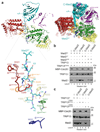
Extended Data Figure 7. TRIP13 interacts with p31comet through a composite interface formed of monomers D and E.
a, Details of the interaction between TRIP13 and p31comet. Left: schematic of TRIP13-p31comet interactions. Right: structure showing details of the main electrostatic contacts between TRIP13 and p31comet. Above: Schematic of the domain architecture of TRIP13 and p31comet. A row of aspartates on α7 engages the conserved safety belt motif residues Arg233 and Arg237 of p31comet. The adjacent Lys162 contacts a Glu-rich loop (111-121) in TRIP13 that is disordered in previous TRIP13 crystal structures ,. In our structure the Glu-rich loop lies directly above pore loop-1. Glu104 and Asp105 of the TRIP13NTD-ATPase domain linker, immediately preceding the Glu-rich loop, contact Arg227 and Lys229 of the p31comet safety-belt, agreeing with the importance of Lys229 for TRIP13-p31comet interactions in vitro and in vivo . On monomer E, the same acidic patch of α7 of the large AAA+ domain contacts basic residues at the N-terminus of α3 of p31comet. b, Details of the interaction of the p31comet α3-4 loop with TRIP13 subunit E. Seven basic residues shown were deleted and the mutant p31comet-α3-4 was tested in Mad2 remodelling assays and for assembly of a TRIP13:p31comet:C-Mad2 complex. c, Multiple sequence alignment of the p31comet α3-4 loop. d, Deletion of the nine N-terminal residues of Mad2 (Mad2Δ9), and mutation of the p31comet α3-4 loop (p31comet-α3-4loop) do not disrupt TRIP13:p31comet:C-Mad2 complex assembly. Coomassie-stained gel showing the gel filtration fraction of wild type and relevant mutant TRIP13:p31comet:C-Mad2 complexes purified by size exclusion chromatography. Experiment in d was performed in triplicate with similar results. See Supplementary Fig. 1 for gel source data.
Extended Data Figure 8
Extended Data Figure 8. Conservation of TRIP13pore loops and Mad2NT.
Multiple sequence alignment of (a) TRIP13 pore loop-1 and (b) pore loop-2 and the (c) N-terminal region of Mad2.
Extended Data Figure 9
Extended Data Figure 9. Models of the TRIP13:p31comet:C-Mad2 complexes in basal state 0 and basal state 1 (pre and post the first catalytic cycle).
a, Basal state 0 (similar to Fig. 4a). Superscripts on TRIP13 subunit labels denote basal state. b, Showing the conformational change of the TRIP13 hexamer post one catalytic cycle (Basal state 1) superimposed on the p31comet:C-Mad2 substrate prior to catalysis (as in basal state 0). The upward movement of the E and F subunits clashes with p31comet and C-Mad2. Pore loop residues of E and F subunits shift by 30 Å and 13 Å, respectively. c, close up view of (b) showing the clash between the F1 and E1 subunits of TRIP13 in basal state 1 with C-Mad2 and p31comet, respectively (as in basal state 0). d, Basal state 1 with TRIP13 and p31comet:C-Mad2 in the remodelled conformation (as in (Fig. 4b)) and now with the whole of the p31comet:C-Mad2:Cdc20 substrate repositioned onto the new C1 and D1 interface and the αA helix unwound by one turn. e, as in (d) but rotated 60° to show the p31comet:C-Mad2:Cdc20 substrate in the same view as in (a). f-j: Panels showing TRIP13 and only C-Mad2NT and connection to the C-Mad2 αA helix and proposed unwinding of the C-Mad2 αA helix. f, BS0 with view as in (e). g, TRIP13 adopts the BS1 conformation as in (e) with the whole p31comet:C-Mad2 substrate repositioned onto the C1-D1 interface. Note that the αA helix of C-Mad2 has not been unwound and C-Mad2NT (residues 2-12) is shifted along with the globular domain of C-Mad2 (C-Mad2GD). h, BS1 with C-Mad2NT in the BS0 position (i.e. unshifted relative to BS0) and αA of C-Mad2GD as p31comet:C-Mad2 is repositioned on the C1-D1 interface (but the αA helix is not unwound). This shows the 11 Å break between the Cα-atoms of Thr12 of C-Mad2NT and Leu13 of C-Mad2GD. C-Mad2NT is shifted down the TRIP13 pore two residues relative to (g). i, Close up view of the C-Mad2NT and connection to the αA helix. The view is the same as in (h) and it superimposes the position of C-Mad2NT as in the BS0 and BS1 states showing a 7.6 Å distance between the Cα-atoms of Thr12 of C-MadNT in the two states. This indicates the extent of required unwinding of the αA helix. j, The first turn of the αA helix unwinds to reconnect Thr12 of C-Mad2NT with Leu13 of the C-Mad2 αA helix. Model building was guided by insights from cryo-EM structures of the AAA+ ATPases VAT , Vps4 and Hsp104 that indicate a processive hand-over-hand mechanism in which the AAA+ motor translocates along the axis of the substrate. ATP hydrolysis propagates a sequential conformational change within the hexameric ring that converts the conformation of each subunit to that of its clockwise neighbour (viewed from p31comet).
Figure 1
Figure 1. Overall structures of the apo and TRIP13:p31-substrate complexes.
Side and top views of the cryo-EM structure model of TRIP13 in the apo state (a) and in complex with p31comet:C-Mad2 (class 2: basal state) (b). Relevant regions of TRIP13, p31comet and C-Mad2 are indicated.
Figure 2
Figure 2. Interaction between p31comet and TRIP13.
a, Surface conservation is displayed for TRIP13 monomers D and E, conservation score colour code is indicated below. A portion of p31comet is show in cartoon representation. NTD: N-terminal domain. b, Electrostatic surface representation for the TRIP13 – p31comet interacting surfaces (surface potential at ±5kT/e), colour code is displayed below. Top: TRIP13 electrostatic surface with a portion of C-Mad2 show in cartoon representation. Monomers D and E form a continuous acidic patch that is unique to this monomer pairing due to the conformation of loop 111-121 of monomer E. Bottom: p31comet electrostatic surface as viewed by TRIP13. Cartoons to the left provides an overall perspective. The surface area buried at the p31comet –TRIP13 interface is 2,392 Å2. c, Deletion of conserved basic residues of the p31comet α3-4 loop (Extended Data Fig. 7b) severely disrupts TRIP13-p31comet-catalysed Mad2 remodelling. Right: experimental design for Cdc20:C-Mad2 complex disassembly reaction. Left: Western blot showing input (i) and size exclusion fractions corresponding to monomeric O-Mad2 (lanes 2 and 4) and Extended Data Fig. 2c. Experiment in c was in triplicate with similar results. For gel source data see Supplementary Fig. 1.
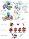
Figure 3
Figure 3. Interaction between C-Mad2NT and the TRIP13 pore loops.
a, Top: Overview of the TRIP13:p31-substrate complex showing Mad2NT inserted into the TRIP13 pore (left) with corresponding EM density map on right. Bottom: Detailed representation of Mad2NT residues interacting with TRIP13 pore loop residues. b, Western blot showing input (i) and O-Mad2 fractions of the Cdc20:C-Mad2 complex disassembly by TRIP13E253Q mutant (lanes 1-2) and TRIP13wt (lanes 3-8). Mad2 levels and loading controls for TRIP13 and Cdc20 are detected with their respective antibodies. Mutants of Mad2NT (lanes 5-8) affect the levels of O-Mad2 released from the Cdc20:C-Mad2 complex. c, Mutation of D269 and E272 of pore loop-2 of TRIP13 ablates TRIP13-p31comet-catalysed Mad2 remodelling (and Extended Data Fig. 2c). Experiments in b, c were in triplicate with similar results.
Figure 4
Figure 4. Sequential catalytic cycles of TRIP13 remodel Mad2.
a, b, Overall views of TRIP13:p31-substrate complex: (a) before catalysis (pre-catalytic: basal state 0) and (b) after the first catalytic cycle (basal state 1). TRIP13 subunit superscripts denote the catalytic cycle. Thr12 of Mad2NT is coloured yellow indicating the boundary with αA in basal state 0. After the first catalytic cycle one turn of the αA helix unwinds (compare (c) and (d)). This disrupts contacts between the N-terminus of the αA helix and the β8’-β8” hairpin. The N-terminal region of C-Mad2 that differs between O-Mad2 and C-Mad2 is in blue (Extended Data Fig. 6b,c). Further structural details are in Extended Data Fig. 9.
Figure 5
Figure 5. Differences between basal and activated states of the TRIP13-p31-substrate.
a, Two views comparing the basal state (grey) and activated state (colour code as in Fig. 1) of the TRIP13:p31-substrate (superimposition on C-Mad2). Left insert panel illustrates displacement of reference pore loop residues between basal and active states. The displacement grows from the p31comet-contacting monomer D to the F monomer, with F featuring the largest displacement of 5 Å. b, The conformational changes of monomer F pore loops and the C-Mad2 β2-β3 hairpin in the active state are shown relative to their positions in the basal state (grey). c, Schematic of the proposed mechanism of TRIP13-p31comet-catalysed remodelling of O-Mad2 (and Supplementary Video 2). The remodelled structural elements of Mad2 are indicated. EA and FA: activated conformations of monomers E and F, respectively. The O-Mad2 products dissociate from p31comet.

References

    1. Musacchio A. The Molecular Biology of Spindle Assembly Checkpoint Signaling Dynamics. Current biology : CB. 2015;25:R1002–1018. - PubMed
    1. Alfieri C, Zhang S, Barford D. Visualizing the complex functions and mechanisms of the anaphase promoting complex/cyclosome (APC/C) Open biology. 2017;7 - PMC - PubMed
    1. Kulukian A, Han JS, Cleveland DW. Unattached kinetochores catalyze production of an anaphase inhibitor that requires a Mad2 template to prime Cdc20 for BubR1 binding. Developmental cell. 2009;16:105–117. - PMC - PubMed
    1. Faesen AC, et al. Basis of catalytic assembly of the mitotic checkpoint complex. Nature. 2017;542:498–502. - PMC - PubMed
    1. Ji Z, Gao H, Jia L, Li B, Yu H. A sequential multi-target Mps1 phosphorylation cascade promotes spindle checkpoint signaling. eLife. 2017;6 doi: 10.7554/eLife.22513. - DOI - PMC - PubMed

Publication types

MeSH terms