. 2018 Jun 19:11:214.
doi: 10.3389/fnmol.2018.00214. eCollection 2018.

Endocannabinoid Signaling at Hypothalamic Steroidogenic Factor-1/Proopiomelanocortin Synapses Is Sex- and Diet-Sensitive

Affiliations

Endocannabinoid Signaling at Hypothalamic Steroidogenic Factor-1/Proopiomelanocortin Synapses Is Sex- and Diet-Sensitive

Carolina Fabelo et al. Front Mol Neurosci. .

Abstract

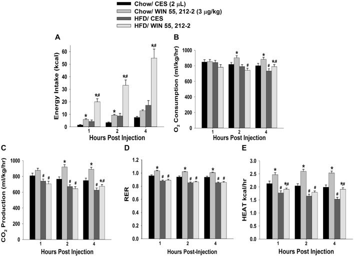
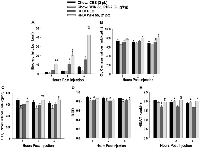
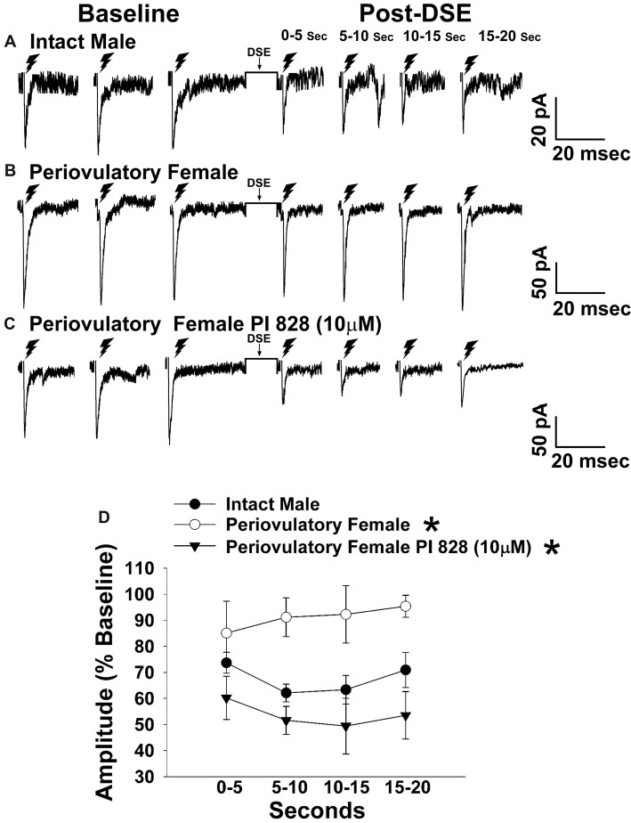
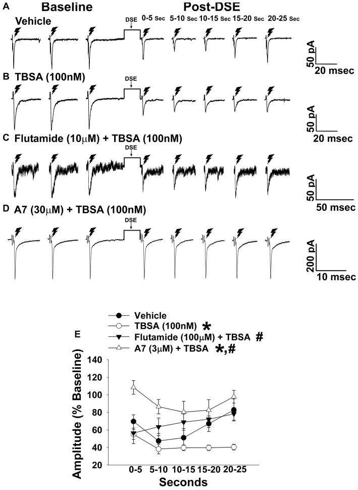
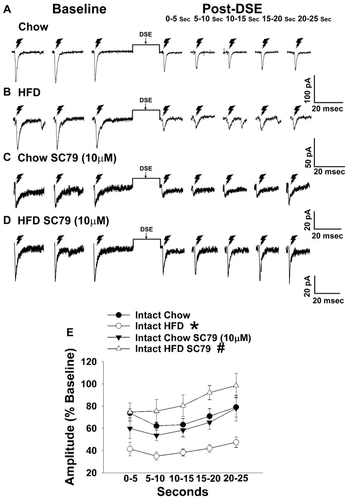
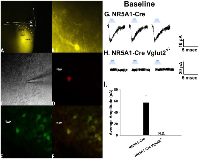
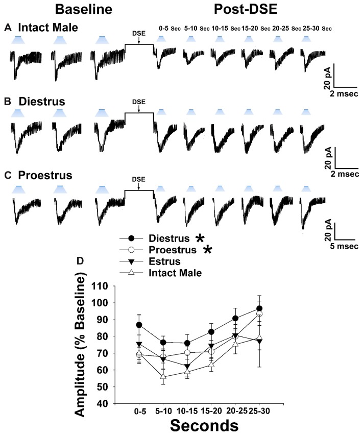
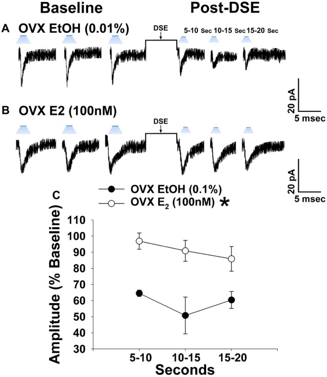
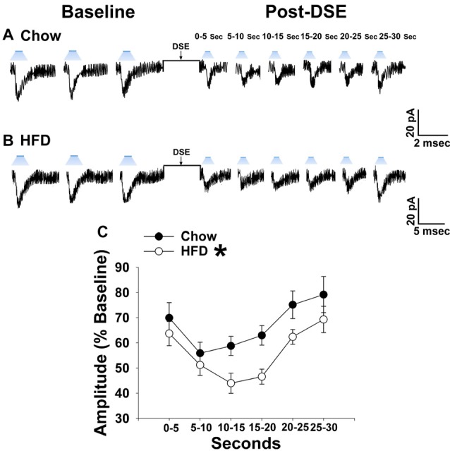
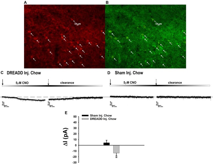
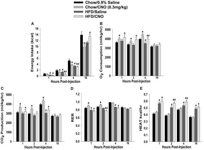
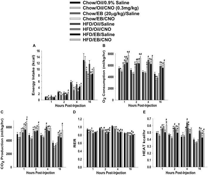
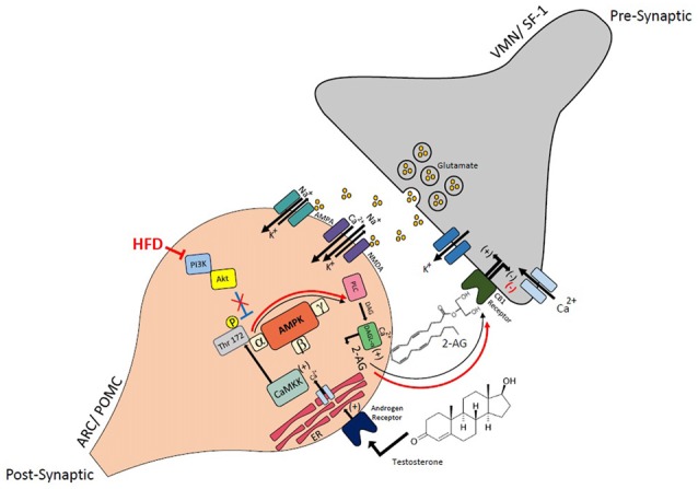
We tested the hypotheses that steroidogenic factor (SF)-1 neurons in the hypothalamic ventromedial nucleus (VMN) provide sexually disparate, endocannabinoid (EC)- and diet-sensitive glutamatergic input onto proopiomelanocortin (POMC) neurons. Electrophysiological recordings were performed in hypothalamic slices from intact and castrated guinea pigs, along with in vitro optogenetic experiments in intact male as well as cycling and ovariectomized female NR5A1-Cre mice. In slices from castrated male and female guinea pigs, depolarized-induced suppression of excitation (DSE) time-dependently reduced the amplitude of evoked excitatory postsynaptic currents (eEPSCs) in POMC neurons generated by electrically stimulating the dorsomedial VMN. Androgen stimulation rapidly enhanced this DSE, which was also found in insulin-resistant, high-fat diet (HFD)-fed males. By contrast, retrograde signaling at VMN/ARC POMC synapses was markedly attenuated in periovulatory females. HFD potentiated central cannabinoid-induced hyperphagia in both males and females, but exerted differential influences on cannabinoid-induced increases in energy expenditure. In NR5A1-Cre mice, the reduction in light-evoked EPSC amplitude caused by postsynaptic depolarization in cycling females was modest in comparison to that seen in intact males. Estradiol attenuated the DSE in light-evoked EPSC amplitude in slices from ovariectomized females. Moreover, the retrograde inhibition of transmission was further accentuated in HFD-fed males. Chemogenetic activation of SF-1 neurons suppressed appetite and increased energy expenditure in males, effects which were attenuated by HFD. Conversely, energy expenditure was increased in estradiol- but not vehicle-treated ovariectomized females. Together with our previous studies indicating that DSE in POMC neurons is EC-mediated, these findings indicate that VMN SF-1/ARC POMC synapses represent a sexually differentiated, EC- and diet-sensitive anorexigenic component within the hypothalamic energy balance circuitry.

Keywords: endocannabinoids; estradiol; insulin; obesity; proopiomelanocortin; sex differences; steroidogenic factor-1; testosterone.


Figures

Figure 1
Figure 1
High-fat diet (HFD) potentiates cannabinoid-induced changes in energy intake and but not energy expenditure, in male guinea pigs. Bars represent means and vertical lines 1 SEM of the cumulative energy intake (A), O2 consumption (B), CO2 production (C), respiratory exchange ratio (RER; D) and metabolic heat production (E), which were measured 1, 2 and 4 h after administration of the cannabinoid receptor agonist WIN 55,212-2 (3 μg; I.3.V) or its cremephor/ethanol/0.9% saline (CES) vehicle (2 μL; I.3.V). *P < 0.05 relative to CES treated animals; multifactorial analysis of variance (ANOVA)/least significant difference (LSD); n = 5. #P < 0.05 relative to chow-fed animals; repeated measures, multifactorial ANOVA/LSD; n = 5.
Figure 2
Figure 2
Activation of hypothalamic cannabinoid receptors increases energy intake and various measures of energy expenditure in HFD- but not chow-fed periovulatory female guinea pigs. Bars represent means and vertical lines 1 SEM of the cumulative energy intake (A), O2 consumption (B), CO2 production (C), respiratory exchange ratio (RER; D) and metabolic heat production (E), which were measured 1, 2 and 4 h after administration of WIN 55,212-2 (3 μg; I.3.V) or its CES vehicle (2 μL; I.3.V). *P < 0.05 relative to CES treated animals; multifactorial ANOVA/LSD; n = 5. #P < 0.05 relative to chow-fed animals; repeated measures, multifactorial ANOVA/LSD; n = 5.
Figure 3
Figure 3
The postsynaptic depolarization-induced reduction in excitatory input onto proopiomelanocortin (POMC) neurons generated by stimulation of the dorsomedial ventromedial nucleus (VMN) is larger in males than in periovulatory females, which is reversed by the phosphatidylinositol-3-kinase (PI3K) inhibitor PI 828. Membrane current traces illustrating the baseline evoked excitatory postsynaptic current(eEPSC) amplitude, and the time-dependent reduction in amplitude at various time points after the depolarized-induced suppression of excitation (DSE) stimulus, in arcuate nucleus (ARC) POMC neurons are shown for the intact male (A; n = 9) periovulatory female (B; n = 6) and a slice from a periovulatory female pretreated with PI 828 (10 μM; (C) n = 4). The composite line graph (D) further illustrates the PI3K-dependent sex differences in the retrograde inhibition of excitatory input from the dorsomedial VMN onto ARC POMC neurons under baseline conditions. Lines represent means and lines 1 SEM. *P < 0.05; rank-transformed multifactorial ANOVA/LSD; n = 4–9.
Figure 4
Figure 4
The membrane-impermeant form of testosterone rapidly intensifies endocannabinoid (EC)-mediated reduction in eEPSC amplitude, which is blocked by a classical androgen receptor antagonist, as well as via inhibition of calcium/calmodulin-dependent protein kinase. Current membrane traces showing the decrease in eEPSC amplitude caused by postsynaptic depolarization seen in slices treated with either vehicle (A; n = 6) or testosterone 4-androsten-17 μ;-ol-3-one-3 carboxymethyloxime bovine serum albumin (TBSA; 100 nM; B; n = 8). Bath application of flutamide (10 μM) attenuated the rapid potentiating effects of TBSA (100 nM; C; n = 5) on the reduction in eEPSC amplitude, as did the calcium/calmodulin antagonist A7 administered via the internal solution (30 μM; D; n = 5). The composite line graph (E) shows the enhanced retrograde inhibition caused by TBSA. *P < 0.05 relative to vehicle treated slices; rank-transformed multifactorial ANOVA/LSD. #P < 0.05 relative to slices treated with TBSA alone; rank-transformed multifactorial ANOVA/LSD.
Figure 5
Figure 5
HFD intensifies the reduction in eEPSC amplitude caused by postsynaptic depolarization, which is abrogated by Akt activation. The reduction in eEPSC amplitude caused by the DSE seen in chow-fed animals (A; n = 9) is further amplified in HFD-fed animals (B; n = 5). The reduction in eEPSC amplitude caused by postsynaptic depolarization seen during recordings in slices taken from chow-fed animals is unaffected by pretreatment with the Akt activator SC79 (10 μM; C; n = 6). However, the heightened retrograde signaling observed in slices from HFD-fed animals (B) was completely abolished with SC79 (D; n = 5). The composite line graph further illustrates the heightened postsynaptic depolarization-induced reduction in eEPSC amplitude caused by the HFD, and the impact of reduced PI3K/Akt signaling on the augmented EC tone (E). Lines represent means and vertical lines 1 SEM. *P < 0.05 relative to chow-fed animals; rank-transformed multifactorial ANOVA/LSD. #P < 0.05 relative to vehicle-treated slices; rank-transformed multifactorial ANOVA/LSD.
Figure 6
Figure 6
Photostimulation elicits glutamatergic EPSCs at VMN steroidogenic factor (SF-1)/ARC POMC synapses. (A) ChR2 labeling in the VMN of a male NR5A1-Cre mouse visualized at ×4 with enhanced yellow fluorescent protein (eYFP) 2 weeks after injection with a ChR2-containing virus. Of note is the gradient extending from the dorsomedial VMN (VMHDM) to the ventrolateral VMN (VMHVL) that is characteristic of the distribution of VMN SF-1 neurons. (B) Magnified view of the outlined approximate recorded area in the ARC from (A) showing labeling of ChR2-containing fibers in the ARC visualized with eYFP. (C) An infrared, differential interference contrast image taken of an ARC neuron in close proximity to the eYFP-labeled fibers seen in (B). (D) Biocytin labeling of the cell in (A) visualized with streptavidin/Alexa Fluor 546. (E) An antibody directed against cocaine- and amphetamine-regulated transcript (CART), a phenotypic marker of POMC neurons, immunolabels the cell in (C) as visualized with Alexa Fluor 488. (F) A composite overlay of the biocytin/CART labeling seen in the cell in (A). Unless otherwise indicated, all photomicrographs were taken at ×40.A 25-ms stimulus of blue light subsequently delivered to the cell in (C–F) elicited robust light-evoked EPSCs (leEPSCs; G) that were indiscernible in recordings from NR5A1-creVglut2−/– animals (H). This is further illustrated in the composite bar graph in (I) Bars represent means and lines 1 SEM of the EPSC amplitude measured in slices from NR5A1-Cre (n = 22) and NR5A1-CreVglut2−/– (n = 3) animals.
Figure 7
Figure 7
Sex differences in the retrograde inhibition of glutamatergic input at VMN SF-1/ARC POMC synapses in NR5A1-Cre mice. The representative traces of membrane current show that the reduction in leEPSC amplitude caused by postsynaptic depolarization is comparatively larger in gonadally intact males (A; n = 22) than in diestrus (B; n = 19) and proestrus (C; n = 21) females. This can also be seen from the composite line graph (D). Lines represent means and lines 1 SEM. *P < 0.05; rank-transformed multifactorial ANOVA/LSD.
Figure 8
Figure 8
E2 significantly attenuates retrograde EC inhibition of leEPSC amplitude in POMC neurons from ovariectomized NR5A1-cre mice. LeEPSC amplitude is reduced post-DSE in recordings from EtOH-treated slices (0.01%; A; n = 5). However when other slices are treated with E2 (100 nM; n = 7), the degree of reduction is significantly reduced (B). These points are further illustrates by the data shown in the line graph in (C) Bars represent means and vertical lines 1 SEM. *P < 0.05; rank-transformed multifactorial ANOVA/LSD.
Figure 9
Figure 9
The postsynaptic depolarization-induced reduction in leEPSC amplitude seen in chow-fed males is even further reduced in HFD-fed animals. (A) Representative membrane current trace illustrating that the eEPSC amplitude is reduced post DSE in chow-fed animals (n = 22). However, this effect is significantly enhanced in animals fed a HFD (B; n = 19). A composite line graph showing how the reduction in leEPSC amplitude caused by postsynaptic depolarization is significantly augmented (C). Lines represent means and vertical lines 1 SEM. *P < 0.05; rank-transformed multifactorial ANOVA/LSD.
Figure 10
Figure 10
Chemostimulation of VMN SF-1 neurons in male NR5A1-cre mice reversibly stimulates POMC neurons in designer receptor exclusively activated by designer drugs (DREADD)-injected but not sham-injected animals. The color photomicrograph shows the expression of the Gq-M3-DREADD (visualized with mCherry, A) in SF-1 neurons (visualized with AF488, B). (C) Representative traces of membrane current showing that clozapine-N-oxide (CNO) (5 μM) induced a reversible inward current in identified POMC neurons from DREADD-injected animals (n = 13) that was altogether absent in cells from sham-injected animals (D; n = 4). The composite bar graph in (E) further illustrates the stimulatory effect of CNO in POMC neurons from DREADD-injected animals but not sham-injected animals. Bars represent means and lines 1 SEM of the CNO-induced change in membrane current. *P < 0.05; Student’s t-test.
Figure 11
Figure 11
Chemostimulation of VMN SF-1 neurons in male NR5A1-cre mice reduces cumulative energy intake and increases energy expenditure in a diet-sensitive manner. CNO (0.3 mg/kg) significantly decreased cumulative energy intake (A), and increased O2 consumption (B), CO2 production (C) and RER (D) in chow-fed animals, all of which was significantly attenuated in HFD-fed animals. Conversely, CNO increased metabolic heat production in HFD-fed animals (E). Bars represent means and lines 1 SEM of the cumulative food intake, O2 consumption, CO2 production, RER and metabolic heat production seen in chow- and HFD-fed NR5A1-Cre mice treated with either CNO (0.3 mg/kg; chow: n = 8; HFD: n = 5), or its filtered 0.9% saline vehicle (chow: n = 7; HFD: n = 5). *P < 0.05 relative to vehicle-treated animals; multifactorial ANOVA/LSD. #P < 0.05 relative to chow-fed animals; multifactorial ANOVA/LSD.
Figure 12
Figure 12
Chemostimulation of VMN SF-1 neurons in ovariectomized NR5A1-cre mice alters cumulative energy intake (A), O2 consumption (B), CO2 production (C), RER (D) and metabolic heat production (E) in an EB- and diet-sensitive manner. CNO (0.3 mg/kg) significantly decreased energy intake in chow-fed oil-treated animals, and to a larger extent in HFD-fed animals. It also increased energy expenditure in chow- and HFD-fed, EB-treated animals. Bars represent means and lines 1 SEM of the cumulative food intake, O2 consumption, CO2 production, and RER seen in chow- and HFD-fed NR5A1-Cre mice treated with either CNO (0.3 mg/kg; oil/chow: n = 5; oil/HFD: n = 6; EB/chow: n = 6; EB/HFD: n = 5), or its filtered 0.9% saline vehicle (oil/chow: n = 8; oil/HFD: n = 6; EB/chow: n = 6; EB/HFD: n = 6). *P < 0.05 relative to saline-treated animals; multifactorial ANOVA/LSD; n = 5–8. #P < 0.05 relative to chow-fed animals; multifactorial ANOVA/LSD; n = 5–8. P < 0.05 relative to oil-treated animals; multifactorial ANOVA/LSD.
Figure 13
Figure 13
Schematic illustrating the connectivity and EC signaling occurring at VMN SF-1/ARC POMC synapses in males, and how that signaling is rapidly modulated by androgens and influenced by diet. The magnified view of the VMN SF-1/ARC POMC synapse illustrates how testosterone activates a putative membrane androgen receptor to increase intracellular calcium. This increase in intracellular calcium activates calmodulin-dependent protein kinase kinase (CaMKK), which then phosphorylates threonine 172 on the α subunit of the AMPK complex to activate the enzyme. This activation leads to an increased production of 2-arachidonylglycerol (2-AG) through diacylglycerol lipase (DAGL)α, and the 2-AG released can retrogradely activate presynaptic CB1 receptors. This inhibits calcium entry, which decreases glutamatergic input. The resultant relief from the anorexigenic signal leads to hyperphagia. AMPK activity is inhibited by PI3K/Akt signaling, and this inhibition is reduced with HFD-induced obesity/insulin resistance. This further elevates AMPK activity and subsequently EC tone at VMN SF-1/ARC POMC synapses.

Similar articles

Cited by

References

    1. Andrews W. W., Advis J. P., Ojeda S. R. (1981). The maturation of estradiol-negative feedback in female rats: evidence that the resetting of the hypothalamic “gonadostat” does not precede the first preovulatory surge of gonadotropins. Endocrinology 109, 2022–2031. 10.1210/endo-109-6-2022 - DOI - PubMed
    1. Appleyard S. M., Hayward M., Young J. I., Butler A. A., Cone R. D., Rubinstein M., et al. . (2003). A role for endogenous β-endorphin in energy homeostasis. Endocrinology 144, 1753–1760. 10.1210/en.2002-221096 - DOI - PubMed
    1. Asarian L., Geary N. (2006). Modulation of appetite by gonadal steroid hormones. Philos. Trans. R. Soc. Lond. B Biol. Sci. 361, 1251–1263. 10.1098/rstb.2006.1860 - DOI - PMC - PubMed
    1. Asarian L., Geary N. (2013). Sex differences in the physiology of eating. Am. J. Physiol. Regul. Integr. Comp. Physiol. 305, R1215–R1267. 10.1152/ajpregu.00446.2012 - DOI - PMC - PubMed
    1. Balthasar N., Coppari R., McMinn J., Liu S. M., Lee C. E., Tang V., et al. . (2004). Leptin receptor signaling in POMC neurons is required for normal body weight homeostasis. Neuron 42, 983–991. 10.1016/j.neuron.2004.06.004 - DOI - PubMed