. 2018 Jun 20:12:37.
doi: 10.3389/fninf.2018.00037. eCollection 2018.

Differential Path-Length Factor's Effect on the Characterization of Brain's Hemodynamic Response Function: A Functional Near-Infrared Study

Affiliations

Differential Path-Length Factor's Effect on the Characterization of Brain's Hemodynamic Response Function: A Functional Near-Infrared Study

Muhammad A Kamran et al. Front Neuroinform. .

Abstract

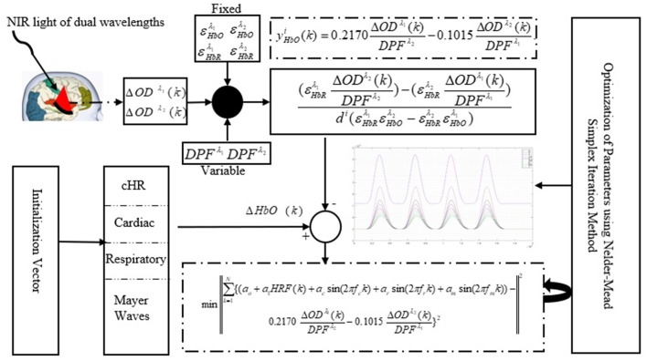
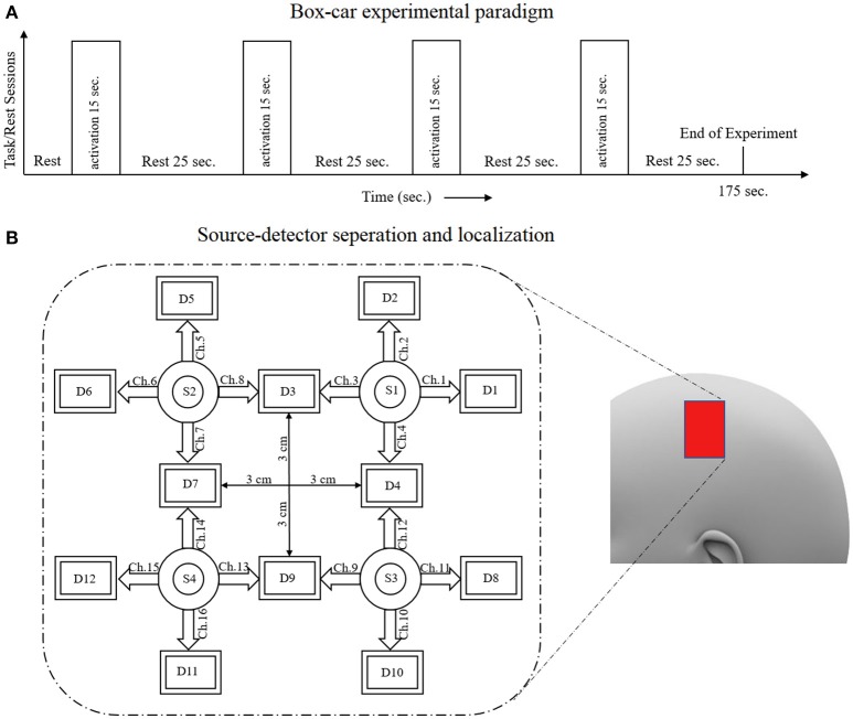
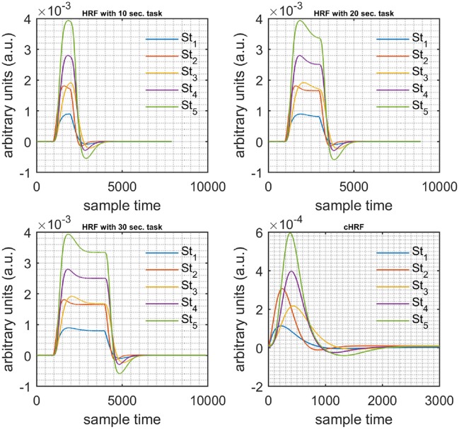
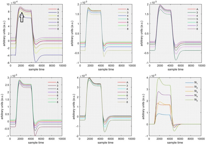
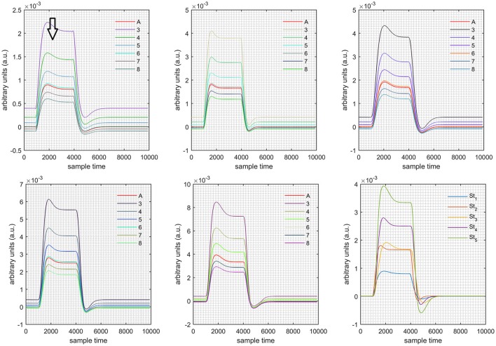
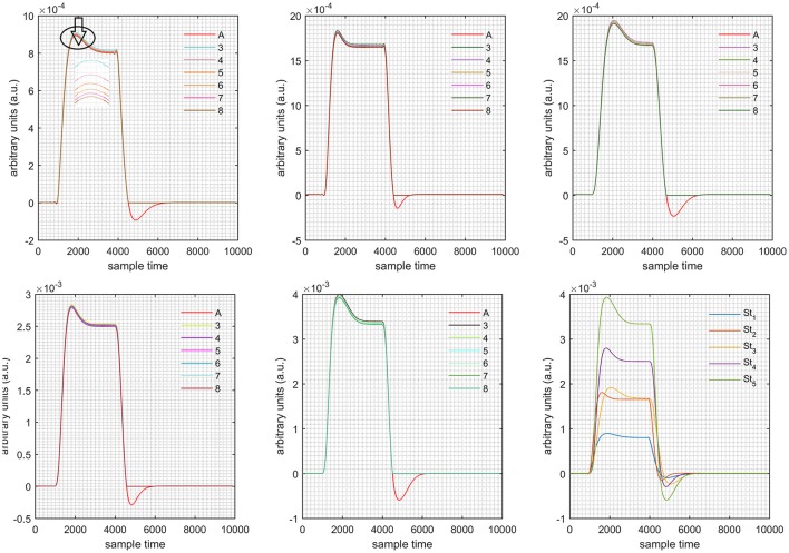
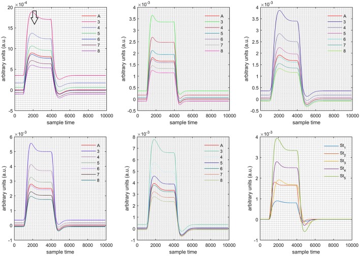
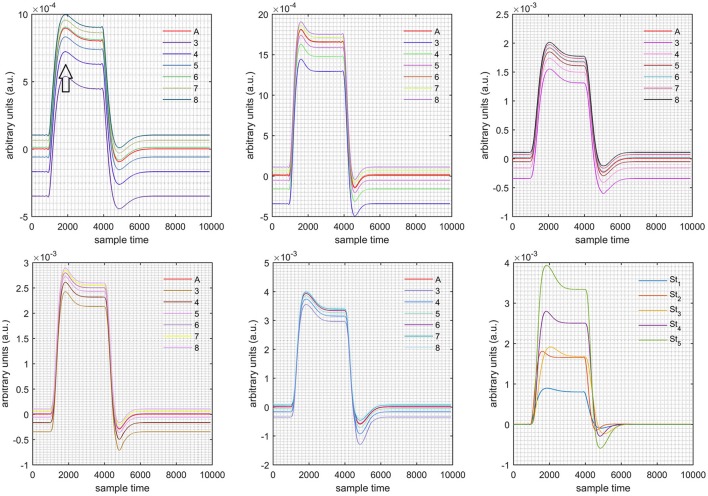
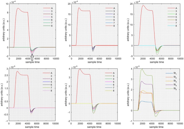
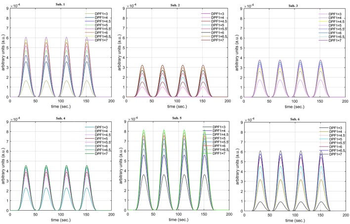
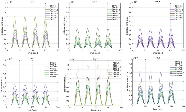
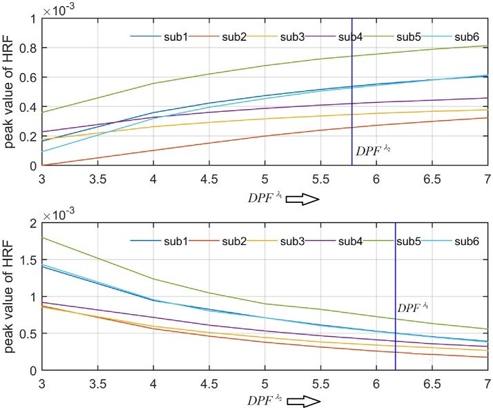
Functional near-infrared spectroscopy (fNIRS) has evolved as a neuro-imaging modality over the course of the past two decades. The removal of superfluous information accompanying the optical signal, however, remains a challenge. A comprehensive analysis of each step is necessary to ensure the extraction of actual information from measured fNIRS waveforms. A slight change in shape could alter the features required for fNIRS-BCI applications. In the present study, the effect of the differential path-length factor (DPF) values on the characteristics of the hemodynamic response function (HRF) was investigated. Results were compiled for both simulated data sets and healthy human subjects over a range of DPF values from three to eight. Different sets of activation durations and stimuli were used to generate the simulated signals for further analysis. These signals were split into optical densities under a constrained environment utilizing known values of DPF. Later, different values of DPF were used to analyze the variations of actual HRF. The results, as summarized into four categories, suggest that the DPF can change the main and post-stimuli responses in addition to other interferences. Six healthy subjects participated in this study. Their observed optical brain time-series were fed into an iterative optimization problem in order to estimate the best possible fit of HRF and physiological noises present in the measured signals with free parameters. A series of solutions was derived for different values of DPF in order to analyze the variations of HRF. It was observed that DPF change is responsible for HRF creep from actual values as well as changes in HRF characteristics.

Keywords: differential path-length factor; functional near-infrared spectroscopy; hemodynamic response; optical brain imaging; optimal cortical model.


Figures

Figure 1
Figure 1
Schematic of algorithm.
Figure 2
Figure 2
(A) Experimental paradigm, (B) source-detector localization and separation.
Figure 3
Figure 3
Simulated data with initial rest (10 s) different stimulus (St1-St5) and task durations (10, 20, and 30 s) followed by 30 s rest, 10 s activation (top left), 20 s activation (top right), 30 s activation (bottom left), and canonical hemodynamic response to all stimuli (bottom right).
Figure 4
Figure 4
Results for both positive cases (Case: 1) with variation in value of DPFλ1 (3–8) under stimulation St1-St5 (top left, top middle, top right, bottom left, and bottom middle).
Figure 5
Figure 5
Results for both negative cases (Case: 2) with variation in value of DPFλ1 (3–8) under stimulation St1-St5 (top left, top middle, top right, bottom left, and bottom middle).
Figure 6
Figure 6
Results for first positive and second negative cases (Case: 3) with variation in value of DPFλ1 (3–8) under stimulation St1-St5 (top left, top middle, top right, bottom left, and bottom middle).
Figure 7
Figure 7
Results for first negative and second positive cases (Case: 4) with variation in value of DPFλ1 (3–8) under stimulation St1-St5 (top left, top middle, top right, bottom left, and bottom middle).
Figure 8
Figure 8
Results for both positive cases (Case: 1) with variation in value of DPFλ2 (3–8) under stimulation St1-St5 (top left, top middle, top right, bottom left, and bottom middle).
Figure 9
Figure 9
Results for both negative cases (Case: 2) with variation in value of DPFλ2 (3–8) under stimulation St1-St5 (top left, top middle, top right, bottom left, and bottom middle).
Figure 10
Figure 10
Results for first positive and second negative cases (Case: 3) with variation in value of DPFλ2 (3–8) under stimulation St1-St5 (top left, top middle, top right, bottom left, and bottom middle).
Figure 11
Figure 11
Results for first negative and second positive cases (Case: 4) with variation in value of DPFλ2 (3–8) under stimulation St1-St5 (top left, top middle, top right, bottom left, and bottom middle).
Figure 12
Figure 12
Results for fNIRS data set with variation in DPFλ1.
Figure 13
Figure 13
Results for fNIRS data set with variation in DPFλ2.
Figure 14
Figure 14
Effects of variations in DPFλ1 and DPFλ2 on peak of HRF for all subjects.

References

    1. Abdelnour A. F., Huppert T. (2009). Real-time imaging of human brain function by near-infrared spectroscopy using an adaptive general linear model. Neuroimage 46, 133–143. 10.1016/j.neuroimage.2009.01.033 - DOI - PMC - PubMed
    1. Cavazza A., Aranyi G., Charles F. (2017). BCI control of heuristic search algorithms. Front. Neuroinform. 11:6 10.3389/fninf.2017.00006 - DOI - PMC - PubMed
    1. Chatterjee S., Phillips J. P., Kyriacou P. A. (2015). Differential pathlength factor estimation for brain-like tissue from a single-layer Monte Carlo model, in Engineering in Medicine and Biology Society (EMBC), 2015 37th Annual International Conference of the IEEE (Milan: ), 3279–3282. - PubMed
    1. Delpy D. T., Cope M., van der Zee P., Arridge S. R., Wray S., Wyatt J. S. (1988). Estimation of optical pathlength through tissue from direct time of flight measurements. Phys. Med. Biol. 33, 1433–1442. 10.1088/0031-9155/33/12/008 - DOI - PubMed
    1. Duncan A., Meek J. H., Clemence M., Elwell C. E., Fallon P., Tyszczuk L., et al. . (1996). Measurement of cranial optical path length as a function of age using phase resolved near infrared spectroscopy. Pediatr. Res. 39, 889–894. 10.1203/00006450-199605000-00025 - DOI - PubMed

LinkOut - more resources