. 2018 Jun 20:9:457.
doi: 10.3389/fneur.2018.00457. eCollection 2018.

Pre-α-pro-GDNF and Pre-β-pro-GDNF Isoforms Are Neuroprotective in the 6-hydroxydopamine Rat Model of Parkinson's Disease

Affiliations

Pre-α-pro-GDNF and Pre-β-pro-GDNF Isoforms Are Neuroprotective in the 6-hydroxydopamine Rat Model of Parkinson's Disease

Anna-Maija Penttinen et al. Front Neurol. .

Abstract

Glial cell line-derived neurotrophic factor (GDNF) is one of the most studied neurotrophic factors. GDNF has two splice isoforms, full-length pre-α-pro-GDNF (α-GDNF) and pre-β-pro-GDNF (β-GDNF), which has a 26 amino acid deletion in the pro-region. Thus far, studies have focused solely on the α-GDNF isoform, and nothing is known about the in vivo effects of the shorter β-GDNF variant. Here we compare for the first time the effects of overexpressed α-GDNF and β-GDNF in non-lesioned rat striatum and the partial 6-hydroxydopamine lesion model of Parkinson's disease. GDNF isoforms were overexpressed with their native pre-pro-sequences in the striatum using an adeno-associated virus (AAV) vector, and the effects on motor performance and dopaminergic phenotype of the nigrostriatal pathway were assessed. In the non-lesioned striatum, both isoforms increased the density of dopamine transporter-positive fibers at 3 weeks after viral vector delivery. Although both isoforms increased the activity of the animals in cylinder assay, only α-GDNF enhanced the use of contralateral paw. Four weeks later, the striatal tyrosine hydroxylase (TH)-immunoreactivity was decreased in both α-GDNF and β-GDNF treated animals. In the neuroprotection assay, both GDNF splice isoforms increased the number of TH-immunoreactive cells in the substantia nigra but did not promote behavioral recovery based on amphetamine-induced rotation or cylinder assays. Thus, the shorter GDNF isoform, β-GDNF, and the full-length α-isoform have comparable neuroprotective efficacy on dopamine neurons of the nigrostriatal circuitry.

Keywords: GDNF; alternative splicing; dopamine; neurodegeneration; neurotrophic factors; splice variant; tyrosine hydroxylase.


Figures

Figure 1
Figure 1
Organization of main human GDNF splice isoforms. (A,B) In GDNF gene line represents introns and boxes represent exons (not in scale). Black boxes represent protein coding areas. Pre-α-pro-GDNF isoform has a full-length 58 amino acid pro-region, whereas the pre-β-pro-GDNF has shorter 32 amino acid pro-region.
Figure 2
Figure 2
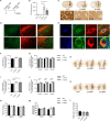
Effects of GDNF isoform overexpression on dopaminergic markers in non-lesioned striatum 3 weeks after AAV-injection. (A) Experimental design. (B) Overexpression levels of GDNF isoforms were confirmed with ELISA [Kruskal-Wallis test H(3) = 15.457, p = 0.001, followed by Bonferroni corrected Mann-Whitney U post-hoc test, **p < 0.01, *p < 0.05, n = 5 in each group]. (C) Representative images of GFP- and GDNF-stained striatal sections. Arrows point to the injected side. 40x magnification of the area is designated by the black box and scale bar is 50 μm. (D) GFP signal was observed in SN reticulata, but not in TH-immunoreactive cells in SNpc. Upper panels show 5x magnification with scale bar 100 μm, lower panels show 20x magnification with scale bar 50 μm. (E) Both GDNF isoforms co-localized with scgII-immunoreactive structures. Blue = dapi, green = scgII, red = GDNF (upper row alpha, lower row beta), scale bar 7.5 μm. (F) Optical density of striatal TH-immunoreactive fibers was similar in all treatment groups (GFP 111 ± 4%, α-GDNF 124 ± 7%, and β-GDNF 118 ± 6% of the intact side, n = 8–10 in each group) (G) Density of TH-immunoreactive fibers was at similar level in all treatment groups throughout the whole striatum (n = 8–10). (H) Representative images of TH-stained striatal sections. Arrows point to the injected side. (I) Overexpression of both GDNF isoforms increased the optical density of striatal DAT-immunoreactive fibers one-way ANOVA F(2, 24) = 11.336, p < 0.001, Fisher's LSD post-hoc test α-GDNF vs. GFP p < 0.001 and β-GDNF vs. GFP p = 0.002, ***p < 0.001, **p < 0.01, n = 8–10]. (J) The effects of GDNF isoforms were consistent throughout whole striatum [one-way ANOVA rostral F(2, 24) = 5.315, p = 0.012, Fisher's LSD post-hoc analysis α-GDNF vs. GFP p = 0.005 and β-GDNF vs. GFP p = 0.026; central F(2, 24) = 11.339, p < 0.0001, Fisher's LSD post-hoc analysis α-GDNF vs. GFP p < 0.0001 and β-GDNF vs. GFP p = 0.002; caudal: F(2, 24) = 7.674, p = 0.003 Fisher's LSD post-hoc analysis α-GDNF vs. GFP p = 0.001 and β-GDNF vs. GFP p = 0.006, ***p < 0.001, **p < 0.01, *p < 0.05 n = 8–10]. (K) Representative images of DAT-stained striatal sections. Arrows point to the injected side. (L,M) Short-term overexpression of GDNF isoforms in non-lesioned striatum did not induce behavioral changes in the cylinder test, as measured by (N) vertical activity (baseline GFP 43 ± 4, α-GDNF 38 ± 3, and β-GDNF 40 ± 3 rearings, 3 weeks after scAAV GFP 37 ± 5, α-GDNF 36 ± 3, and β-GDNF 38±3 rearings) or (M) contralateral paw touches, (n = 8–10 in each group). (L) All animals gained weight in similar manner during the 3 weeks of the experiment (n = 15 in each group). Data is expressed as mean ± SEM.
Figure 3
Figure 3
Effects of GDNF isoform overexpression on dopaminergic markers in non-lesioned striatum 7 weeks after AAV-injection. (A) Experimental design. (B) Optical density of striatal TH-immunoreactive fibers was significantly lower in both isoform treated groups compared to GFP [one-way ANOVA, F(2, 28) = 10.56, p = 0.0004, followed by Bonferroni post-hoc test, ***p < 0.001, **p < 0.01 n = 10–11]. (C) Representative images of TH- (green), GFP-, (red) and GDNF- (red) stained striatal sections from infrared analysis, arrows point to the injected side. (D) No significant changes in contralateral (left) paw touches were observed 3 or 7 weeks after injection of GDNF isoforms [one-way ANOVA F(2, 28) = 0.7678, p = 0.4736]. Data is expressed as mean ± SEM. (E) Animals treated with either α- or β-GDNF isoform gained weight significantly less compared to GFP-treated animals, both 3 and 7 weeks after AAV-injections [two-way ANOVA treatment effect F(2, 58) = 33.044, p < 0.0001; time effect F(1, 58) = 5 2,966, p < 0.0001; treatment × time interaction F(2, 58) = 0.358, p = 0.701]. 3 week time point 1-way ANOVA F(2, 29) = 13.040, p < 0.0001, Fisher's LSD post-hoc test GFP vs. α-GDNF p < 0.001, GFP vs. β-GDNF vs. p = 0.001, and α-GDNF vs. β-GDNF p = 0.294. Seven week time point one-way ANOVA F(2, 29) = 18.689, p < 0.0001, Fisher's LSD post-hoc test GFP vs. α-GDNF p < 0.0001, GFP vs. β-GDNF vs. p = 0.0001, and α-GDNF vs. β-GDNF p = 0.073, ***p < 0.001, n = 10 in each group.
Figure 4
Figure 4
Neither GDNF isoform displayed neuroprotective effects in the rotational assay or the drug-free cylinder test. (A) Experimental design. (B) The rotational behavior was at similar level in all treatment groups (n = 15–16). (C) α-GDNF increased the use of contralateral paw in the pre-lesion cylinder test on week three [one-way ANOVA F(2, 43) = 4.492, p = 0.017, followed by Fisher's LSD post-hoc test **p < 0.01, n = 15–16], but the effect was abolished after 6-OHDA administration. (D) Both GDNF isoforms increased the exploratory activity of the animals before the lesion in the cylinder test [one-way ANOVA F(2, 43) = 3.871, p = 0.028, followed by Fisher's LSD post-hoc test *p < 0.05, n = 15–16 in each group]. Data is expressed as mean ± SEM.
Figure 5
Figure 5
Immunohistochemistry revealed both isoforms to protect TH-immunoreactive cells in the SNpc. (A) Optical density of striatal TH-immunoreactive fibers was decreased in all treatment groups 4 weeks after 6-OHDA administration (n = 14–15). (B) The density of TH-immunoreactive fibers was at the same level through the whole rostro-caudal axis of striatum (n = 14–15). (C) Representative images of striatal TH-immunoreactivity. (D) Optical density of striatal DAT-immunoreactive fibers [one-way ANOVA F(2, 29) = 1.815, p = 0.181, n = 10–11]. (E) Optical density of DAT-immunoreactive fibers was increased in GDNF-treated animals throughout whole striatum [two-way ANOVA treatment effect p = 0.015, One-way ANOVA rostral F(2, 28) = 1.358, p = 0.274; central: F(2, 28) = 1.045, p = 0.375; caudal: F(2, 29) = 2.343, p = 0.114, n = 10–11]. (F) Representative images of striatal DAT-immunoreactivity. (G) Both GDNF isoforms increased the number of TH-immunoreactive cells in the SNpc [one-way ANOVA F(2, 42) = 8.828, p < 0.001, followed by Fisher's LSD analysis, ***p < 0.001, n = 14–16]. (H) The effect was consistent throughout the whole SNpc [One-way ANOVA rostral F(2, 42) = 6.004, p = 0.005, Fisher's LSD post-hoc analysis α-GDNF vs. GFP p = 0.003 and β-GDNF vs. GFP p = 0.008; central F(2, 41) = 8.784, p = 0.001, Fisher's LSD post-hoc analysis α-GDNF vs. GFP p = 0.004 and β-GDNF vs. GFP p < 0.0001; caudal F(2, 41) = 7.214, p = 0.002 Fisher's LSD post-hoc analysis α-GDNF vs. GFP p = 0.001 and β-GDNF vs. GFP p = 0.004, **p < 0.01, ***p < 0.001, n = 14–16]. (I) Representative images of TH-immunoreactivity in the SN. Scale bar 200 μm. Data expressed as mean ± SEM.
Figure 6
Figure 6
Both GDNF isoforms induced sprouting of TH- and DAT-immunoreactive fibers in globus pallidus. Representative images from intact side, GFP, α-GDNF, and β-GDNF treated animals. Scale bar is 500 μm.

References

    1. Lin LF, Doherty DH, Lile JD, Bektesh S, Collins F. GDNF: a glial cell line-derived neurotrophic factor for midbrain dopaminergic neurons. Science (1993) 260:1130–2. 10.1126/science.8493557 - DOI - PubMed
    1. Bourque MJ, Trudeau LE. GDNF enhances the synaptic efficacy of dopaminergic neurons in culture. Eur J Neurosci. (2000) 12:3172–80. 10.1046/j.1460-9568.2000.00219.x - DOI - PubMed
    1. D'anglemont De Tassigny X, Pascual A, Lopez-Barneo J. GDNF-based therapies, GDNF-producing interneurons, and trophic support of the dopaminergic nigrostriatal pathway. Implications for Parkinson's disease. Front Neuroanat. (2015) 9:10. 10.3389/fnana.2015.00010 - DOI - PMC - PubMed
    1. Kearns CM, Gash DM. GDNF protects nigral dopamine neurons against 6-hydroxydopamine in vivo. Brain Res. (1995) 672:104–11. 10.1016/0006-8993(94)01366-P - DOI - PubMed
    1. Sauer H, Rosenblad C, Bjorklund A. Glial cell line-derived neurotrophic factor but not transforming growth factor beta 3 prevents delayed degeneration of nigral dopaminergic neurons following striatal 6-hydroxydopamine lesion. Proc Natl Acad Sci USA. (1995) 92:8935–9. 10.1073/pnas.92.19.8935 - DOI - PMC - PubMed

LinkOut - more resources