

A Comparative Study of Two Blast-Induced Traumatic Brain Injury Models: Changes in Monoamine and Galanin Systems Following Single and Repeated Exposure
- PMID: 29973912
- PMCID: PMC6019469
- DOI: 10.3389/fneur.2018.00479
A Comparative Study of Two Blast-Induced Traumatic Brain Injury Models: Changes in Monoamine and Galanin Systems Following Single and Repeated Exposure
Abstract
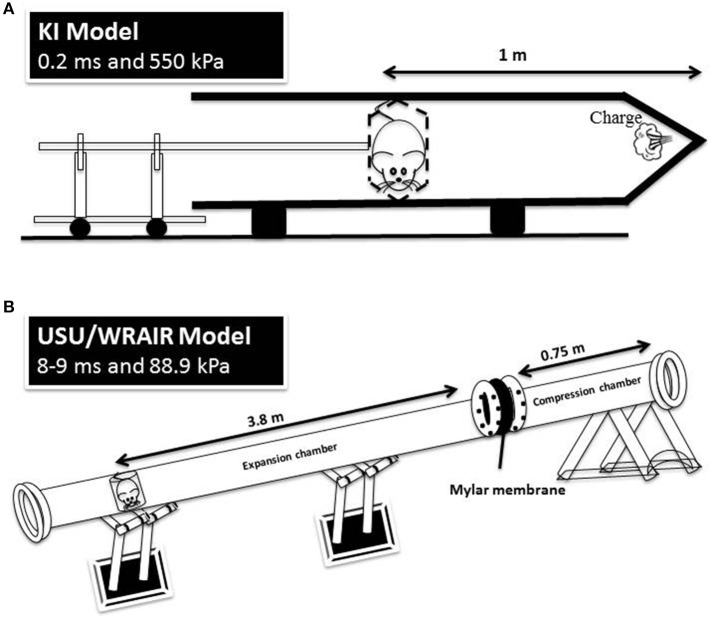
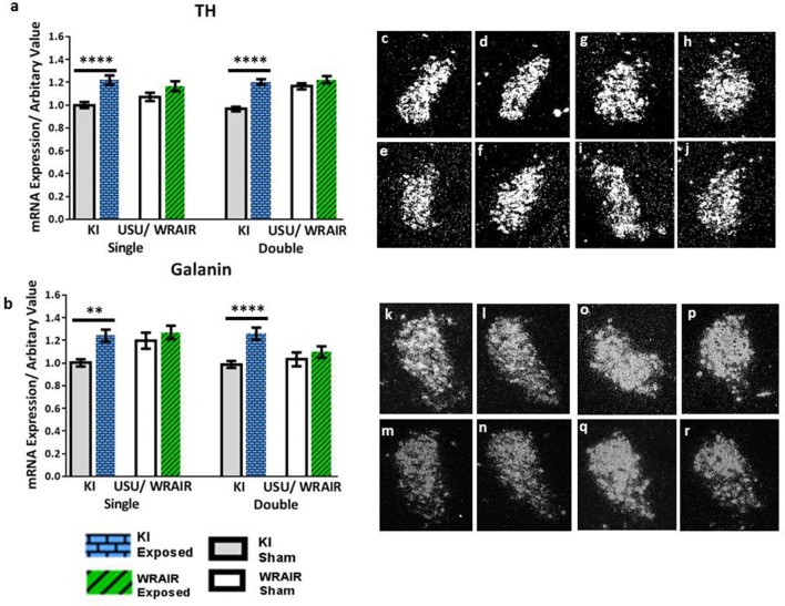
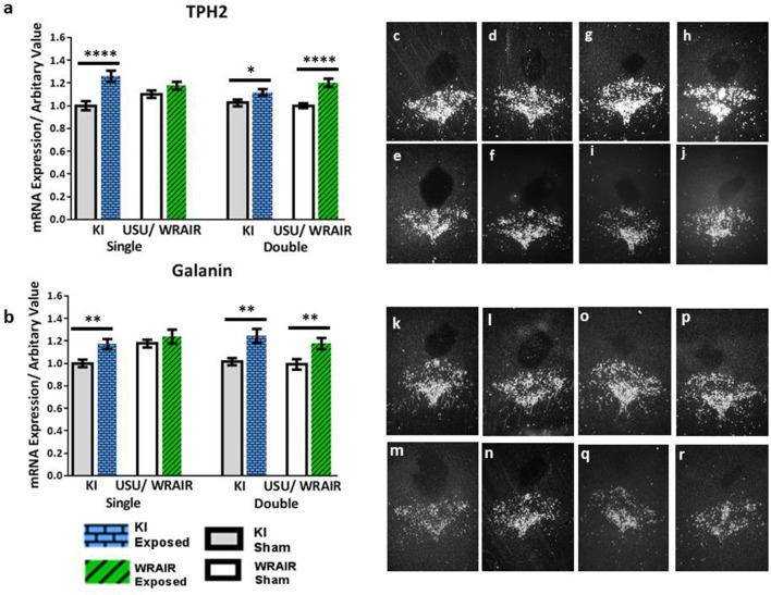
Repeated mild blast-induced traumatic brain injury (rmbTBI), caused by recurrent exposure to low levels of explosive blast, is a significant concern for military health systems. However, the pathobiology of rmbTBI is currently poorly understood. Animal models are important tools to identify the molecular changes of rmbTBI, but comparisons across different models can present their own challenges. In this study, we compared two well-established rodent models of mbTBI, the "KI model" and the "USU/WRAIR model." These two models create different pulse forms, in terms of peak pressure and duration. Following single and double exposures to mild levels of blast, we used in situ hybridization (ISH) to assess changes in mRNA levels of tyrosine hydroxylase (TH), tryptophan hydroxylase (TPH2), and galanin in the locus coeruleus (LC) and dorsal raphe nucleus (DRN). These systems and their transmitters are known to mediate responses to stress and anxiety. We found increased mRNA levels of TH, TPH2 and galanin in the LC and DRN of single-exposed rats relative to sham rats in the KI but not the USU/WRAIR model. Sham mRNA values measured in the USU/WRAIR model were substantially higher than their KI counterparts. Double exposure caused similarly significant increases in mRNA values in the KI model but not the USU/WRAIR model, except TPH2 and galanin levels in the DRN. We detected no cumulative effect of injury in either model at the used inter-injury interval (30 min), and there were no detectable neuropathological changes in any experimental group at 1 day post-injury. The apparent lack of early response to injury as compared to sham in the USU/WRAIR model is likely caused by stressors (e.g., transportation and noise), associated with the experimental execution, that were absent in the KI model. This study is the first to directly compare two established rodent models of rmbTBI, and to highlight the challenges of comparing findings from different animal models. Additional studies are needed to understand the role of stress, dissect the effects of psychological and physical injuries and to identify the window of increased cerebral vulnerability, i.e., the inter-injury interval that results in a cumulative effect following repeated blast exposure.
Keywords: animal models; anxiety; catecholamines; dorsal raphe nucleus; locus coeruleus; neuropeptide; post-traumatic stress disorder; transmitter coexistence.
Figures
Similar articles
-
Sex-Specific Differences in Rodents Following a Single Primary Blast Exposure: Focus on the Monoamine and Galanin Systems.Front Neurol. 2020 Oct 15;11:540144. doi: 10.3389/fneur.2020.540144. eCollection 2020. Front Neurol. 2020. PMID: 33178100 Free PMC article.
-
Neurotransmitter Systems in a Mild Blast Traumatic Brain Injury Model: Catecholamines and Serotonin.J Neurotrauma. 2015 Aug 15;32(16):1190-9. doi: 10.1089/neu.2014.3669. Epub 2015 Apr 1. J Neurotrauma. 2015. PMID: 25525686 Free PMC article.
-
Expression of galanin and its receptors are perturbed in a rodent model of mild, blast-induced traumatic brain injury.Exp Neurol. 2016 May;279:159-167. doi: 10.1016/j.expneurol.2016.02.019. Epub 2016 Feb 27. Exp Neurol. 2016. PMID: 26928087
-
[Mild traumatic brain injury and postconcussive syndrome: a re-emergent questioning].Encephale. 2012 Sep;38(4):329-35. doi: 10.1016/j.encep.2011.07.003. Epub 2011 Aug 31. Encephale. 2012. PMID: 22980474 Review. French.
-
Brain injuries from blast.Ann Biomed Eng. 2012 Jan;40(1):185-202. doi: 10.1007/s10439-011-0424-0. Epub 2011 Oct 20. Ann Biomed Eng. 2012. PMID: 22012085 Review.
Cited by
-
Explosive-driven double-blast exposure: molecular, histopathological, and behavioral consequences.Sci Rep. 2020 Oct 15;10(1):17446. doi: 10.1038/s41598-020-74296-2. Sci Rep. 2020. PMID: 33060648 Free PMC article.
-
Modification of the Marmarou model in developing countries.Ann Med Surg (Lond). 2020 Jul 22;57:109-113. doi: 10.1016/j.amsu.2020.07.029. eCollection 2020 Sep. Ann Med Surg (Lond). 2020. PMID: 32742649 Free PMC article.
-
Blood-Based Lateral-Flow Immunoassays Dipstick Test for Damaged Mitochondrial Electron Transport Chain in Pyruvate Treated Rats with Combined Blast Exposure and Hemorrhagic Shock.J Clin Med. 2025 Jan 24;14(3):754. doi: 10.3390/jcm14030754. J Clin Med. 2025. PMID: 39941423 Free PMC article.
-
A Systematic Review on Traumatic Brain Injury Pathophysiology and Role of Herbal Medicines in its Management.Curr Neuropharmacol. 2023;21(12):2487-2504. doi: 10.2174/1570159X21666230126151208. Curr Neuropharmacol. 2023. PMID: 36703580 Free PMC article.
-
Investigation of the direct and indirect mechanisms of primary blast insult to the brain.Sci Rep. 2021 Aug 6;11(1):16040. doi: 10.1038/s41598-021-95003-9. Sci Rep. 2021. PMID: 34362935 Free PMC article.
References
LinkOut - more resources
Full Text Sources
Other Literature Sources
Research Materials