Review
. 2018 Sep;11(3):456-474.
doi: 10.1007/s12178-018-9509-9.

Current Concepts in Talar Neck Fracture Management

Affiliations
Review

Current Concepts in Talar Neck Fracture Management

Colin Whitaker et al. Curr Rev Musculoskelet Med. 2018 Sep.

Abstract

Purpose of the review: Talar neck fractures are a rare but potentially devastating injury, which require a comprehensive understanding of the unique osteology, vasculature, and surrounding anatomy to recognize pathology and treat correctly. The purpose of this article is to describe both classic and current literature to better understand the evolution of talar neck fracture management.

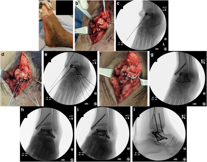
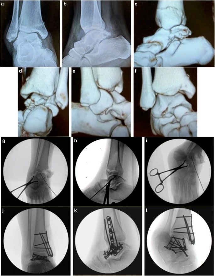
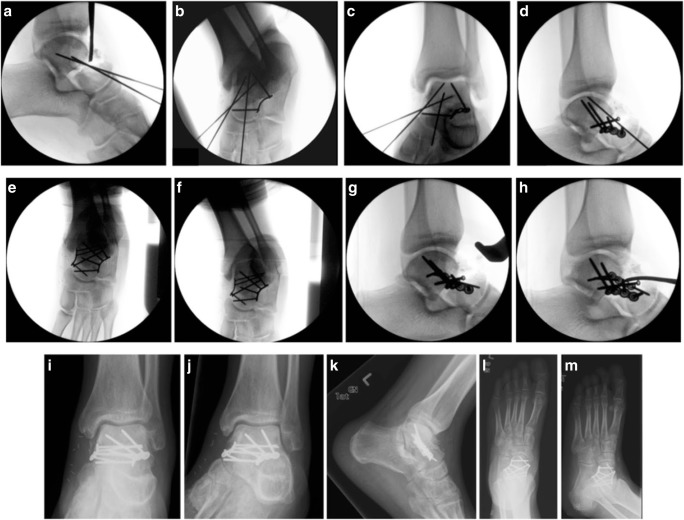
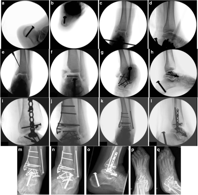
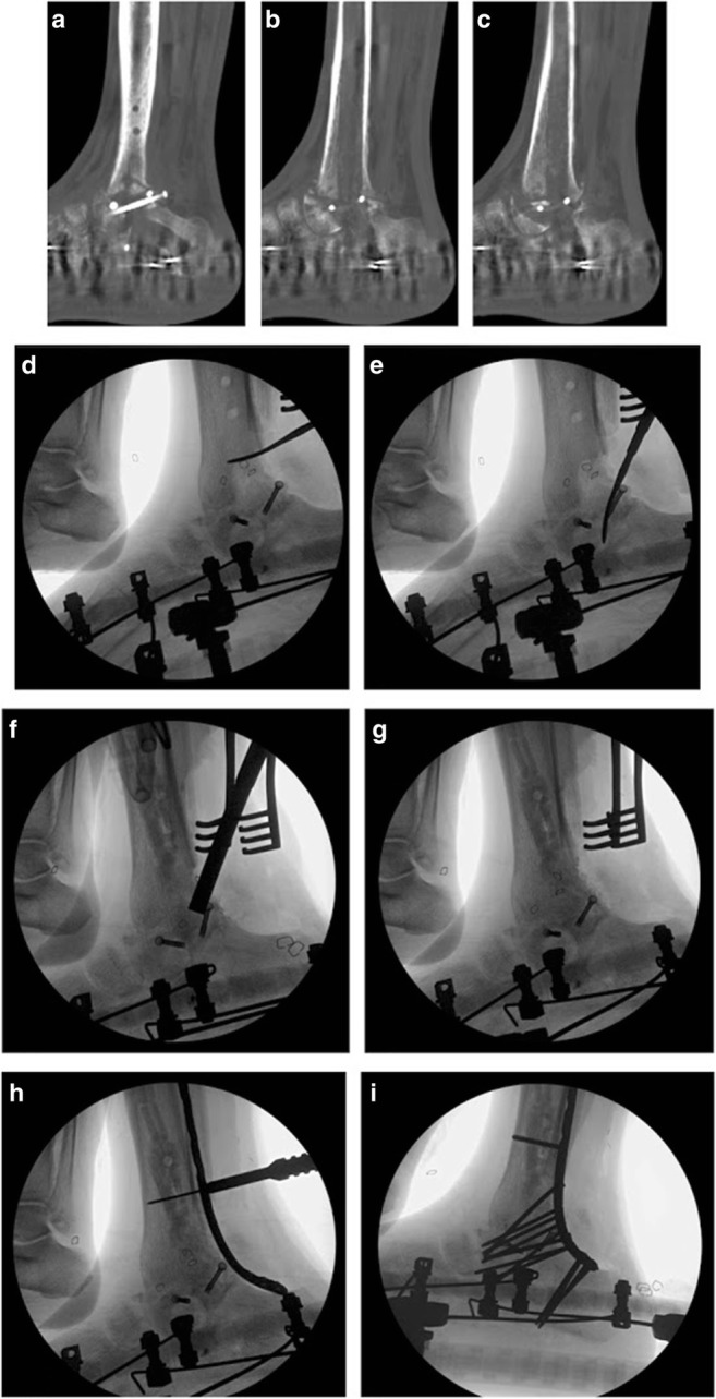
Recent findings: Urgent reduction of displaced fractures and dislocations remains the standard of care to protect the soft tissue envelope and neurovascular structures. Delayed definitive fixation has proven to be safe. CT is the imaging modality of choice to fully identify the fracture pattern and associated injuries. Anatomic reduction and restoration of the peritalar articular surfaces are the pillars of talar neck fracture treatment. Dual incision approach with plate and screw fixation has become the modern surgical strategy of choice to accomplish these goals. Although complications such as osteonecrosis (ON) and posttraumatic arthritis (PTA) can still occur at high rates, treatment should be dictated by patient symptoms. Talar neck fractures pose treatment challenges with both initial injury and potential sequelae. Future research will determine whether modern treatment algorithms will decrease complication rate and improve patient outcome.

Keywords: Complications; Outcomes; Review; Talar neck fracture; Treatment.


Conflict of interest statement

Conflict of interest

The authors declare that they have no conflict of interest.

Human and animal rights and informed consent

This article does not contain any studies with human or animal subjects performed by any of the authors.

Figures

Fig. 1
Fig. 1
A comminuted Hawkins type III talar neck injury after a fall from two stories in a 18-year-old male with poorly controlled type I diabetes. Figure 1a–c: injury ankle radiographs. Figure 1d–f: postreduction radiographs showing tibiotalar and subtalar joints are now reduced. Figure 1g–i: postreduction 3-D CT scan reconstructions. Note the significant medial and lateral talar neck comminution. Comminution is also seen within the medial aspect of the subtalar joint. The avulsion type fracture of the medial malleolus is better visualized
Fig. 2
Fig. 2
Images of a 19-year-old female with a Hawkins III talar neck injury secondary to a motor vehicle collision. Figure 2a–b: initial radiographs displaying fracture at the talar neck-body junction with medial malleolus fracture. The tibiotalar and subtalar joints are dislocated and the talonavicular joint is reduced. The talar body is posteromedially dislocated but detail is obscured with the overlying splint material. Figure 2c–d: 3-D CT reconstructions demonstrating complete posteromedial dislocation of the talar body. Figure 2e–g: this patient was taken urgently to the OR given the irreducible nature of the injury. Immediate postoperative radiographs after surgery. Dual incision approach was used with mini-fragment lateral plate fixation and medial screw fixation of the talar neck, and a 2.7-mm mini-fragment screw fixation of the medial malleolus fracture
Fig. 3
Fig. 3
Images of a 71-year-old female with rheumatoid arthritis who suffered injuries to the left tibial plafond, calcaneus, and talar neck and body after being struck by a motor vehicle. Figure 3a–c: initial radiographs displaying injuries. Figure 3d–f: representative 2-D CT reconstruction images demonstrating full extent of all the injuries. Note the comminution of the medial talar neck and body. There is also medial plafond impaction
Fig. 4
Fig. 4
Intraoperative images of surgical treatment of a talar neck and body fracture in 42-year-old male who fell from a ladder. Figure 4a: anteromedial incision. Figure 4b–g: medial malleolar osteotomy. Two 2.0-mm K-wires are placed perpendicular to the planned osteotomy. Figure 4h: medial malleolus displaced inferiorly with a dental pick to expose the medial talar body fracture and dome. Figure 4i: mini-fragment buttress plate fixation along the medial talar body. Figure 4j: AP fluoroscopic ankle view of medial talar body buttress fixation. A K-wire is placed within the one of the predrilled holes of the medial malleolus osteotomy for temporary reduction to obtain appropriate fluoroscopic imaging. Figure 4k: lateral fluoroscopic view of medial talar body buttress fixation. A K-wire has been placed along the medial talar neck for initial talar neck preliminary fixation
Fig. 5
Fig. 5
Intraoperative images of the surgical treatment of a talar neck and body fracture in a 42-year-old male who fell from a ladder continued from Fig. 4. Figure 5a: anterolateral incision. Figure 5b: exposure of the lateral talar dome (TD), body, lateral process (LP), and lateral talar neck (TN). Note the comminution in the talar neck. Figure 5c: Canale fluoroscopic view of talar neck prior to lateral neck reduction and plate placement. Note the K-wire placement along the medial talar neck. Figure 5d–e: initial lateral neck reduction and K-wire placement with contoured mini-fragment plate extending from the lateral process of the talus to the distal extent of the talar neck. Figure 5f: view of lateral plate fixation with all K-wires removed. A comminuted fragment along the lateral neck which was used for initial reduction evaluation was removed (CF). Figure 5g–h: final fluoroscopic views after replacement of the medial K-wire with a mini-fragment screw for medial strut support of the neck and two 2.7-mm mini-fragment screws final fixation of the medial malleolus osteotomy. Note the gap seen in Fig. 5h (but not in 5i) at the medial malleolus osteotomy as a result of the thickness of the saw blade used to make the osteotomy
Fig. 6
Fig. 6
Images of a 22-year-old female with a Hawkins IIa talar neck fracture with medial talar body extension, and associated medial plafond injury after a severe twisting injury to her ankle. Figure 6a–b: Initial injury AP and LAT ankle radiographs. Figure 6c–f: 3-D CT reconstructions demonstrating posteromedial talar body comminution, subluxation of the subtalar joint, and lateral talar neck fracture. Figure 6g–l: intraoperative fluoroscopy showing open reduction internal fixation through dual incision approach. Medial plafond injury was carefully displaced along its fracture plane to access the medial talar body and dome. Anatomic reduction was obtained of the lateral neck with point to point reduction forceps based on cortical reads. Final fixation of the talar neck was completed with lateral mini-fragment locking plate fixation and supplemental retrograde mini-fragment screws as a medial strut. The medial talar body was addressed with a buttress mini-fragment locking plate. Fixation of the plafond injury was done with a combination of 2.7-mm screws and a mini-fragment locking plate
Fig. 7
Fig. 7
Images of the Hawkins III talar neck fracture in the 18-year-old male shown in Fig. 1. Figure 7a: patient had delayed definitive fixation 10 days after the injury and reduction once his soft tissue envelope was amenable for fixation. Lateral ankle fluoroscopic view of initial reduction of talar neck fracture with K-wires through dual incisions. Note significant dorsal comminution and resultant bone loss as indicated by periosteal elevator. Figure 7b–d: Canale, ankle mortise, and lateral ankle fluoroscopic views of a subsequent lateral talar neck 2.7-mm mini-fragment locking plate application and placement of K-wires along medial talar neck. Figure 7e–f: due to the significant medial and lateral comminution and to maintain appropriate alignment, a 2.4-mm mini-fragment locking plate was placed strategically on the inferior medial talar neck. Note the thickness of the medial plate in relation to the lateral plate to prevent possible impingement in the medial tibiotalar joint gutter. One of the medial K-wires was replaced with a 2.7-mm mini-fragment screw as supplemental strut fixation. Also note the differences in imaging with different angles of ankle eversion with Canale views obtained in Fig. 7e, f. Figure 7g–h: LAT and oblique LAT ankle fluoroscopic views demonstrating the significant dorsal comminution. This defect was filled with a viable cellular bone matrix. Figure 7i–m: ankle and foot radiographs 5-month postsurgery demonstrating excellent maintained reduction, healing of the talar neck fracture, and filling in of the dorsal bony defect
Fig. 8
Fig. 8
Images of left tibial plafond, calcaneus, and talar neck and body injuries of the 71-year-old female shown in Fig. 3. Figure 8a–d: patient had initial percutaneous fixation of the tongue type calcaneus fracture and placement of a spanning external fixator for her plafond and talar neck/body injuries as her soft tissue envelope was not amenable for definitive fixation. Figure 8e–h: patient was taken for definitive fixation 8 days later for when her soft tissue envelope improved. The medial plafond injury fracture plane was used to gain access to the medial talar body and dome. Initial medial to lateral screw fixation of the talar dome was performed. The talar neck was addressed with lateral mini-fragment locking plate fixation and medial column medial column mini-fragment screw fixation. Figure 8i–l: after reducing the marginal impaction of the medial plafond injury, fixation was completed with mini-fragment locking plate and screw fixation. The fibular fracture was percutaneously fixed with retrograde IM screw fixation. Figure 8m–q: ankle and foot radiographs 5-month postsurgery demonstrating excellent maintained reduction, healing of the all fractures, and no signs of osteonecrosis. Calcium phosphate bone graft substitute was used to backfill the plafond impaction injury and is seen on the ankle radiographs
Fig. 9
Fig. 9
Images of an infected nonunion in a 63-year-old male with poorly controlled type II diabetes and hepatitis C who suffered fractures of his right plafond, talar neck, and posteromedial talar body dislocation. Figure 9a: the patient presented with a chronic draining lateral wound 7 months after initial surgery where fibular excision, talus fixation, and primary tibiotalar and subtalar fusion with a TTC IM nail was performed. Figure 9b–d: presenting radiographs showing nonunion of the tibiotalar and subtalar joints with radiolucency around the TTC nail and screws. Figure 9e–g: representative sagittal CT reconstructions confirming nonunion of tibiotalar and subtalar joints with significant radiolucency around TTC IM nail screws. Patient had bone biopsy confirming pan sensitive Staph lugdunesis infection. Figure 9h–i: patient wished to undergo limb salvage surgery. After 4 months of IV and oral antibiotics, the lateral draining wound healed. The patient was taken to the OR for first stage chronic infection treatment. The TTC IM nail was removed, irrigation and debridement of the nail tract with reamer-irrigation aspirator (RIA) system was performed, IM antibiotic impregnated calcium sulfate (CaSO4) bullets were placed, and a circular frame fixator was used for stability. Figure 9i–l: clinical pictures of circular frame fixator with negative pressure wound therapy dressings over incisions that were used to remove TTC IM nails screws
Fig. 10
Fig. 10
Images of second stage tibiotalarcalcaneal fusion in the 63-year-old male from Fig. 9. Figure 10a–c: after 3 months of IV and oral antibiotics, the patient’s clinical exam and laboratory markers returned to normal. CT scan confirmed only partial bony bridging from distal tibia to calcaneus through what was left of the talus. Figure 10d–e: second stage chronic infection treatment. Intraoperative fluoroscopy showing bony defect after excision of all nonviable bone and tissue. Figure 10f–g: intraoperative fluoroscopy showing placement of antibiotic impregnated CaSO4 bullets in the tibial canal and viable cellular bone matrix at the TTC nonunion site. Figure 10h–i: posterior plate TTC fusion using a medial distal tibia locking plate contoured to fit from the tibial shaft to the calcaneus. Circular frame fixator was left on postoperatively to protect the plate fixation

References

    1. Santavirta S, Seitsalo S, Kiliuoto O, Myllynen P. Fracture of the talus. J Trauma. 1984;24(11):986–989. doi: 10.1097/00005373-198411000-00011. - DOI - PubMed
    1. Hawkins L. Fractures of the neck of the talus. J Bone Joint Surg Am. 1970;52(5):991–1002. doi: 10.2106/00004623-197052050-00013. - DOI - PubMed
    1. Kenwright J, Taylor RG. Major injuries of the talus. J Bone Joint Surg Br. 1970;52(1):36–48. doi: 10.1302/0301-620X.52B1.36. - DOI - PubMed
    1. Lorentzen JE, Christensen SB, Krogsøe O, Sneppen O. Fractures of the neck of the talus. Acta Orthop Scand. 1977;48(1):115–120. doi: 10.3109/17453677708985121. - DOI - PubMed
    1. Canale ST, Kelly FB. Fractures of the neck of the talus. Long-term evaluation of seventy-one cases. J Bone Joint Surg Am. 1978;60(2):143–156. doi: 10.2106/00004623-197860020-00001. - DOI - PubMed

LinkOut - more resources