. 2018 Jul 4;8(1):59.
doi: 10.1186/s13550-018-0399-z.

Discriminating radiation injury from recurrent tumor with [18F]PARPi and amino acid PET in mouse models

Affiliations

Discriminating radiation injury from recurrent tumor with [18F]PARPi and amino acid PET in mouse models

Patrick L Donabedian et al. EJNMMI Res. .

Abstract

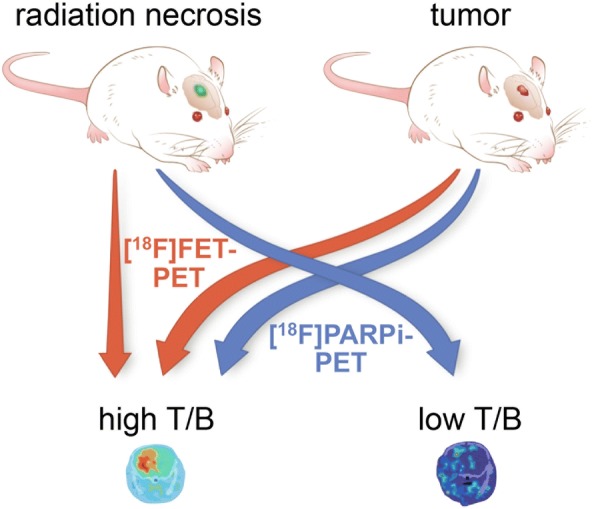
Background: Radiation injury can be indistinguishable from recurrent tumor on standard imaging. Current protocols for this differential diagnosis require one or more follow-up imaging studies, long dynamic acquisitions, or complex image post-processing; despite much research, the inability to confidently distinguish between these two entities continues to pose a significant dilemma for the treating clinician. Using mouse models of both glioblastoma and radiation necrosis, we tested the potential of poly(ADP-ribose) polymerase (PARP)-targeted PET imaging with [18F]PARPi to better discriminate radiation injury from tumor.

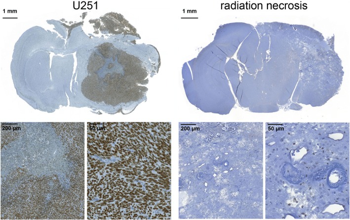
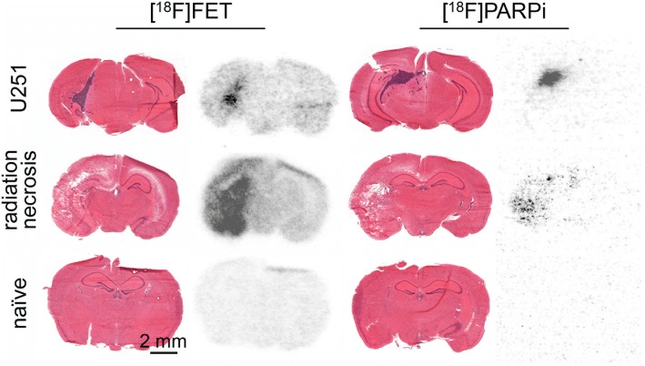
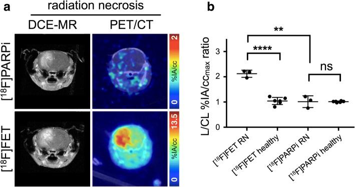
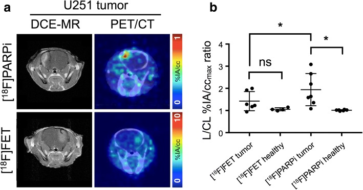
Results: In mice with experimental radiation necrosis, lesion uptake on [18F]PARPi-PET was similar to contralateral uptake (1.02 ± 0.26 lesion/contralateral %IA/ccmax ratio), while [18F]FET-PET clearly delineated the contrast-enhancing region on MR (2.12 ± 0.16 lesion/contralateral %IA/ccmax ratio). In mice with focal intracranial U251 xenografts, tumor visualization on PARPi-PET was superior to FET-PET, and lesion-to-contralateral activity ratios (max/max, p = 0.034) were higher on PARPi-PET than on FET-PET.

Conclusions: A murine model of radiation necrosis does not demonstrate [18F]PARPi avidity, and [18F]PARPi-PET is better than [18F]FET-PET in distinguishing radiation injury from brain tumor. [18F]PARPi-PET can be used for discrimination between recurrent tumor and radiation injury within a single, static imaging session, which may be of value to resolve a common dilemma in neuro-oncology.

Keywords: Amino acid PET; Biomarkers; PARP1; PET/CT; Radiation injury; Radiation necrosis.


Conflict of interest statement

Ethics approval

All animal experiments were performed in accordance with protocols approved by the Institutional Animal Care and Use Committees of Memorial Sloan Kettering Cancer Center (MSK) or Washington University and followed the National Institutes of Health guidelines for animal welfare.

Competing interests

The authors declare that they have no competing interests.

Publisher’s Note

Springer Nature remains neutral with regard to jurisdictional claims in published maps and institutional affiliations.

Figures

Fig. 1
Fig. 1
Experimental setup and hypothesis for imaging radiation necrosis and orthotopic brain tumors with [18F]FET-PET and [18F]PARPi-PET. Due to the molecular properties of each lesion and tracer, [18F]FET-PET will generate high lesion-to-background contrast in both tumor and radiation necrosis, while [18F]PARPi-PET will generate high contrast only in tumor
Fig. 2
Fig. 2
Anti-PARP1 immunohistochemistry. Staining of transaxial formalin-fixed, paraffin-embedded sections of mice implanted with U251 tumors (left group) mice with experimental radiation necrosis (right group) reveals high PARP1 expression in the nuclei of tumor cells and low PARP1 expression elsewhere in healthy brain and radiation necrosis
Fig. 3
Fig. 3
Hematoxylin and eosin stains and autoradiography. Hematoxylin and eosin stains (left) and digital storage phosphor autoradiography (right) of whole transaxial sections of mice with implanted U251 tumors (top row), experimental radiation necrosis (middle row), and naïve mice (bottom row), injected with [18F]FET (left column) or [18F]PARPi (right column). Autoradiographic scans have been contrast-adjusted for visibility. Mice were sacrificed 2.5 h post-injection of radiotracer
Fig. 4
Fig. 4
PET imaging of experimental murine radiation necrosis. a (left column) DCE-MR and (right column) fused PET/CT transaxial slices of mice with experimental radiation necrosis, injected with (top row) [18F]PARPi and (bottom row) [18F]FET. b Lesioned-to-contralateral hemisphere %IA/ccmax ratios for mice in different groups. **Significant at p < 0.005; ****significant at p < 0.0001
Fig. 5
Fig. 5
PET imaging of mice with focal intracranial U251 cell xenografts. a (left column) DCE-MR and (right column) fused PET/CT transaxial slices of mice with U251 tumor, injected with (top row) [18F]PARPi and (bottom row) [18F]FET. b Lesioned-to-contralateral hemisphere %IA/ccmax ratios for mice in different groups. *Significant at p < 0.05

References

    1. Ricard D, Idbaih A, Ducray F, Lahutte M, Hoang-Xuan K, Delattre JY. Primary brain tumours in adults. Lancet. 2012;379:1984–1996. doi: 10.1016/S0140-6736(11)61346-9. - DOI - PubMed
    1. Kohutek ZA, Yamada Y, Chan TA, Brennan CW, Tabar V, Gutin PH, et al. Long-term risk of radionecrosis and imaging changes after stereotactic radiosurgery for brain metastases. J Neuro-Oncol. 2015;125:149–156. doi: 10.1007/s11060-015-1881-3. - DOI - PMC - PubMed
    1. Valk PE, Dillon WP. Radiation injury of the brain. Am J Neuroradiol. 1991;12:45–62. - PMC - PubMed
    1. Remler MP, Marcussen WH, Tiller-Borsich J. The late effects of radiation on the blood brain barrier. Int J Radiat Oncol Biol Phys. 1986;12:1965–1969. doi: 10.1016/0360-3016(86)90133-1. - DOI - PubMed
    1. Walker AJ, Ruzevick J, Malayeri AA, Rigamonti D, Lim M, Redmond KJ, et al. Postradiation imaging changes in the CNS: how can we differentiate between treatment effect and disease progression? Future Oncol. 2014;10:1277–1297. doi: 10.2217/fon.13.271. - DOI - PMC - PubMed

LinkOut - more resources