

Signalling through Src family kinase isoforms is not redundant in models of thrombo-inflammatory vascular disease
- PMID: 29974666
- PMCID: PMC6111872
- DOI: 10.1111/jcmm.13721
Signalling through Src family kinase isoforms is not redundant in models of thrombo-inflammatory vascular disease
Abstract
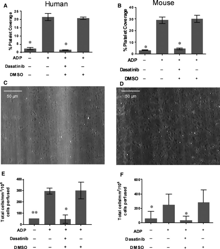
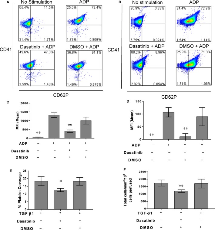
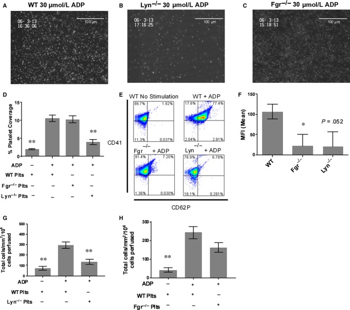
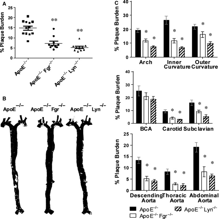
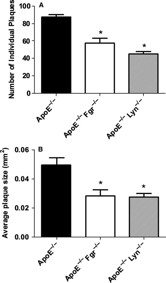
The Src family kinases (SFK) are a group of signalling molecules with important regulatory functions in inflammation and haemostasis. Leucocytes and platelets express multiple isoforms of the SFKs. Previous studies used broad-spectrum pharmacological inhibitors, or murine models deficient in multiple SFK isoforms, to demonstrate the functional consequences of deficiencies in SFK signalling. Here, we hypothesized that individual SFK operate in a non-redundant fashion in the thrombo-inflammatory recruitment of monocyte during atherosclerosis. Using in vitro adhesion assays and single SFK knockout mice crossed with the ApoE-/- model of atherosclerosis, we find that SFK signalling regulates platelet-dependent recruitment of monocytes. However, loss of a single SFK, Fgr or Lyn, reduced platelet-mediated monocyte recruitment in vitro. This translated into a significant reduction in the burden of atherosclerotic disease in Fgr-/- /ApoE-/- or Lyn-/- /ApoE-/- animals. SFK signalling is not redundant in thrombo-inflammatory vascular disease and individual SFK may represent targets for therapeutic intervention.
Keywords: Src family kinases; atherosclerosis; inflammation; monocytes; platelets.
© 2018 The Authors. Journal of Cellular and Molecular Medicine published by John Wiley & Sons Ltd and Foundation for Cellular and Molecular Medicine.
Figures
Similar articles
-
Distinct and overlapping functional roles of Src family kinases in mouse platelets.J Thromb Haemost. 2012 Aug;10(8):1631-45. doi: 10.1111/j.1538-7836.2012.04814.x. J Thromb Haemost. 2012. PMID: 22694307 Free PMC article.
-
Map3k8 Modulates Monocyte State and Atherogenesis in ApoE-/- Mice.Arterioscler Thromb Vasc Biol. 2017 Feb;37(2):237-246. doi: 10.1161/ATVBAHA.116.308528. Epub 2016 Nov 17. Arterioscler Thromb Vasc Biol. 2017. PMID: 27856455
-
Hyperreactivity of junctional adhesion molecule A-deficient platelets accelerates atherosclerosis in hyperlipidemic mice.Circ Res. 2015 Feb 13;116(4):587-99. doi: 10.1161/CIRCRESAHA.116.304035. Epub 2014 Dec 3. Circ Res. 2015. PMID: 25472975
-
Src family kinases: regulation of their activities, levels and identification of new pathways.Biochim Biophys Acta. 2008 Jan;1784(1):56-65. doi: 10.1016/j.bbapap.2007.08.012. Epub 2007 Aug 22. Biochim Biophys Acta. 2008. PMID: 17905674 Review.
-
Regulation of phagocyte migration and recruitment by Src-family kinases.Cell Mol Life Sci. 2008 Jul;65(14):2175-90. doi: 10.1007/s00018-008-8005-6. Cell Mol Life Sci. 2008. PMID: 18385944 Free PMC article. Review.
Cited by
-
Epigenetic mechanisms underlying variation of IL-6, a well-established inflammation biomarker and risk factor for cardiovascular disease.Atherosclerosis. 2025 Aug;407:120219. doi: 10.1016/j.atherosclerosis.2025.120219. Epub 2025 May 20. Atherosclerosis. 2025. PMID: 40480020 Free PMC article.
-
The (Patho)Biology of SRC Kinase in Platelets and Megakaryocytes.Medicina (Kaunas). 2020 Nov 24;56(12):633. doi: 10.3390/medicina56120633. Medicina (Kaunas). 2020. PMID: 33255186 Free PMC article. Review.
-
Xinmailong Modulates Platelet Function and Inhibits Thrombus Formation via the Platelet αIIbβ3-Mediated Signaling Pathway.Front Pharmacol. 2019 Aug 23;10:923. doi: 10.3389/fphar.2019.00923. eCollection 2019. Front Pharmacol. 2019. PMID: 31507419 Free PMC article.
-
GeneLab Database Analyses Suggest Long-Term Impact of Space Radiation on the Cardiovascular System by the Activation of FYN Through Reactive Oxygen Species.Int J Mol Sci. 2019 Feb 3;20(3):661. doi: 10.3390/ijms20030661. Int J Mol Sci. 2019. PMID: 30717456 Free PMC article.
-
Src-family Protein Tyrosine Kinases: A promising target for treating Cardiovascular Diseases.Int J Med Sci. 2021 Jan 14;18(5):1216-1224. doi: 10.7150/ijms.49241. eCollection 2021. Int J Med Sci. 2021. PMID: 33526983 Free PMC article. Review.
References
-
- Ross R. Cell biology of atherosclerosis. Annu Rev Physiol. 1995;57:791‐804. - PubMed
-
- Diacovo T, Roth S, Buccola J, Bainton D, Springer T. Neutrophil rolling, arrest, and transmigration across activated, surface‐adherent platelets via sequential action of P‐selectin and the beta 2‐integrin CD11b/CD18. Blood. 1996;88:146‐157. - PubMed
-
- Butler LM, Metson‐Scott T, Felix J, et al. Sequential adhesion of platelets and leukocytes from flowing whole blood onto a collagen‐coated surface: requirement for a GpVI‐binding site in collagen. Thromb Haemost. 2007;97:814‐821. - PubMed
-
- da Costa Martins P, van den Berk N, Ulfman LH, Koenderman L, Hordijk PL, Zwaginga JJ. Platelet‐monocyte complexes support monocyte adhesion to endothelium by enhancing secondary tethering and cluster formation. Arterioscler Thromb Vasc Biol. 2004;24:193‐199. - PubMed
-
- Huo Y, Schober A, Forlow SB, et al. Circulating activated platelets exacerbate atherosclerosis in mice deficient in apolipoprotein E. Nat Med. 2003;9:61‐67. - PubMed
Publication types
MeSH terms
Substances
Grants and funding
LinkOut - more resources
Full Text Sources
Other Literature Sources
Medical
Molecular Biology Databases
Miscellaneous
