

Endothelial Cells May Have Tissue-Specific Origins
- PMID: 29974893
- PMCID: PMC6028056
Endothelial Cells May Have Tissue-Specific Origins
Abstract
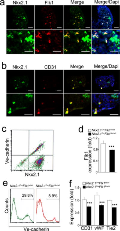
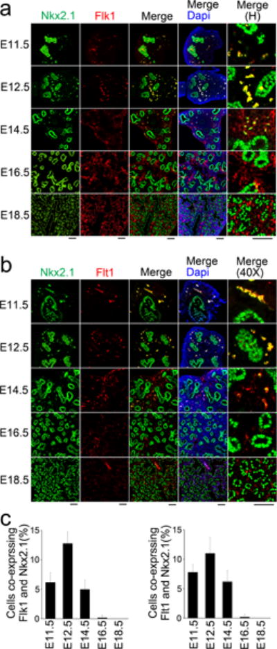
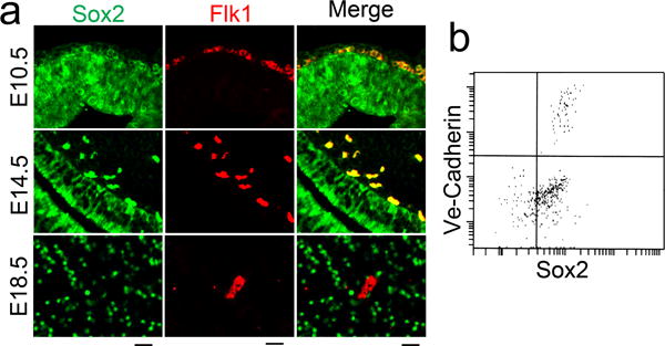
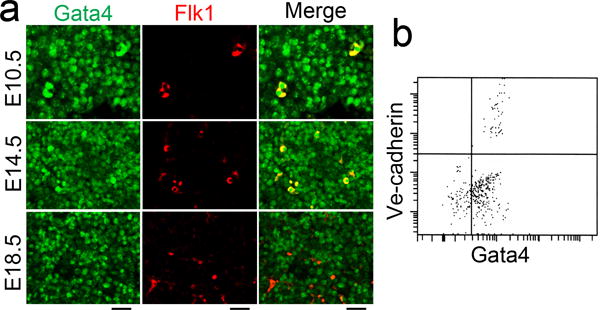
Endothelial heterogeneity reflects many functions performed by endothelial cells (ECs) in various tissues. However, the origin of this heterogeneity is unclear. Here, we report that tissue-specific ECs in lungs, brain and liver co-expressed the lineage markers of their coordinating tissue-specific cells at very early stages. Specifically, we found that the pulmonary EC population was significantly suppressed after pulmonary epithelial-specific (Nkx2.1-Cre mediated) deletion of fetal liver kinase-1 (Flk1). Together, the results suggest that tissues-specific ECs may originate from the same progenitor cells as tissue-specific cells.
Keywords: Differentiation; Endothelial cells; Heterogeneity; Tissue-specific.
Conflict of interest statement
The authors have declared that no conflict of interest exists.
Figures
Similar articles
-
[Study on Flk1+ Cells during Mouse Early Embryogenesis by Lineage Tracing].Zhongguo Shi Yan Xue Ye Xue Za Zhi. 2019 Jun;27(3):942-949. doi: 10.19746/j.cnki.issn.1009-2137.2019.03.050. Zhongguo Shi Yan Xue Ye Xue Za Zhi. 2019. PMID: 31204959 Chinese.
-
Effects of cellular origin on differentiation of human induced pluripotent stem cell-derived endothelial cells.JCI Insight. 2016 Jun 2;1(8):e85558. doi: 10.1172/jci.insight.85558. JCI Insight. 2016. PMID: 27398408 Free PMC article.
-
Single-Cell Transcriptome Atlas of Murine Endothelial Cells.Cell. 2020 Feb 20;180(4):764-779.e20. doi: 10.1016/j.cell.2020.01.015. Epub 2020 Feb 13. Cell. 2020. PMID: 32059779
-
Endothelial cell activation of the smooth muscle cell phosphoinositide 3-kinase/Akt pathway promotes differentiation.J Vasc Surg. 2005 Mar;41(3):509-16. doi: 10.1016/j.jvs.2004.12.024. J Vasc Surg. 2005. PMID: 15838487
-
Probing antiphospholipid-mediated thrombosis: the interplay between anticardiolipin antibodies and endothelial cells.Lupus. 2003;12(7):539-45. doi: 10.1191/961203303lu398oa. Lupus. 2003. PMID: 12892395 Review.
Cited by
-
Organ-Specific Endothelial Cell Differentiation and Impact of Microenvironmental Cues on Endothelial Heterogeneity.Int J Mol Sci. 2022 Jan 27;23(3):1477. doi: 10.3390/ijms23031477. Int J Mol Sci. 2022. PMID: 35163400 Free PMC article. Review.
-
Physiological and Pathological Remodeling of Cerebral Microvessels.Int J Mol Sci. 2022 Oct 21;23(20):12683. doi: 10.3390/ijms232012683. Int J Mol Sci. 2022. PMID: 36293539 Free PMC article. Review.
-
The potential role of 3D-bioprinting in xenotransplantation.Curr Opin Organ Transplant. 2019 Oct;24(5):547-554. doi: 10.1097/MOT.0000000000000684. Curr Opin Organ Transplant. 2019. PMID: 31385888 Free PMC article. Review.
-
Vascular Niche in Lung Alveolar Development, Homeostasis, and Regeneration.Front Bioeng Biotechnol. 2019 Nov 12;7:318. doi: 10.3389/fbioe.2019.00318. eCollection 2019. Front Bioeng Biotechnol. 2019. PMID: 31781555 Free PMC article. Review.
-
Skip is essential for Notch signaling to induce Sox2 in cerebral arteriovenous malformations.Cell Signal. 2020 Apr;68:109537. doi: 10.1016/j.cellsig.2020.109537. Epub 2020 Jan 10. Cell Signal. 2020. PMID: 31927035 Free PMC article.
References
Grants and funding
LinkOut - more resources
Full Text Sources
Other Literature Sources