

Lung eosinophilia induced by house dust mites or ovalbumin is modulated by nicotinic receptor α7 and inhibited by cigarette smoke
- PMID: 29975102
- PMCID: PMC6230881
- DOI: 10.1152/ajplung.00230.2018
Lung eosinophilia induced by house dust mites or ovalbumin is modulated by nicotinic receptor α7 and inhibited by cigarette smoke
Abstract
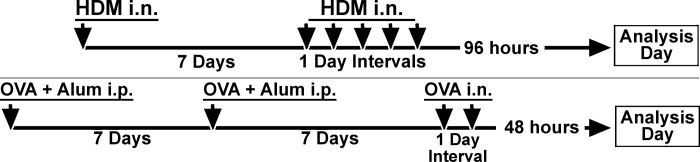
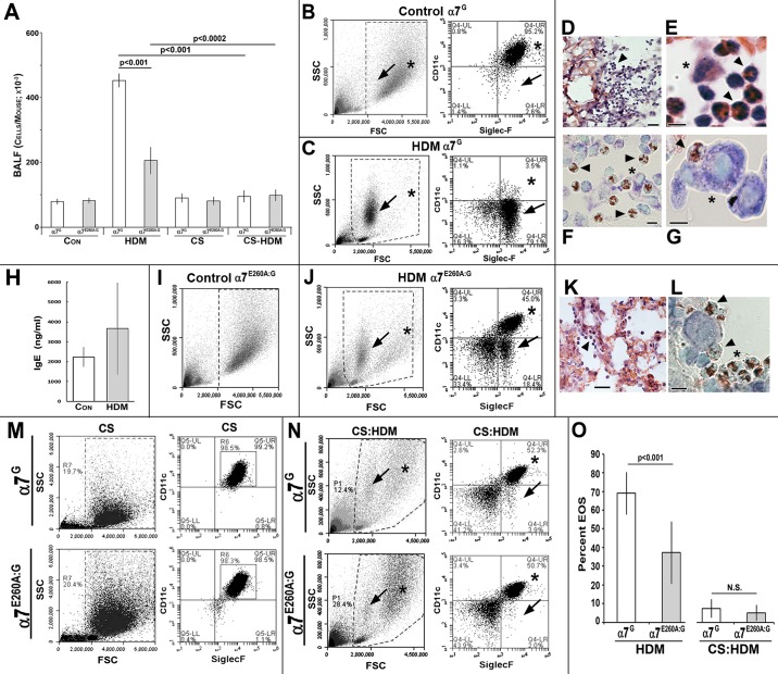
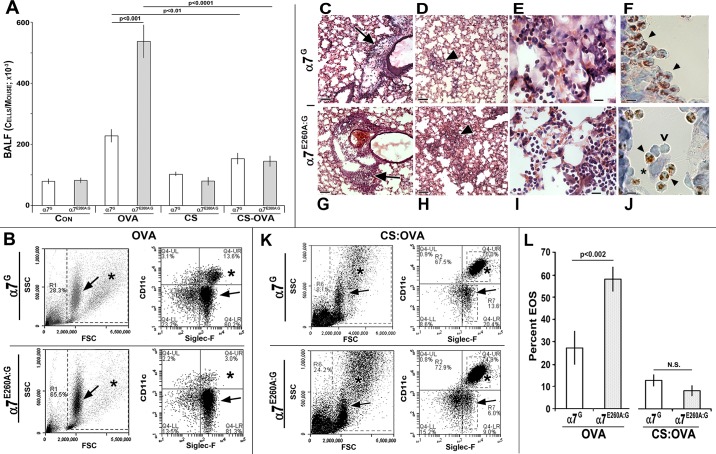
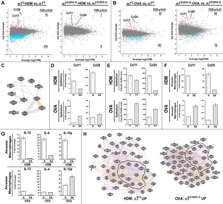
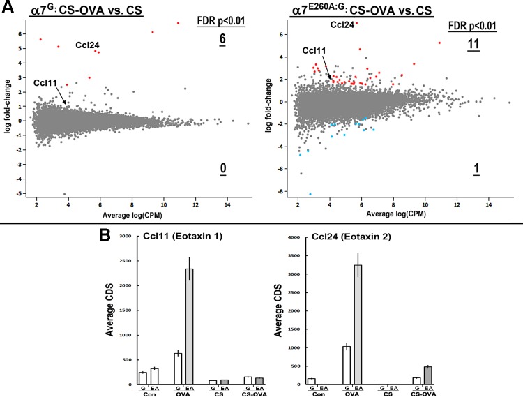
Eosinophilia (EOS) is an important component of airway inflammation and hyperresponsiveness in allergic reactions including those leading to asthma. Although cigarette smoking (CS) is a significant contributor to long-term adverse outcomes in these lung disorders, there are also the curious reports of its ability to produce acute suppression of inflammatory responses including EOS through poorly understood mechanisms. One possibility is that proinflammatory processes are suppressed by nicotine in CS acting through nicotinic receptor α7 (α7). Here we addressed the role of α7 in modulating EOS with two mouse models of an allergic response: house dust mites (HDM; Dermatophagoides sp.) and ovalbumin (OVA). The influence of α7 on EOS was experimentally resolved in wild-type mice or in mice in which a point mutation of the α7 receptor (α7E260A:G) selectively restricts normal signaling of cellular responses. RNA analysis of alveolar macrophages and the distal lung epithelium indicates that normal α7 function robustly impacts gene expression in the epithelium to HDM and OVA but to different degrees. Notable was allergen-specific α7 modulation of Ccl11 and Ccl24 (eotaxins) expression, which was enhanced in HDM but suppressed in OVA EOS. CS suppressed EOS induced by both OVA and HDM, as well as the inflammatory genes involved, regardless of α7 genotype. These results suggest that EOS in response to HDM or OVA is through signaling pathways that are modulated in a cell-specific manner by α7 and are distinct from CS suppression.
Keywords: chemokine; cigarette smoke; eosinophils; eotaxin; house dust mites; nicotine; nicotinic receptor α7; ovalbumin.
Figures
Similar articles
-
Lung epithelial response to cigarette smoke and modulation by the nicotinic alpha 7 receptor.PLoS One. 2017 Nov 8;12(11):e0187773. doi: 10.1371/journal.pone.0187773. eCollection 2017. PLoS One. 2017. PMID: 29117258 Free PMC article.
-
Inhaled aerosolized nicotine suppresses the lung eosinophilic response to house dust mite allergen.Am J Physiol Lung Cell Mol Physiol. 2020 Oct 1;319(4):L683-L692. doi: 10.1152/ajplung.00227.2020. Epub 2020 Jul 29. Am J Physiol Lung Cell Mol Physiol. 2020. PMID: 32726138 Free PMC article.
-
Cigarette smoke differentially affects eosinophilia and remodeling in a model of house dust mite asthma.Am J Respir Cell Mol Biol. 2011 Oct;45(4):753-60. doi: 10.1165/rcmb.2010-0404OC. Epub 2011 Feb 11. Am J Respir Cell Mol Biol. 2011. PMID: 21317378
-
Nicotinic receptors in airway disease.Am J Physiol Lung Cell Mol Physiol. 2024 Feb 1;326(2):L149-L163. doi: 10.1152/ajplung.00268.2023. Epub 2023 Dec 12. Am J Physiol Lung Cell Mol Physiol. 2024. PMID: 38084408 Free PMC article. Review.
-
Prebiotics improve parameters indicative of allergy-induced asthma in murines: a systematic review with meta-analysis.J Sci Food Agric. 2025 May 10. doi: 10.1002/jsfa.14325. Online ahead of print. J Sci Food Agric. 2025. PMID: 40346864 Review.
Cited by
-
Neurotransmitter and neuropeptide regulation of mast cell function: a systematic review.J Neuroinflammation. 2020 Nov 25;17(1):356. doi: 10.1186/s12974-020-02029-3. J Neuroinflammation. 2020. PMID: 33239034 Free PMC article.
-
Nicotine affects mitochondrial structure and function in human airway smooth muscle cells.Am J Physiol Lung Cell Mol Physiol. 2023 Dec 1;325(6):L803-L818. doi: 10.1152/ajplung.00158.2023. Epub 2023 Nov 7. Am J Physiol Lung Cell Mol Physiol. 2023. PMID: 37933473 Free PMC article.
-
Nicotinic Acetylcholine Receptors in the Respiratory Tract.Molecules. 2021 Oct 9;26(20):6097. doi: 10.3390/molecules26206097. Molecules. 2021. PMID: 34684676 Free PMC article. Review.
-
Asthma without borders.Am J Physiol Lung Cell Mol Physiol. 2020 May 1;318(5):L1001-L1003. doi: 10.1152/ajplung.00114.2020. Epub 2020 Apr 1. Am J Physiol Lung Cell Mol Physiol. 2020. PMID: 32233787 Free PMC article. No abstract available.
-
Age-Associated Tooth Loss and Oral Microbial Dysbiosis in a Mouse Genetic Model of Chronic Nicotine Exposure.Front Immunol. 2020 Oct 7;11:575200. doi: 10.3389/fimmu.2020.575200. eCollection 2020. Front Immunol. 2020. PMID: 33117372 Free PMC article.
References
-
- Botelho FM, Llop-Guevara A, Trimble NJ, Nikota JK, Bauer CM, Lambert KN, Kianpour S, Jordana M, Stämpfli MR. Cigarette smoke differentially affects eosinophilia and remodeling in a model of house dust mite asthma. Am J Respir Cell Mol Biol 45: 753–760, 2011. doi:10.1165/rcmb.2010-0404OC. - DOI - PubMed
-
- Braun-Fahrländer C, Riedler J, Herz U, Eder W, Waser M, Grize L, Maisch S, Carr D, Gerlach F, Bufe A, Lauener RP, Schierl R, Renz H, Nowak D, von Mutius E; Allergy and Endotoxin Study Team . Environmental exposure to endotoxin and its relation to asthma in school-age children. N Engl J Med 347: 869–877, 2002. doi:10.1056/NEJMoa020057. - DOI - PubMed
Publication types
MeSH terms
Substances
Grants and funding
LinkOut - more resources
Full Text Sources
Other Literature Sources
Molecular Biology Databases