

Human Herpes Virus 6 (HHV-6)-associated Lymphadenitis: Pitfalls in Diagnosis in Benign and Malignant Settings
- PMID: 29975251
- PMCID: PMC6133726
- DOI: 10.1097/PAS.0000000000001121
Human Herpes Virus 6 (HHV-6)-associated Lymphadenitis: Pitfalls in Diagnosis in Benign and Malignant Settings
Abstract
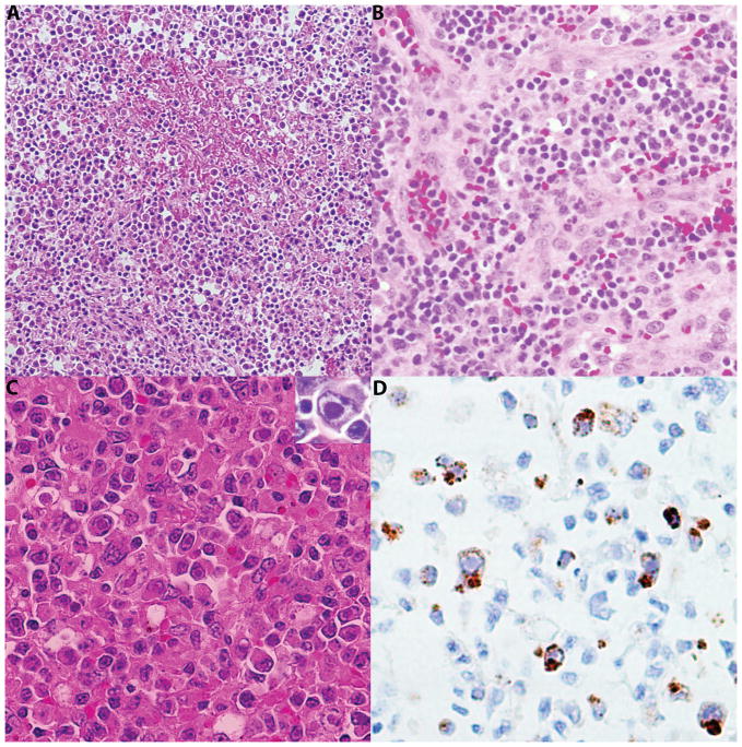
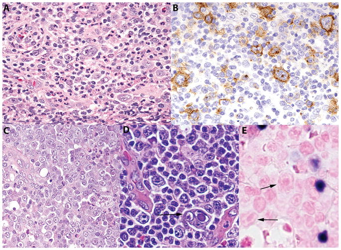
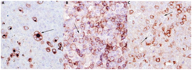
Human herpes virus 6 (HHV-6) is a member of the β-herpesvirinae subfamily. Most people acquire HHV-6 primary infection early in life and reactivation may occur, most often in immunocompromised individuals, leading to various clinical manifestations. HHV-6 infected cells may be identified in lymph nodes in both reactive and neoplastic conditions. Cases were retrieved from the hematopathology consultation service archives at National Institutes of Health from 2003 to 2017 in which infection by HHV-6 had been documented by immunohistochemical stains to HHV-6 gp60/110 envelope glycoprotein. Five cases of reactive lymphadenitis and 3 cases of lymphoma; 2 angioimmunoblastic T-cell lymphoma and 1 classic Hodgkin lymphoma, positive for HHV-6 were identified. The reactive lymph nodes showed marked paracortical hyperplasia and admixed large atypical lymphoid cells exhibiting pleomorphic nuclei, vesicular chromatin, and prominent eosinophilic intranuclear inclusions. Vascular proliferation and necrosis were also present, raising suspicion of peripheral T-cell lymphoma. The 3 cases of lymphoma showed similar viral inclusions, in addition to the characteristic features diagnostic of the lymphoma. Staining for HHV-6 was positive with a membranous and Golgi pattern and was restricted to cells with evident inclusions on hematoxylin and eosin. HHV-6 infected cells were positive for CD3 and CD4. HHV-6 lymphadenitis can present with morphologic atypia creating a diagnostic pitfall for lymphoma. In such cases, careful attention to the characteristic viral inclusions can lead to immunohistochemical analysis highlighting the replicating virus. In cases of lymphoma, identification of the inclusions is key in detecting the associated infection as well as in avoiding misinterpretation of the lymphoma subtype.
Conflict of interest statement
Disclosures: The author(s) have no conflicts of interest or funding to disclose
Figures
Similar articles
-
Human herpesvirus-6-associated acute lymphadenitis in immunocompetent adults.Mod Pathol. 2004 Nov;17(11):1427-33. doi: 10.1038/modpathol.3800179. Mod Pathol. 2004. PMID: 15494709 Free PMC article.
-
HHV-6-associated acute lymphadenitis in immunocompetent patients: a case report and review of literature.Int J Clin Exp Pathol. 2014 May 15;7(6):3413-7. eCollection 2014. Int J Clin Exp Pathol. 2014. PMID: 25031769 Free PMC article. Review.
-
Acute viral lymphadenitis mimicking low-grade peripheral T-cell lymphoma. A clinicopathological study of nine cases.APMIS. 2001 Jun;109(6):419-27. doi: 10.1034/j.1600-0463.2001.090603.x. APMIS. 2001. PMID: 11506473
-
Lymphadenitis and lymphoproliferative lesions associated with the human herpes virus-6 (HHV-6).Virchows Arch B Cell Pathol Incl Mol Pathol. 1991;61(3):179-87. doi: 10.1007/BF02890420. Virchows Arch B Cell Pathol Incl Mol Pathol. 1991. PMID: 1685279
-
Human Herpesvirus Type 8-associated Large B-cell Lymphoma: A Nonserous Extracavitary Variant of Primary Effusion Lymphoma in an HIV-infected Man: A Case Report and Review of the Literature.Clin Lymphoma Myeloma Leuk. 2016 Jun;16(6):311-21. doi: 10.1016/j.clml.2016.03.013. Epub 2016 Apr 1. Clin Lymphoma Myeloma Leuk. 2016. PMID: 27234438 Free PMC article. Review.
Cited by
-
The Possible Role of Pathogens and Chronic Immune Stimulation in the Development of Diffuse Large B-Cell Lymphoma.Biomedicines. 2024 Mar 14;12(3):648. doi: 10.3390/biomedicines12030648. Biomedicines. 2024. PMID: 38540261 Free PMC article. Review.
-
Human Herpesvirus-6 Infectious Meningitis With Lymphadenitis in an Immunocompetent Adult.Brain Behav. 2025 Jun;15(6):e70590. doi: 10.1002/brb3.70590. Brain Behav. 2025. PMID: 40444527 Free PMC article.
-
Angioimmunoblastic T-cell lymphoma: a concise overview encompassing the pathogenetic, pathological, clinical, therapeutical characteristics, and recent advances.Clin Exp Med. 2025 Jun 25;25(1):218. doi: 10.1007/s10238-025-01754-4. Clin Exp Med. 2025. PMID: 40560424 Free PMC article. Review.
-
Neutrophil and Eosinophil Extracellular Traps in Hodgkin Lymphoma.Hemasphere. 2021 Sep 1;5(9):e633. doi: 10.1097/HS9.0000000000000633. eCollection 2021 Sep. Hemasphere. 2021. PMID: 34485830 Free PMC article.
References
-
- Salahuddin SZ, Ablashi DV, Markham PD, et al. Isolation of a new virus, HBLV, in patients with lymphoproliferative disorders. Science (New York, NY) 1986;234:596–601. - PubMed
-
- Yamanishi K, Okuno T, Shiraki K, et al. Identification of human herpesvirus-6 as a causal agent for exanthem subitum. Lancet (London, England) 1988;1:1065–1067. - PubMed
-
- Ablashi DV, Salahuddin SZ, Josephs SF, et al. HBLV (or HHV-6) in human cell lines. Nature. 1987;329:207. - PubMed
-
- Merelli E, Sola P, Barozzi P, et al. An encephalitic episode in a multiple sclerosis patient with human herpesvirus 6 latent infection. Journal of the neurological sciences. 1996;137:42–46. - PubMed
Publication types
MeSH terms
Substances
Grants and funding
LinkOut - more resources
Full Text Sources
Other Literature Sources
Research Materials
