. 2019 Jan;33(1):239-253.
doi: 10.1096/fj.201701350RR. Epub 2018 Jul 5.

A suboptimal maternal diet combined with accelerated postnatal growth results in an altered aging profile in the thymus of male rats

Affiliations

A suboptimal maternal diet combined with accelerated postnatal growth results in an altered aging profile in the thymus of male rats

Jane L Tarry-Adkins et al. FASEB J. 2019 Jan.

Abstract

Reduced fetal nutrition and rapid postnatal growth accelerates the aging phenotype in many organ systems; however, effects on the immune system are unclear. We addressed this by studying the thymus from a rat model of developmental programming. The recuperated group was generated by in utero protein restriction, followed by cross-fostering to control-fed mothers, and were then compared with controls. Fat infiltration and adipocyte size increased with age ( P < 0.001) and in recuperated thymi ( P < 0.05). Cortex/medulla ratio decreased with age ( P < 0.001) and decreased ( P < 0.05) in 12-mo recuperated thymi. Age-associated decreases in thymic-epithelial cell ( P < 0.01) and thymocyte markers ( P < 0.01) were observed in both groups and was decreased ( P < 0.05) in recuperated thymi. These data demonstrate effects of developmental programming upon thymic involution. The recuperated group had longer thymic telomeres than controls ( P < 0.001) at 22 d and at 3 mo, which was associated with increased expression of telomere-length maintenance molecules [telomerase RNA component ( Terc; P < 0.01), P23 ( P = 0.02), and Ku70 and Ku80 ( P < 0.01)]. By 12 mo, recuperated offspring had shorter thymic telomeres than controls had ( P < 0.001) and reduced DNA damage-response markers [( DNA-PKcs, Mre11 ( P < 0.01), Xrcc4 ( P = 0.02), and γ-H2ax ( P < 0.001], suggesting failure of earlier compensatory responses. Our results suggest that low birth weight with rapid postnatal growth results in premature thymic maturation, resulting in accelerated thymic aging. This could lead to increased age-associated vulnerability to infection.-Tarry-Adkins, J. L., Aiken, C. E., Ashmore, T. J., Fernandez-Twinn, D. S., Chen, J.-H., Ozanne, S. E. A suboptimal maternal diet combined with accelerated postnatal growth results in an altered aging profile in the thymus of male rats.

Keywords: developmental programming; immunosenescence; involution.


Conflict of interest statement

The authors thank James Warner, Gregory Strachan, and M. S. Martin-Gronert (University of Cambridge Metaboloc Research Laboratories and MRC Metabolic Diseases Unit, Wellcome Trust-MRC Institute of Metabolic Science) for technical assistance. This work was supported by The British Heart Foundation (Grants PG/09/037/27387 and FS/09/029/27902), the Medical Research Council (MRC; Grant MC_UU_12012/4), and by an Isaac Newton Trust/Wellcome Institutional Strategic Support Fund (ISSF)/University of Cambridge Joint Research Grant. S.E.O. is a member of the MRC Metabolic Diseases Unit. The authors declare no conflicts of interest.

Figures

Figure 1
Figure 1
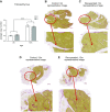
The effect of in utero protein restriction, accelerated postnatal growth, and aging upon total thymus area (A), thymic cortex to medulla area ratio (B), cortex to total thymus area ratio (C), 3-mo control (D) (representative image), 3-mo recuperated (E) (representative image), 12-mo control (F) (representative image), and 12-mo recuperated (G) (representative image). Results are expressed as means ± sem; n = 8/group. C, control; R, recuperated. *P < 0.05, ***P < 0.001.
Figure 2
Figure 2
The effect of in utero protein restriction, accelerated postnatal growth, and aging upon fibrosis in the thymus of 3- and 12-mo-old, male rats. Fibrosis/thymus area (A), 3-mo control (B) (representative image), 3-mo recuperated (C) (representative image), 12-mo control (D) (representative image), and 12-mo recuperated (E) (representative image) results are shown. Results are expressed as means ± sem; n = 8/group. C, control; R, recuperated . ***P < 0.001.
Figure 3
Figure 3
The effect of in utero protein restriction, accelerated postnatal growth, and aging upon the expression of markers of fat accumulation in the thymus of 3- and 12-mo-old, male rats. Total adipose area (A), average adipocyte count (B), average adipocyte area (C), and Lep (D) and Glut4 (E) mRNA expression are shown. Results are expressed as means ± sem. C, control; R, recuperated; n = 8/group. For gene expression data, overall effect of maternal diet (P mat diet) <0.06, and overall effect of age (P age) <0.01. *P < 0.05, ***P < 0.001 for histologic data.
Figure 4
Figure 4
The effect of in utero protein restriction, accelerated postnatal growth, and aging upon the expression of TEC and thymocyte markers in 3- and 12 mo-old male rats. A) Geometric mean of markers of TEC lineage. B) Geometric mean of markers of thymocyte lineage. C) Ratio of TEC to thymocyte markers. Results are expressed as means ± sem; n = 8/group. C, control; R, recuperated. Interaction between maternal diet and age (P int) <0.05. Overall effect of maternal diet <0.01. Overall effect of age <0.01. TEC/thymocyte ratio is expressed as a geometric mean.
Figure 5
Figure 5
The effect of in utero protein restriction, accelerated postnatal growth, and aging upon the thymic telomere length in 22-d, 3-mo, and 12-mo male rats. 145-48.5kb (A), 48.5-8.6kb (B), 8.6-4.2kb (C), and 4.2-1.3kb (D). Results are expressed as median ± interquartile range and total range, excluding outliers. C, control; R, recuperated; n = 8/group.
Figure 6
Figure 6
The effect of in utero protein restriction, accelerated postnatal growth, and aging upon the expression of telomerase complex molecules in the thymus of 3- and 12-mo-old male rats. Tert1 (A), Terc (B), Hsp90 (C), and P23 (D). Results are expressed as means ± sem; n = 8/group. C, control; R, recuperated. P adj-int <0.01; P mat diet <0.03 and 0.01; P age <0.01 and 0.05.
Figure 7
Figure 7
The effect of in utero protein restriction, accelerated postnatal growth, and aging upon the expression of shelterin complex proteins in the thymus of 3- and 12-mo-old male rats. Trf1 (A), Trf2 (B), Tin2 (C), Pot1 (D), Ku70 (E), and Ku80 (F). Results are expressed as means ± sem. C, control; R, recuperated; n = 8/group. P int <0.01, 0.02, and 0.05. P mat <0.01; P age <0.01.
Figure 8
Figure 8
The effect of in utero protein restriction, accelerated postnatal growth, and aging upon the expression of markers of DNA damage repair in the thymus of 3- and 12-mo-old male rats. DNA-PKcs (A), Mre11 (B), Xrcc4 (C), and γH2ax (D). Results are expressed as means ± sem; n = 8/group. C, control; R, recuperated. Padj-int < 0.01 and 0.03. P mat diet <0.01, 0.01, and 0.02; P age <0.01.
Figure 9
Figure 9
Correlations of thymic telomere length analysis vs. gene expression of γH2ax in 3- and 12-mo-old male rats. A) γH2ax vs. 145–48.5 kb. B) γH2ax vs. 48.5–8.6 kb. C) γH2ax vs. 8.6–4.2 kb. D) γH2ax vs. 4.2–1.3 kb. Results are expressed as means ± sem; n = 8/group. C, control; R, recuperated.
Figure 10
Figure 10
The effect of in utero protein restriction, accelerated postnatal growth, and aging upon the expression of sources of nonmitochondrial ROS and antioxidant-defense capacity in the thymus of 3- and 12-mo-old male rats. Xo (A), MnSOD (B), CuZnSOD (C), ECSOD (D), Catalase (E). Results are expressed as means ± sem; n = 8.group. C, control; R, recuperated. P int <0.01; P age <0.01.

References

    1. Aw D., Silva A. B., Palmer D. B. (2007) Immunosenescence: emerging challenges for an ageing population. Immunology 120, 435–446 - PMC - PubMed
    1. Tarry-Adkins J. L., Ozanne S. E. (2017) Nutrition in early life and age-associated diseases. Ageing Res. Rev. 39, 96–105 - PubMed
    1. Aspinall R., Andrew D. (2000) Thymic involution in aging. J. Clin. Immunol. 20, 250–256 - PubMed
    1. Hales C. N., Barker D. J. (2001) The thrifty phenotype hypothesis. Br. Med. Bull. 60, 5–20 - PubMed
    1. Miller J. F. (1961) Immunological function of the thymus. Lancet 2, 748–749 - PubMed

Publication types