. 2018 Jul 5;16(7):e2005710.
doi: 10.1371/journal.pbio.2005710. eCollection 2018 Jul.

Drosophila melanogaster establishes a species-specific mutualistic interaction with stable gut-colonizing bacteria

Affiliations

Drosophila melanogaster establishes a species-specific mutualistic interaction with stable gut-colonizing bacteria

Inês S Pais et al. PLoS Biol. .

Abstract

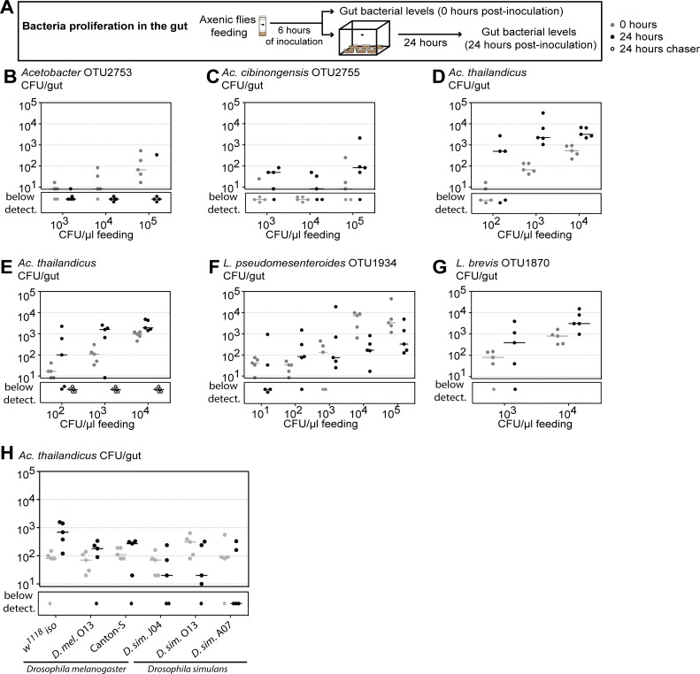
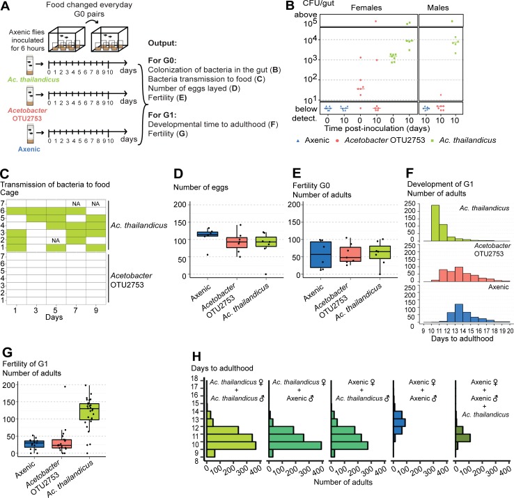
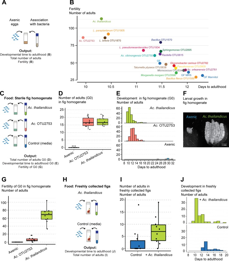
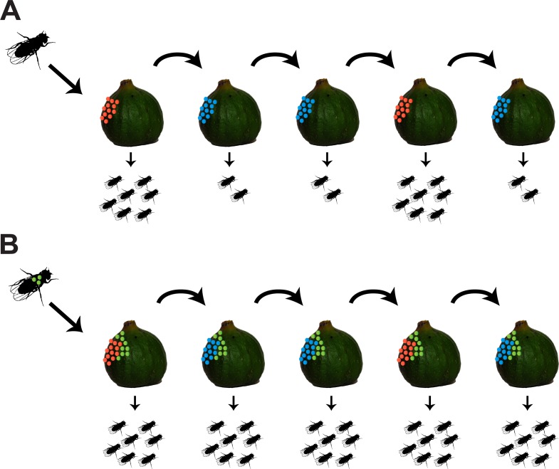
Animals live together with diverse bacteria that can impact their biology. In Drosophila melanogaster, gut-associated bacterial communities are relatively simple in composition but also have a strong impact on host development and physiology. It is generally assumed that gut bacteria in D. melanogaster are transient and their constant ingestion with food is required to maintain their presence in the gut. Here, we identify bacterial species from wild-caught D. melanogaster that stably associate with the host independently of continuous inoculation. Moreover, we show that specific Acetobacter wild isolates can proliferate in the gut. We further demonstrate that the interaction between D. melanogaster and the wild isolated Acetobacter thailandicus is mutually beneficial and that the stability of the gut association is key to this mutualism. The stable population in the gut of D. melanogaster allows continuous bacterial spreading into the environment, which is advantageous to the bacterium itself. The bacterial dissemination is in turn advantageous to the host because the next generation of flies develops in the presence of this particularly beneficial bacterium. A. thailandicus leads to a faster host development and higher fertility of emerging adults when compared to other bacteria isolated from wild-caught flies. Furthermore, A. thailandicus is sufficient and advantageous when D. melanogaster develops in axenic or freshly collected figs, respectively. This isolate of A. thailandicus colonizes several genotypes of D. melanogaster but not the closely related D. simulans, indicating that the stable association is host specific. This work establishes a new conceptual model to understand D. melanogaster-gut microbiota interactions in an ecological context; stable interactions can be mutualistic through microbial farming, a common strategy in insects. Moreover, these results develop the use of D. melanogaster as a model to study gut microbiota proliferation and colonization.


Conflict of interest statement

The authors have declared that no competing interests exist.

Figures

Fig 1
Fig 1. Wild-caught Drosophila melanogaster have a stable gut microbiota.
Single 3–6-day-old w1118 iso males were kept in the same vial during 10 days (A) or exposed to a stability protocol by being passed to new vials twice a day (B, C). (A, B) Ten individuals were analyzed each day and total number of CFUs per gut was determined by bacterial plating. Bacterial levels between Day 0 and Day 10 increase in (A) and decrease in (B) (lmm, p < 0.001 for both). Supporting data can be found in S1 and S2 Data. (C) Relative amount of 16S rRNA bacterial gene was measured by qPCR in 10 individual guts from each day, using the host gene Rpl32 as a reference gene. The relative amount of 16S rRNA gene decreases between days (lmm, p < 0.001). Supporting data can be found in S3 Data. (D) Single 3–6-day-old w1118 iso males were placed in food vials for 24 hours and then discarded. Bacterial levels on the food were determined at this point (Day 1) and after incubating the vials for a further 9 days (Day 10). Bacterial levels were also assessed in control vials, not exposed to flies (Day 10 control). Five vials were analyzed for each condition and total number of CFUs per vial was determined by bacterial plating. Bacterial levels increase between Day 1 and Day 10 (lm, p < 0.001). Supporting data can be found in S4 Data. (E) Bacterial levels from wild-caught flies at the day of collection (Day 0) and after 10 days of the stability protocol (Day 10). Ten individuals were analyzed for each day and total number of CFUs per gut was determined by plating. Bacterial levels on the flies significantly decrease with time (lmm, p = 0.004). Supporting data can be found in S5 Data. (A–E) Each dot represents an individual gut or vial and lines represent medians. Statistical analyses were performed together with replicate experiments shown in S1 Fig. CFU, colony-forming unit; lm, linear model; lmm, linear mixed model; w1118 iso, w1118 DrosDel isogenic strain; qPCR, quantitative PCR.
Fig 2
Fig 2. Higher diversity of gut bacterial communities in wild-caught Drosophila melanogaster.
Bacterial OTUs present in the gut of laboratory (1–20) and wild-caught (21–40) flies before (Day 0) and after being exposed to the stability protocol (Day 10). Gut homogenates from flies represented in Fig 1B and 1E were plated in different culture media, and representative colonies of each morphological type were sequenced. Each column represents one individual gut. Bacterial levels are represented on a gray scale from 100 to 107 CFUs per gut. Colonies of different Lactobacillus, Acetobacteriaceae, or Enterobactereaceae were not possible to distinguish by morphological type and are grouped together. The presence of Lactobacillus species and Leuconostoc pseudomesenteroides in wild-caught flies is not independent (Pearson’s chi-squared test, p = 0.014). Frequencies of the different OTUs in these groups are represented on Fig 3B, 3D, 3F and 3H and S3B Fig. Supporting data can be found in S6 Data. CFU, colony-forming unit; OTU, operational taxonomic unit.
Fig 3
Fig 3. Wild-caught flies maintain particular Acetobacter species in the gut.
Total levels of Acetobactereaceae (A, E) and Lactobacillus (C, G) in laboratory w1118 iso (A, C) and in wild-caught flies (E, G) before (Day 0) and after 10 days of the stability protocol (Day 10). Each dot represents one individual gut and lines represent medians. Levels of Acetobactereaceae decrease between days in both types of flies (lm, p ≤ 0.002 for both). Changes in levels of Lactobacillus are not significant in both (lm, p ≤ 0.302). Frequencies of sequenced colonies of Acetobactereaceae (B, F) and Lactobacillus (D, H) in w1118 iso (B, D) and in wild-caught flies (F, H). Numbers on the top of the bars correspond to the number of flies carrying each OTU, from a total of 10 flies (B, D, F, H). *Acetobacter thailandicus was initially identified as A. indonesiensis OTU2758 based on partial sequence of 16S rRNA gene; see Materials and methods and S1 Text. Supporting data can be found in S6 Data. Ac., Acetobacter; CFU, colony-forming unit; G., Gluconobacter; L., Lactobacillus; lm, linear model; OTU, operational taxonomic unit; w1118 iso, w1118 DrosDel isogenic strain.
Fig 4
Fig 4. Acetobacter thailandicus stably persists in the foregut of Drosophila melanogaster.
(A–E) Stability of different bacteria in monoassociation. Single 3–6-day-old w1118 iso males from monoassociated stocks, with Acetobacter OTU2753 (B), A. cibinongensis (C), A. thailandicus, (D) or Leuconostoc pseudomesenteroides (E), were exposed to the stability protocol in cages, as shown in the scheme (A). Number of CFUs in individual guts was assessed by plating at Days 0, 1, 2, 5, and 10 of the protocol. Stability of different bacteria was analyzed by fitting the data to an exponential decay model represented in S5I Fig. Supporting data can be found in S7 Data. (F–N) Localization of A. thailandicus in the gut. (G–H) Number of CFUs in each gut compartment from w1118 iso males monoassociated with A. thailandicus before (G) and after (H) 5 days of the stability protocol. Guts were dissected and cut according to the scheme in (F). Each dot represents one gut or one gut fragment and lines represent medians; statistical analyses were performed together with replicate experiments shown in S6 Fig. Supporting data can be found in S8 Data. (I, J) Fluorescent in situ hybridization with Cy3 labeled Bacteria 16S rRNA universal probe EUB338 in the gut of males monoassociated with A. thailandicus, after 5 days of the stability protocol. A. thailandicus persists in the crop duct (I′), the crop (I″), and the proventriculus (J and J′). In the crop, A. thailandicus cells are observed close to the chitin (I″). Chitin autofluorescence is indicated by white arrowheads and bacteria by white arrows. (K, L) Live/dead staining in the crop (K) and proventriculus (L) of males monoassociated with A. thailandicus 9 days after the stability protocol. Live bacteria stained with SYTO9 (green) and dead bacteria with propidium iodide (red). (I–L) DNA was stained with Hoechst. (M–N) Aggregates of A. thailandicus observed by transmission electron microscopy in the lumen of the proventriculus. Some cells present membrane invaginations (mi) and seem to be dividing. Cells appear to be attached to each other by external appendages such as fimbriae (f). A pili-like structure is also present (p). Extracellular vesicles are found between cells (v). Scale bar corresponds to 200 μm in (K), 50 μm in (I, J, L), 10 μm in (I′, I″, J′), 5 μm in the zoom in panels of (K, L), and 0.5 μm in (M, N). CFU, colony-forming unit; Cy3, cyanine 3 dye; mi, membrane invagination; OTU, operational taxonomic unit; p, pili; v, vesicles; w1118 iso, w1118 DrosDel isogenic strain.
Fig 5
Fig 5. Acetobacter thailandicus and A. cibinongensis proliferate in the gut of Drosophila melanogaster.
(A–G) Proliferation of different bacteria in the gut of D. melanogaster. Three- to six-day-old axenic w1118 iso males were inoculated for 6 hours, with different concentrations of Acetobacter OTU2753 (B), A. cibinongensis (C), A. thailandicus (D, E), Leuconostoc pseudomesenteroides (F), and Lactobacillus brevis (G). Bacterial levels were assessed 0 and 24 hours postinoculation. During this period, males were singly placed in cages, as shown in scheme (A). In (E), axenic chaser males were placed in cages together with males inoculated with A. thailandicus. At 24 hours, bacterial levels were assessed for both males. Bacterial levels between 0 and 24 hours decrease in flies inoculated with Acetobacter OTU2753 (lmm, p < 0.001), increase in flies inoculated with A. cibinongensis, A. thailandicus, and L. brevis (p = 0.024, p < 0.001, and p = 0.046, respectively), and do not significantly change in flies inoculated with L. pseudomesenteroides (p = 0.158). Supporting data can be found in S9 and S10 Data. (H) Proliferation of A. thailandicus in the gut of D. melanogaster and D. simulans. Three- to six-day-old D. melanogaster or D. simulans males were inoculated for 6 hours with A. thailandicus (104 CFU/μL). Bacterial levels were assessed 0 and 24 hours postinoculation. During this period, males were singly placed in bottles. Three different genetic backgrounds for D. melanogaster (w1118 iso, D. mel. O13, and Canton-S) and for D. simulans (D. sim. J04, D. sim. O13, and D. sim. A07) were tested. Bacterial levels in the gut increase in D. melanogaster and decrease in D. simulans (lmm, p < 0.001). Supporting data can be found in S13 Data. Five individuals were analyzed for each condition, per replicate, and total number of CFUs per gut was determined by plating. Each dot represents one gut and the lines represent medians. Statistical analysis was performed together with replicate experiments shown in S10 Fig and S12C–S12E Fig. CFU, colony-forming unit; lmm, linear mixed model; w1118 iso, w1118 DrosDel isogenic strain.
Fig 6
Fig 6. Acetobacter thailandicus stable association with Drosophila melanogaster is mutualistic.
(A) Axenic 1–3-day-old w1118 iso males and females (G0) were in contact with an inoculum of 105 CFU/μL of Acetobacter OTU2753, A. thailandicus, or sterile Mannitol (Axenic) for 6 hours. Two males and one female were placed per cage, with 6–7 cages for each condition, during 10 days, with food changed daily. This experimental setup corresponds to data shown in panels B–G. (B) Bacterial levels in single guts of females at time 0 (0 days) and 10 days postinoculation and in males 10 days postinoculation, analyzed by plating. Bacterial levels between the two time points increased in females inoculated with A. thailandicus and decreased in females inoculated with Acetobacter OTU2753 (Mann–Whitney test, p < 0.001 and p = 0.048, respectively). Supporting data can be found in S15 Data. (C) Presence of bacteria on the food collected from cages at days 1, 3, 5, 7, and 9 of the protocol, analyzed by plating. Filled rectangles represent presence of bacteria. NA stands for samples that were not analyzed. A. thailandicus is transmitted to the food with higher frequency than Acetobacter OTU2753 (glm-binomial, p < 0.001). Supporting data can be found in S16 Data. (D–G) Effect of bacterial association on the fitness of D. melanogaster. Total number of eggs laid by flies inoculated, or not, with different Acetobacter (D) and total number of adults that emerged from these eggs (E). Total number of eggs or adults is not different between conditions (lmm, p > 0.484 for all comparisons). (F) Developmental time to adulthood of the progeny (G1) of flies inoculated, or not, with different Acetobacter. Developmental time to adulthood is faster in progeny from flies inoculated with A. thailandicus than in the other two conditions and in progeny from flies inoculated with Acetobacter OTU2753 compared to progeny from axenic flies (lmm, p < 0.001 for these comparisons). Supporting data can be found in S17 and S18 Data. (G) Fertility of G1 was assessed by placing two males and one female of G1 per vial, flipping them every other day for 10 days, and analyzing total number of emerged adults. Fifteen or more couples were made per condition. Fertility is higher in progeny from flies inoculated with A. thailandicus compared with the other two conditions (lmm, p < 0.001 for both comparisons) and not different in the comparison between the progeny of flies inoculated with Acetobacter OTU2753 or axenic (p = 0.592). Supporting data can be found in S19 Data. (H) One male and one female 1–2-day-old w1118 iso, either axenic or monoassociated with A. thailandicus, were placed in vials and flipped every other day for 10 days. To one set of vials with axenic parents, A. thailandicus was added on the eggs after passing the parents. Developmental time to adulthood of the progeny was assessed. Ten couples were made per condition. There are no differences on developmental time to adulthood if either or both parents are monoassociated with A. thailandicus (lmm, p > 0.412 for all these comparisons). Progeny from couples in which either or both parents are monoassociated and progeny from axenic flies in which A. thailandicus culture is added on the eggs develop faster than progeny from axenic flies (lmm, p < 0.001 for all these comparisons). Supporting data can be found in S20 Data. (B) Each dot represents one gut and lines represent medians. (D, E, and G) Each dot represents the total progeny of one female. All statistical analyses were done together with replicate experiments shown in S14 and S15 Figs. CFU, colony-forming unit; glm-binomial, generalized linear model with binomial distribution; lmm, linear mixed model fit; NA, not analyzed; w1118 iso, w1118 DrosDel isogenic strain.
Fig 7
Fig 7. Acetobacter thailandicus is beneficial in the context of other wild bacteria and natural food substrates.
(A) w1118 iso eggs were associated with different bacteria isolated from the gut of wild-caught Drosophila melanogaster. As controls, axenic eggs that had no treatment (GF) or in which sterile media were added (GF MRS and GF Mannitol) were used. (B) For each bacterium, estimates of developmental time to adulthood of these eggs are plotted against estimates of their fertility. These estimates derive from the statistical analysis of data presented in S16C–S16F Fig. There is a negative correlation between developmental time and fertility (Pearson correlation −0.91, p < 0.001). Supporting data can be found in S21 and S22 Data. (C) Thirty axenic w1118 iso eggs were placed in vials containing sterilized fig homogenate. A. thailandicus, Acetobacter OTU2753, or sterile culture media were added on the top of the eggs. Ten vials were used per condition. Total number of adults that emerged (D) and developmental time to adulthood (E) was determined. More eggs inoculated with A. thailandicus and Acetobacter OTU2753 developed to adulthood and faster than axenic eggs (lmm, p < 0.001 for both comparisons). Supporting data can be found in S23 Data. (F) Larvae 5 days postinoculation with A. thailandicus or sterile media in fig homogenate. (G) Progeny of flies developed in fig homogenate, with and without the addition of Acetobacter species. One male and one female were collected from G0 of each condition and placed per vial containing fig homogenate for 10 days, with vials flipped every other day. A. thailandicus and Acetobacter OTU2753 conditions have 10 replicates, but only 3 from axenic eggs were possible to perform. Flies that developed with A. thailandicus had more progeny than flies that developed with Acetobacter OTU2753 or sterile media (lmm, p < 0.001). Supporting data can be found in S24 Data. (H) Fifty axenic w1118 iso eggs were placed in vials containing freshly collected nonsterile figs. A. thailandicus culture or sterile media (Control) was added on the top of the eggs. The total number of adults that emerged (I) and their developmental time to adulthood (J) was analyzed. Ten vials were analyzed per condition. There were more adults emerging from vials inoculated with A. thailandicus (lmm, p = 0.010). Developmental time to adulthood was faster in eggs inoculated with A. thailandicus in this experimental replicate but was not significantly different in the other replicate represented on S18K Fig (lmm, p < 0.001 and p = 0.557, respectively). Supporting data can be found in S25 Data. Statistical analyses from (D–J) were done together with replicate experiments shown in S18 Fig. GF, germ free (axenic); lmm, linear mixed model fit; MRS, de Man, Rogosa and Sharpe broth; w1118 iso, w1118 DrosDel isogenic strain.
Fig 8
Fig 8. Model for an ecological advantage of a stable association between Drosophila melanogaster and beneficial gut bacteria.
(A) In the absence of stable gut bacteria, the fitness of D. melanogaster is dependent on the presence of more (red) or less (blue) beneficial bacteria in the food substrate. (B) Carrying a stable population of beneficial bacteria (green) in the gut allows constant bacterial inoculation of food substrate and consequent association with the next host generation. This leads to a higher fitness of this next generation.

Similar articles

Cited by

References

    1. McFall-Ngai M, Hadfield MG, Bosch TCG, Carey HV, Domazet-Lošo T, Douglas AE, et al. Animals in a bacterial world, a new imperative for the life sciences. Proc Natl Acad Sci USA. 2013;110: 3229–3236. 10.1073/pnas.1218525110 - DOI - PMC - PubMed
    1. Sasaki T, Ishikawa H. Production of Essential Amino Acids from Glutamate by Mycetocyte Symbionts of the Pea Aphid, Acyrthosiphon pisum. J Insect Physiol. 1995;41: 41–46.
    1. Douglas AE. Reproductive failure and the free amino acid pools in pea aphids (Acyrthosiphon pisum) lacking symbiotic bacteria. J Insect Physiol. 1996;42: 247–255. 10.1016/0022-1910(95)00105-0 - DOI
    1. Akman L, Yamashita A, Watanabe H, Oshima K, Shiba T, Hattori M, et al. Genome sequence of the endocellular obligate symbiont of tsetse flies, Wigglesworthia glossinidia. Nat Genet. 2002;32: 402–407. 10.1038/ng986 - DOI - PubMed
    1. Pais R, Lohs C, Wu Y, Wang J, Aksoy S. The obligate mutualist Wigglesworthia glossinidia influences reproduction, digestion, and immunity processes of its host, the tsetse fly. Appl Environ Microbiol. 2008;74: 5965–5974. 10.1128/AEM.00741-08 - DOI - PMC - PubMed

Publication types

LinkOut - more resources