

Genetics instability of wtAAV2 genome and AAV promoter activities in the Baculovirus/Sf9 cells system
- PMID: 29975713
- PMCID: PMC6033426
- DOI: 10.1371/journal.pone.0199866
Genetics instability of wtAAV2 genome and AAV promoter activities in the Baculovirus/Sf9 cells system
Abstract
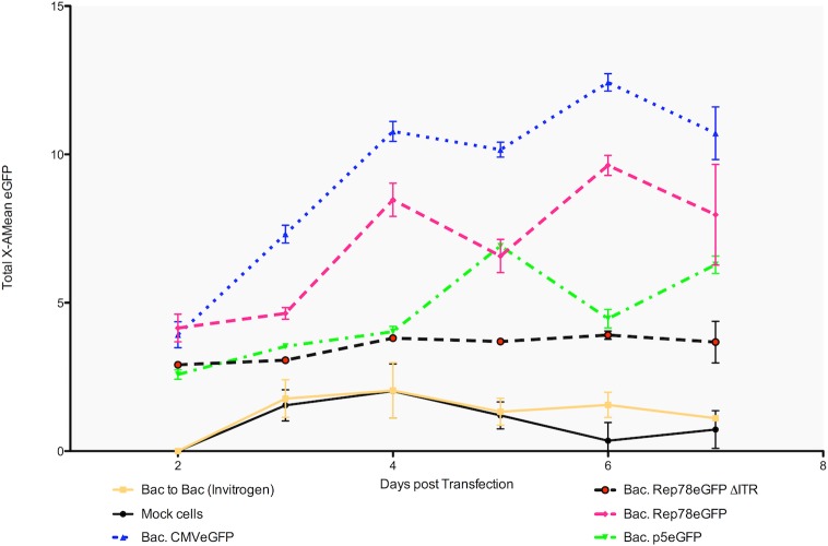
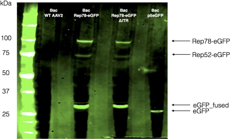
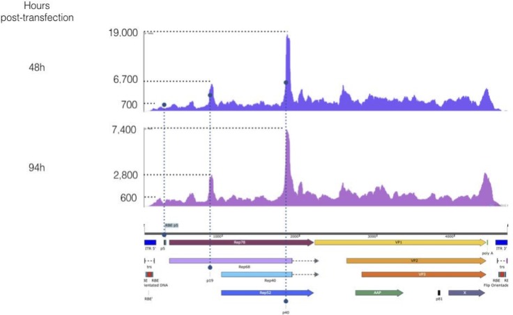
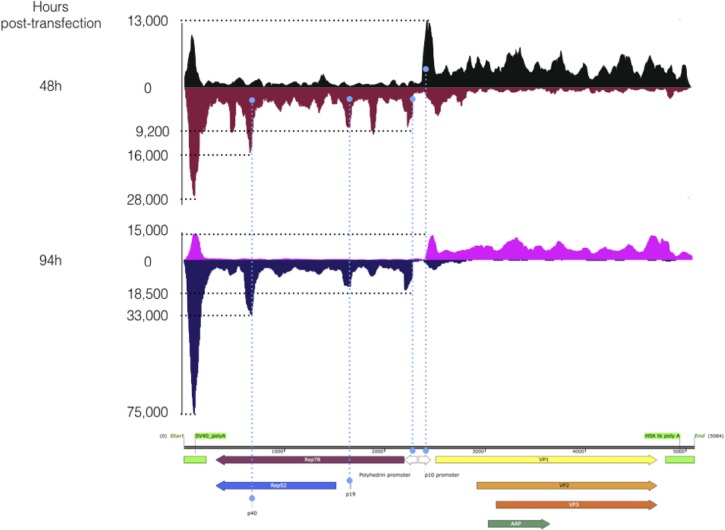
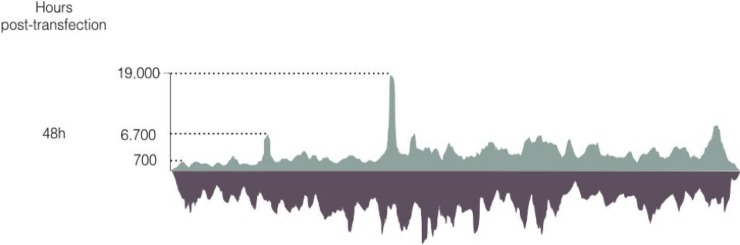
The human Adeno-Associated Virus serotype 2 (wtAAV2) is a common non-pathological virus and its recombinant form (rAAV) is widely used as gene therapy vector. Although rAAVs are routinely produced in the Baculovirus/Sf9 cell system, wtAAV2 has never been studied in this context. We tried to produce wtAAV2 in the baculovirus/Sf9 cell system hypothesizing that the wtAAV2 may be considered as a normal recombinant AAV transgene. Through our attempts to produce wtAAV2 in Baculovirus/Sf9, we found that wtAAV2 p5 promoter, which controls the expression of large Rep proteins in mammalian cells, was active in this system. p5 promoter activity in the baculovirus/Sf9 cell system leads to the expression of Rep78 that finally excises wtAAV2 genome from the baculovirus genome during the earliest phases of baculovirus stock production. Via p5 promoter expression kinetics and strand specific RNA-Seq analysis of wtAAV2, rAAV and Rep2/Cap2 cassettes in the baculovirus context we could demonstrate that wtAAV2 native promoters, p5, p19 and p40 are all active in the context of the baculovirus system and lead to the expression of different proteins and peptides. In addition, this study has proven that the baculovirus brings at least some of the helper functions needed in the AAV replication/life cycle.
Conflict of interest statement
The affiliation with FinVector Vision Thérapies Oy, Synpromics Ltd., and Genethon does not alter our adherence to PLOS ONE policies on sharing data and materials.
Figures
Similar articles
-
Impact of Inverted Terminal Repeat Integrity on rAAV8 Production Using the Baculovirus/Sf9 Cells System.Hum Gene Ther Methods. 2017 Oct;28(5):277-289. doi: 10.1089/hgtb.2016.133. Hum Gene Ther Methods. 2017. PMID: 28967288 Free PMC article.
-
Production of recombinant AAV vectors encoding insulin-like growth factor I is enhanced by interaction among AAV rep regulatory sequences.Virol J. 2009 Jan 7;6:3. doi: 10.1186/1743-422X-6-3. Virol J. 2009. PMID: 19128486 Free PMC article.
-
A novel method using baculovirus-mediated gene transfer for production of recombinant adeno-associated virus vectors.J Gen Virol. 2001 Sep;82(Pt 9):2051-2060. doi: 10.1099/0022-1317-82-9-2051. J Gen Virol. 2001. PMID: 11514714
-
A review of alternative promoters for optimal recombinant protein expression in baculovirus-infected insect cells.Protein Expr Purif. 2021 Oct;186:105924. doi: 10.1016/j.pep.2021.105924. Epub 2021 Jun 1. Protein Expr Purif. 2021. PMID: 34087362 Free PMC article. Review.
-
Molecular design for recombinant adeno-associated virus (rAAV) vector production.Appl Microbiol Biotechnol. 2018 Feb;102(3):1045-1054. doi: 10.1007/s00253-017-8670-1. Epub 2017 Dec 4. Appl Microbiol Biotechnol. 2018. PMID: 29204900 Free PMC article. Review.
Cited by
-
Origins of truncated supplementary capsid proteins in rAAV8 vectors produced with the baculovirus system.PLoS One. 2018 Nov 15;13(11):e0207414. doi: 10.1371/journal.pone.0207414. eCollection 2018. PLoS One. 2018. PMID: 30440025 Free PMC article.
-
Development of an insect cell-based adeno-associated virus packaging cell line employing advanced Rep gene expression control system.Mol Ther Methods Clin Dev. 2022 Oct 28;27:391-403. doi: 10.1016/j.omtm.2022.10.015. eCollection 2022 Dec 8. Mol Ther Methods Clin Dev. 2022. PMID: 36381303 Free PMC article.
-
Monobac System-A Single Baculovirus for the Production of rAAV.Microorganisms. 2021 Aug 24;9(9):1799. doi: 10.3390/microorganisms9091799. Microorganisms. 2021. PMID: 34576695 Free PMC article.
References
-
- Atchison RW, Casto BC, Hammon WM. Adenovirus-Associated Defective Virus Particles. Science. 1965. August 13;149(3685):754–6. - PubMed
-
- Samulski RJ, Muzyczka N. AAV-Mediated Gene Therapy for Research and Therapeutic Purposes. Annu Rev Virol. 2014. November;1(1):427–51. doi: 10.1146/annurev-virology-031413-085355 - DOI - PubMed
-
- Sonntag F, Schmidt K, Kleinschmidt JA. A viral assembly factor promotes AAV2 capsid formation in the nucleolus. Proc Natl Acad Sci USA. National Acad Sciences; 2010. June 1;107(22):10220–5. doi: 10.1073/pnas.1001673107 - DOI - PMC - PubMed
Publication types
MeSH terms
Substances
LinkOut - more resources
Full Text Sources
Other Literature Sources
Molecular Biology Databases
Miscellaneous
