. 2018 Jul 5;13(7):e0199612.
doi: 10.1371/journal.pone.0199612. eCollection 2018.

Quercetin prevents rhinovirus-induced progression of lung disease in mice with COPD phenotype

Affiliations

Quercetin prevents rhinovirus-induced progression of lung disease in mice with COPD phenotype

Mohammad Farazuddin et al. PLoS One. .

Abstract

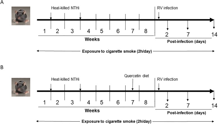
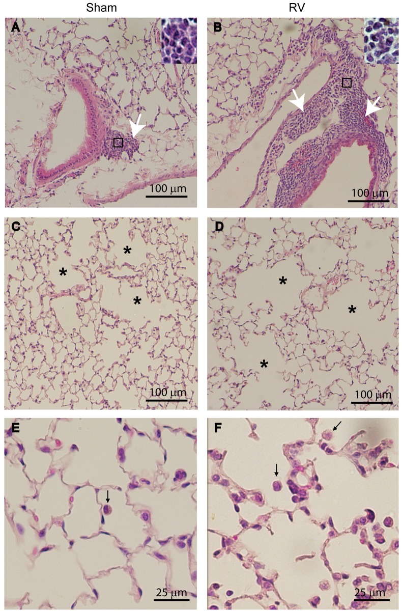
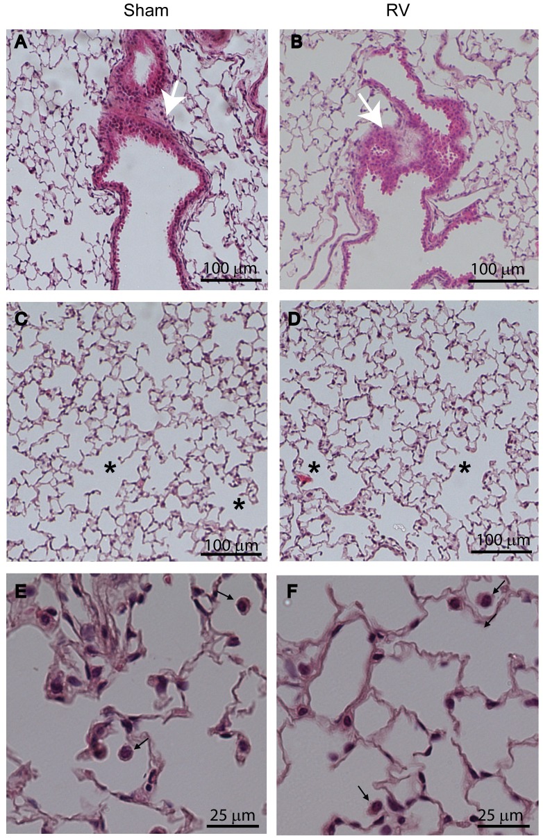
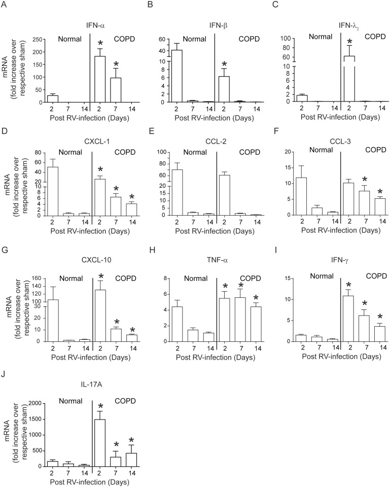
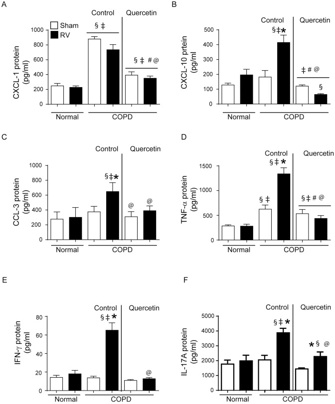
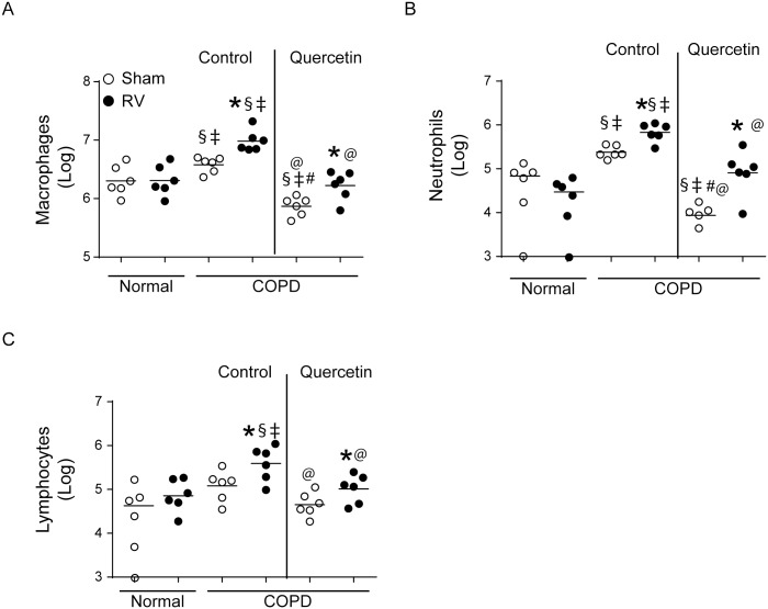
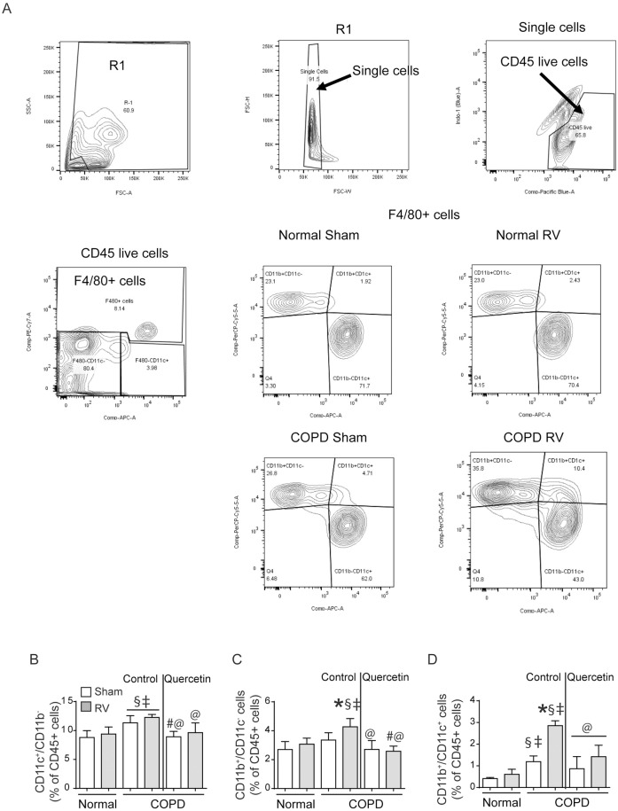
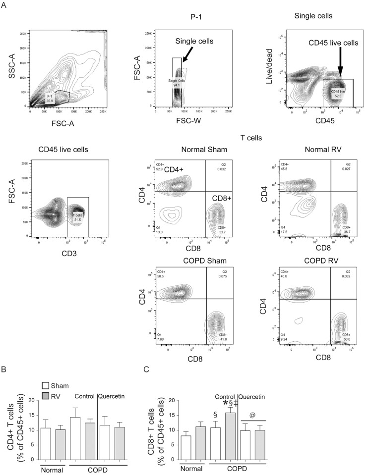
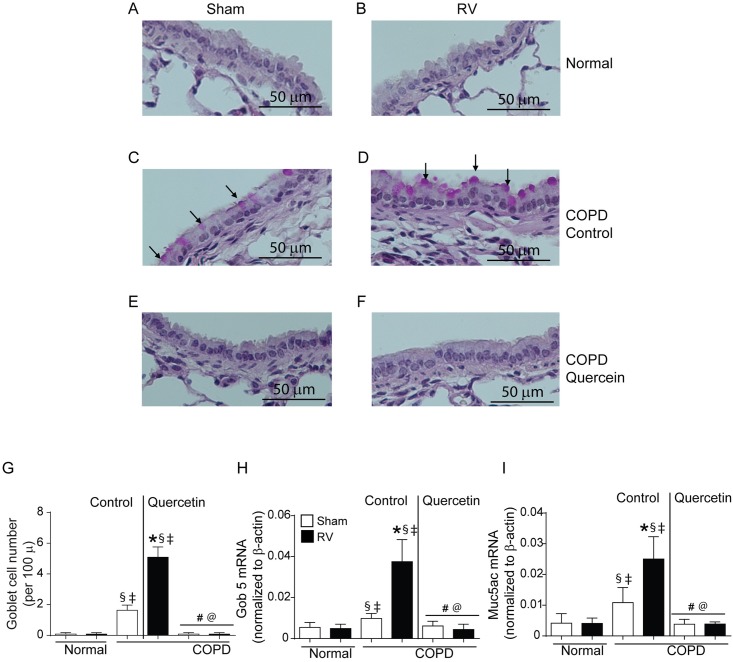
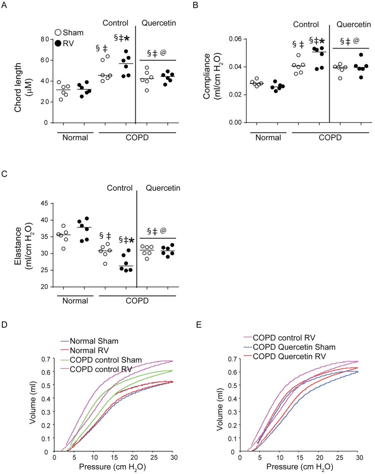
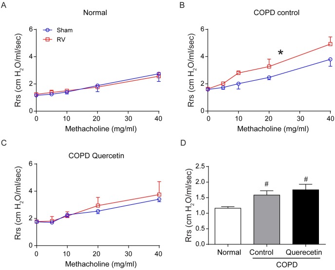
Acute exacerbations are the major cause of morbidity and mortality in patients with chronic obstructive pulmonary disease (COPD). Rhinovirus, which causes acute exacerbations may also accelerate progression of lung disease in these patients. Current therapies reduces the respiratory symptoms and does not treat the root cause of exacerbations effectively. We hypothesized that quercetin, a potent antioxidant and anti-inflammatory agent with antiviral properties may be useful in treating rhinovirus-induced changes in COPD. Mice with COPD phenotype maintained on control or quercetin diet and normal mice were infected with sham or rhinovirus, and after 14 days mice were examined for changes in lung mechanics and lung inflammation. Rhinovirus-infected normal mice showed no changes in lung mechanics or histology. In contrast, rhinovirus-infected mice with COPD phenotype showed reduction in elastic recoiling and increase in lung inflammation, goblet cell metaplasia, and airways cholinergic responsiveness compared to sham-infected mice. Interestingly, rhinovirus-infected mice with COPD phenotype also showed accumulation of neutrophils, CD11b+/CD11c+ macrophages and CD8+ T cells in the lungs. Quercetin supplementation attenuated rhinovirus-induced all the pathologic changes in mice with COPD phenotype. Together these results indicate that quercetin effectively mitigates rhinovirus-induced progression of lung disease in a mouse model of COPD. Therefore, quercetin may be beneficial in the treatment of rhinovirus-associated exacerbations and preventing progression of lung disease in COPD.


Conflict of interest statement

The authors have declared that no competing interests exist.

Figures

Fig 1
Fig 1. Scheme showing exposure of mice to cigarette smoke and heat-killed NTHi, treatment with RV infection and maintenance of mice on quercetin diet.
Fig 2
Fig 2. Mice with COPD phenotype show persistent inflammation after RV infection.
H & E-stained lung sections from sham or RV-infected mice with COPD phenotype illustrate that compared to sham-, RV-infected mice show (A and B) increased inflammation in peribronchiolar and perivascular areas in COPD mice (white arrows), and (C and D) enlarged air spaces in lung parenchyma (asterisks). (E and F), represent higher magnification of parenchyma showing macrophages in the air space (represented by black arrow). Insets in (A and B) represent magnified area marked in rectangle showing predominantly mononuclear inflammatory cells. Images are representative of 6 mice per group from two independent experiments.
Fig 3
Fig 3. Quercetin blocks RV-induced inflammation in mice with COPD phenotype.
H & E-stained lung sections from RV-infected mice with COPD phenotype maintained on quercetin containing diet show (A and B), reduced peribronchial inflammation (white arrows) and (C and D) no enlargement in air space (asterisk). (E and F), represent higher magnifcation of parenchyma showing macrophages in the air space (represented by black arrow). Images are representative of 6 mice per group.
Fig 4
Fig 4. Mice with COPD phenotype show sustained expression of cytokines following RV infection.
Total RNA isolated from the lungs of normal mice and mice with COPD phenotype at 2, 7 and 14 days post-infection was used to determine the mRNA expression of cytokines by qPCR. Data was normalized to house keeping gene, β-actin and expressed as fold expression over respective sham-infected animals. Data represent mean ± SEM calculated from two independent experiments with a total of 6 mice per group (*p≤0.05, different from normal mice at respective time point; ANOVA with Tukey post-hoc test).
Fig 5
Fig 5. Querecetin blocks RV-induced sustained increase in cytokines at protein level.
Supernatants from lung homogenates of sham or RV infected normal and COPD mice were used for detection of cytokines by ELISA. Data represent mean ± SEM calculated from two independent experiments with a total of 6 mice per group (* p≤0.05, different from respective sham; § p≤0.05, different from normal sham; ‡ p≤0.05, different from normal RV; # p≤0.05, different from sham-infected COPD mice on control diet; @ p≤0.05, different from RV-infected COPD mice on control diet ANOVA with Tukey post-hoc test).
Fig 6
Fig 6. Quercetin reduces accumulation of inflammatory cells in RV-infected mice with COPD phenotype.
Cytospins of CD45+ cells isolated from lung digests of sham- or RV-infected mice were stained with DiffQuick and different cell types were counted under microscope. Experiment was performed twice with 3 mice per group. Data represent median and range (* p≤0.05, different from respective sham; § p≤0.05, different from normal sham; ‡ p≤0.05, different from normal RV; # p≤0.05, different from sham-infected COPD mice on control diet; @ p≤0.05, different from RV-infected COPD mice on control diet ANOVA on ranks with Kruskal-Wallis H test).
Fig 7
Fig 7. RV-infected COPD mice show accumulation of intermediate macrophages.
Single cell suspensions from lung digest were stained with antibodies to CD45, F4/80, CD11b and CD11c to detect subtypes of macrophages. (A) illustrates gating strategy used for detection of subtypes of macrophages, (B to D) quantification of CD11c+ alveolar macrophages, CD11b+ interstitial macrophages and CD11b+/CD11c+ intermediate macrophages respectively. Data represent mean ± SEM calculated from two independent experiments with a total of 6 mice per group (* p≤0.05, different from respective sham; § p≤0.05, different from normal sham; ‡ p≤0.05, different from normal RV; @ p≤0.05, different from RV-infected COPD mice on control diet; ANOVA with Tukey post-hoc test).
Fig 8
Fig 8. RV-infected COPD mice show increase in CD8+ T cell population.
Single cell suspensions from lung digest were stained with antibodies to CD45, CD3, CD8 and CD4 to detect subtypes of T cells. (A) illustrates gating strategy used for detection of subtypes of T cells, (B and C) quantification of CD4+ and CD8+ T cells. Data represent mean ± SEM calculated from two independent experiments with a total of 6 mice per group (* p≤0.05, different from respective sham; § p≤0.05, different from normal sham; ‡ p≤0.05, different from normal RV; @ p≤0.05, different from RV-infected COPD mice on control diet; ANOVA with Tukey post-hoc test).
Fig 9
Fig 9. RV-infected mice with COPD phenotype show enhanced goblet cells metaplasia.
Five micron thick paraffin sections were deparaffinized and stained with PAS to visualize goblet cells (arrows). (A and B) normal mice (C and D) mice with COPD phenotype maintained on control diet, (E and F) mice with COPD phenotype maintained quercetin diet. Images are representative of 6 mice per group. (F) quantitation indicate significantly more goblet cells RV-infected mice with COPD phenotype. (G and H) expression of Gob5 and Muc5ac was determined by qPCR using total lung RNA isolated from RV- or sham-infected normal and mice with COPD phenotype and data normalized to β-actin and expressed as fold change over respective sham-infected animals. Data in (G to I) represent mean ± SEM (* p≤0.05, different from respective sham; § p≤0.05, different from normal sham; ‡ p≤0.05, different from normal RV; # p≤0.05, different from sham-infected COPD mice on control diet; @ p≤0.05, different from RV-infected COPD mice on control diet, ANOVA with Tukey post-hoc test).
Fig 10
Fig 10. Quercetin abrogates progression of emphysematous changes in RV-infected mice with COPD phenotype.
(A) Lung sections from sham- or infected mice with COPD phenotype maintained on control or quercetin diet and normal mice were stained with H & E and the diameters of the air spaces were measured in at least 10 random fields per section. (B to D) anesthetized mice intubated and connected to mechanical ventilator were used to measure dynamic compliance and elastance, and pressure-volume relationship. Data (A to C) represent median with range calculated from two independent experiments with 6 mice per group in total (* p≤0.05, different from respective sham; § p≤0.05, different from normal sham; ‡ p≤0.05, different from normal RV; @ p≤0.05, different from RV-infected COPD mice on control diet, ANOVA on ranks with Kruskal-Wallis H test). Data in (D and E) are representative of 6 mice per group.
Fig 11
Fig 11. Quercetin inhibits RV-induced airway hyperreactivity in mice with COPD phenotype.
(A and B) After relevant treatment, mice were anesthetized, intubated and connected to mechanical ventilator and airways responsiveness to nebulized increasing dose of methacholine was determined. (D) mice with COPD phenotype show higher airway resistance at baseline. Data represent mean and SEM calculated from 6 animals (* p≤0.05, different from respective sham, Two-way ANOVA; # p≤0.05, different from normal sham, ANOVA).

References

    1. Adeloye D, Chua S, Lee C, Basquill C, Papana A, Theodoratou E, et al. (2015) Global and regional estimates of COPD prevalence: Systematic review and meta-analysis. J Glob Health 5: 020415 - PMC - PubMed
    1. Donaldson GC, Seemungal TA, Bhowmik A, Wedzicha JA (2002) Relationship between exacerbation frequency and lung function decline in chronic obstructive pulmonary disease. Thorax 57: 847–852. doi: 10.1136/thorax.57.10.847 - DOI - PMC - PubMed
    1. Seemungal TA, Donaldson GC, Paul EA, Bestall JC, Jeffries DJ, Wedzicha JA (1998) Effect of exacerbation on quality of life in patients with chronic obstructive pulmonary disease. Am J Respir Crit Care Med 157: 1418–1422. doi: 10.1164/ajrccm.157.5.9709032 - DOI - PubMed
    1. Langsetmo L, Platt RW, Ernst P, Bourbeau J (2008) Underreporting exacerbation of chronic obstructive pulmonary disease in a longitudinal cohort. Am J Respir Crit Care Med 177: 396–401. doi: 10.1164/rccm.200708-1290OC - DOI - PubMed
    1. Wilkinson TMA, Aris E, Bourne S, Clarke SC, Peeters M, Pascal TG, et al. (2017) A prospective, observational cohort study of the seasonal dynamics of airway pathogens in the aetiology of exacerbations in COPD. Thorax 72: 919–927. doi: 10.1136/thoraxjnl-2016-209023 - DOI - PMC - PubMed

Publication types

MeSH terms