. 2018 Jul 5;18(1):714.
doi: 10.1186/s12885-018-4630-0.

3-O-Acetyloleanolic acid inhibits VEGF-A-induced lymphangiogenesis and lymph node metastasis in an oral cancer sentinel lymph node animal model

Affiliations

3-O-Acetyloleanolic acid inhibits VEGF-A-induced lymphangiogenesis and lymph node metastasis in an oral cancer sentinel lymph node animal model

Jeon Hwang-Bo et al. BMC Cancer. .

Abstract

Background: Sentinel lymph node metastasis is a common and early event in the metastatic process of head and neck squamous cell carcinoma (HNSCC) and is the most powerful prognostic factor for survival of HNSCC patients. 3-O-acetyloleanolic acid (3AOA), a pentacyclic triterpenoid compound isolated from seeds of Vigna sinensis K., has been reported to have potent anti-angiogenesis and anti-tumor activities. However, its effects on tumor-related lymphangiogenesis and lymph node metastasis are not yet understood.

Methods: The in vitro inhibitory effects of 3AOA on VEGF-A-induced lymphangiogenesis were investigated via in vitro experiments using mouse oral squamous cell carcinoma (SCCVII) cells and human lymphatic microvascular endothelial cells (HLMECs). The in vivo inhibitory effects of 3AOA on VEGF-A-induced lymphangiogenesis and sentinel lymph node metastasis were investigated in an oral cancer sentinel lymph node (OCSLN) animal model.

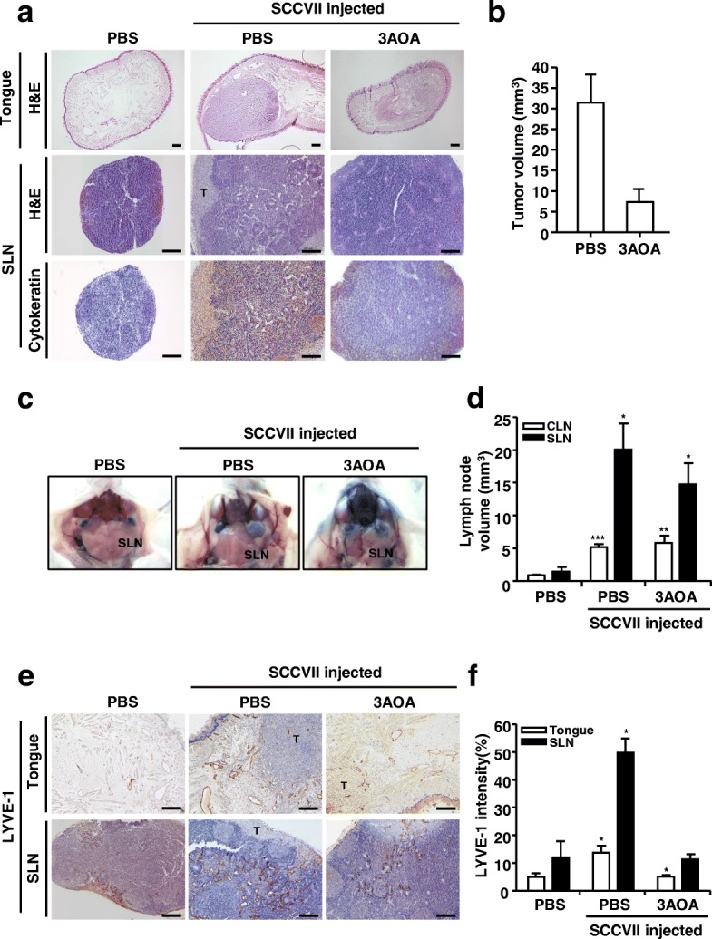
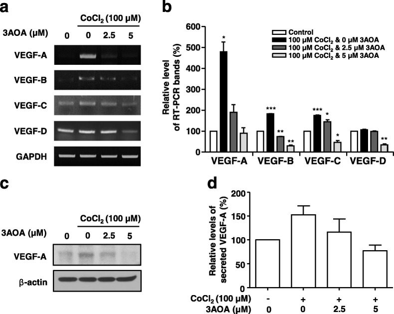
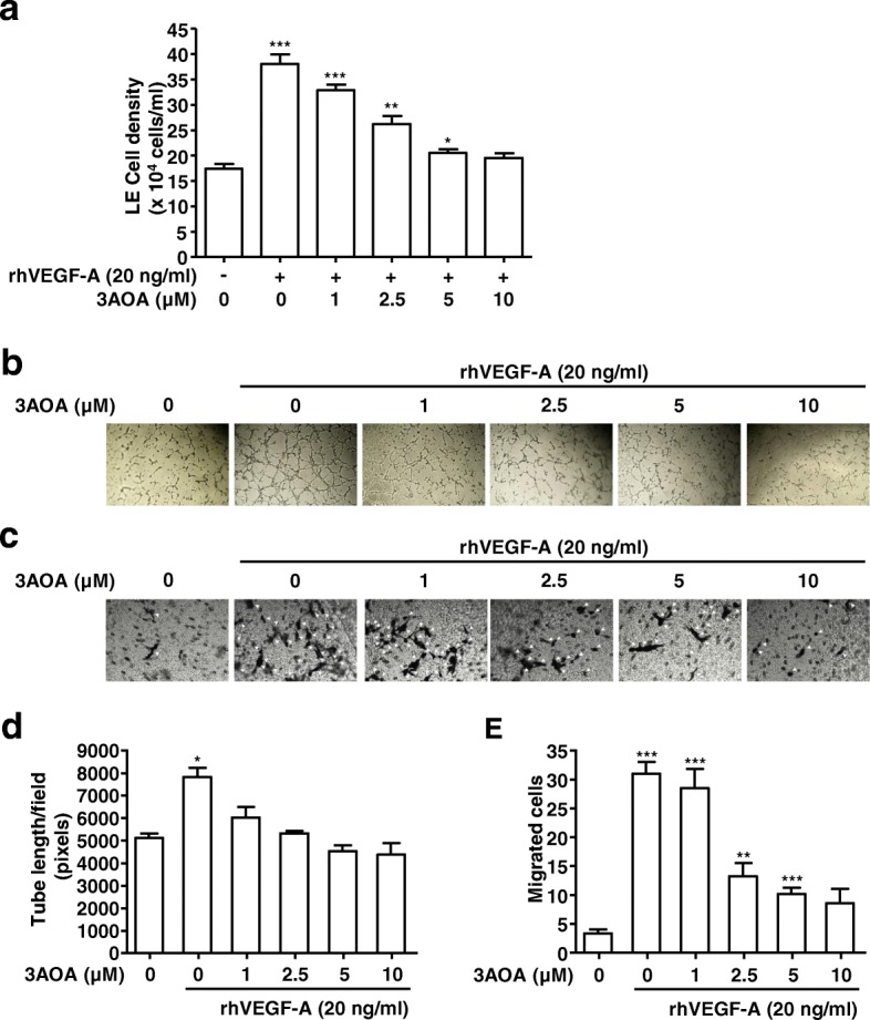
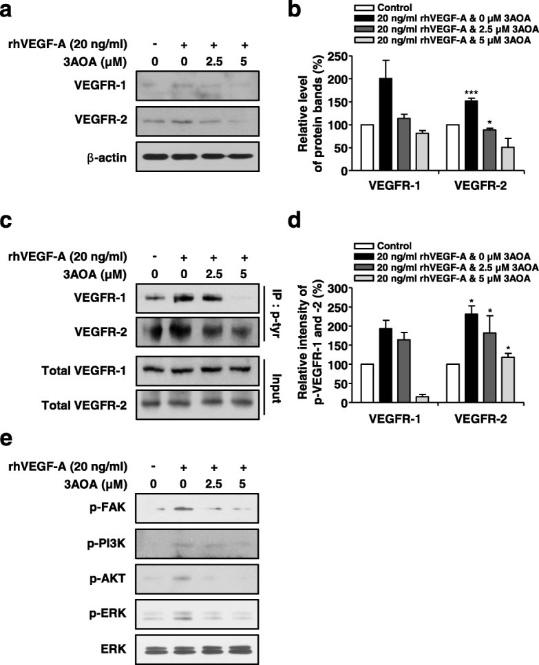
Results: 3AOA inhibited tumor-induced lymphangiogenesis and sentinel lymph node metastasis in an OCSLN animal model, and reduced expression of VEGF-A, a lymphangiogenic factor in hypoxia mimetic agent CoCl2-treated SCCVII cells. 3AOA inhibited proliferation, tube formation, and migration of VEGF-A-treated HLMECs. The lymphatic vessel formation that was stimulated in vivo in a by VEGF-A Matrigel plug was reduced by 3AOA. 3AOA suppressed phosphorylation of vascular endothelial growth factor (VEGFR) -1 and - 2 receptors that was stimulated by VEGF-A. In addition, 3AOA suppressed phosphorylation of the lymphangiogenesis-related downstream signaling factors PI3K, FAK, AKT, and ERK1/2. 3AOA inhibited tumor growth, tumor-induced lymphangiogenesis, and sentinel lymph node metastasis in a VEGF-A-induced OCSLN animal model that was established using VEGF-A overexpressing SCCVII cells.

Conclusion: 3AOA inhibits VEGF-A-induced lymphangiogenesis and sentinel lymph node metastasis both in vitro and in vivo. The anti-lymphangiogenic effects of 3AOA are probably mediated via suppression of VEGF-A/VEGFR-1 and VEGFR-2 signaling in HLMECs, and can be a useful anti-tumor agent to restrict the metastatic spread of oral cancer.

Keywords: 3-O-acetyloleanolic acid; Lymph node metastasis; Lymphangiogenesis; Oral cancer sentinel lymph node animal model; VEGF-A.


Conflict of interest statement

Ethics approval

This study (KHUASP-15-09) was reviewed and approved by the Institutional Animal Care and Use Committee of Kyung Hee University, and animal care and experimental procedures followed the University guidelines for the care and use of laboratory animals. The human cell line, HLMEC cells used in this study, were purchased from Lonza, and the use of widely available cell lines (Lonza, a publicly accessible repository) does not require ethical approval at our institution.

Consent for publication

Not applicable.

Competing interests

The authors declare that they have no competing interests.

Publisher’s Note

Springer Nature remains neutral with regard to jurisdictional claims in published maps and institutional affiliations.

Figures

Fig. 1
Fig. 1
Effects of 3AOA on tumor growth and lymphangiogenesis in an oral cancer sentinel lymph node animal model. a, Tumor growth and metastasis to sentinel lymph node b, Tumor volume c, Image of sentinel lymph nodes d, Sentinel lymph node volume e-f, Lymphatic vessels in tumor and sentinel lymph node sections. Tumor growth of each group was confirmed by hematoxylin and eosin staining of tumor(tongue) sections. And metastasis to sentinel lymph node was confirmed using hematoxylin and eosin staining, and immunohistochemical analysis with anti-cytokeratin antibody of sentinel lymph node sections. Tumor sections were digitized and microscopic images were captured under a 100× objective magnification. Scale bar = 200 μm. Sentinel lymph node sections were digitized and microscopic images were captured under a 200× objective magnification. Scale bar = 200 μm. Lymphatic vessels in tumor and sentinel lymph node sections were determined using the immunohistochemical analysis with anti-LYVE-1. All sections were digitized and microscopic images were captured under a 200× objective magnification. Scale bar = 200 μm. Immunohistochemical intensity values of LYVE-1 from captured images were analyzed by the Image J program and represented as a bar diagrams. Data are presented as a mean ± S.D. (*p < 0.05, **p < 0.01, ***p < 0.001). T = tumor; SLN = Sentinel lymph node
Fig. 2
Fig. 2
Effects of 3AOA on the expression of VEGF family proteins in SCCVII cells treated with CoCl2 . a, cDNAs were generated from total RNAs treated with DNase I, and PCR reaction was performed with specific primers of VEGF-A, -B, -C, −D and GAPDH. b, PCR products from three independent experiments (a) were quantified and represented as a bar diagram. The levels of the VEGF-A, -B, -C, and -D transcripts in the control (3AOA- and CoCl2-untreated cells) were estimated as 100%. c, The protein level of VEGF-A in the intracellular fraction was determined using western blot with anti-VEGF-A antibody. d, The secreted VEGF-A protein level in a conditioned medium was determined using an ELISA assay. The amounts of VEGF-A obtained in three independent experiments were quantified and represented as a bar diagram. The level of VEGF-A in the conditioned medium of the control was estimated as 100%. Data are presented as a mean ± S.D. of three independent experiments (*p < 0.05, **p < 0.01, ***p < 0.001)
Fig. 3
Fig. 3
Effects of 3AOA on proliferation, tube formation, and migration in HLMECs stimulated with rhVEGF-A. a, Proliferation in HLMECs stimulated rhVEGF-A. Cells were detached and counted using a hemocytometer. b and d, Tube formation in HLMECs stimulated rhVEGF-A. Cells were imaged under a inverted phase contrast microscope using a digital single-lens reflex camera. Total tube lengths of a unit area were calculated using the Image J program. c and e, The migrated cells to the underside of membranes were fixed with methanol, stained with hematoxylin solution, and then imaged under a inverted phase contrast microscope using a digital camera. Five digital images per well for (C) were obtained, and the numbers of migrated HLMECs were counted. Each sample was assayed in duplicate. Numbers of migrated HLMECs present in 320 mm2 are presented as a bar diagram. Data are presented as a mean ± S.D. of three independent experiments (*p < 0.05, **p < 0.01, ***p < 0.001)
Fig. 4
Fig. 4
Effects of 3AOA on expressions of VEGFR-1 and VEGFR-2, and activation of VEGFR-1, VEGFR-2 and lymphangiogenesis related downstream signaling factors in rhVEGF-A-treated HLMECs. a-b Expression levels of VEGFR-1 and -2 proteins were determined using Western blot analysis. Amounts of VEGFR-1 and -2 obtained in three independent experiments were quantified and represented as a bar diagram. Levels of VEGFR-1 and -2 in 3AOA- and rhVEGF-A-untreated cells were estimated as 100%. c-d, Cell lysates were immunoprecipitated with anti-phospho-Tyr (anti-p-Tyr). The level of phosphorylated VEGFR-1 and -2 in immunoprecipitates was detected using Western blot analysis with anti-VEGFR-1 and anti-VEGFR-2. Phosphorylation levels of VEGFR-1 and -2 obtained in three independent experiments were quantified and represented as a bar diagram. Phosphorylation levels of VEGFR-1 and -2 in 3AOA- and rhVEGF-A-untreated cells were estimated as 100%. e, HLMECs were serum starved for 6 h, then were treated with different concentrations of 3AOA (0, 2.5, 5 μM) in the presence of rhVEGF-A (20 ng/mL) for 60 min. The phosphorylation levels of FAK, PI3K, AKT, and ERK1/2 were determined using Western blot analysis with anti-p-FAK, anti-p-PI3K, anti-p-AKT, and anti-p-ERK1/2. Data are presented as a mean ± S.D. of three independent experiments (*p < 0.05, **p < 0.01, ***p < 0.001)
Fig. 5
Fig. 5
Effects of 3AOA on VEGF-A-induced lymphatic vessel formation in an in vivo Matrigel plug. a, Matrigel plugs were excised and photographed using a digital camera. The lymphatic vessel density values in Matrigel plug sections were measured using the immunohistochemical analysis with anti-LYVE-1 antibody. All Matrigel sections were digitalized and microscopic images were captured under 200× objective magnification. Scale bar = 200 μm. b, Immunohistochemical intensity values (LYVE-1) from captured images were analyzed by the Image J program and represented as a bar diagram. Data are presented as a mean ± S.D. (*p < 0.05)
Fig. 6
Fig. 6
Effects of 3AOA on tumor growth, and sentinel lymph node enlargement and lymph node metastasis in a VEGF-A-induced oral cancer sentinel lymph node animal model. a, Tumor (tongue) and SLN sections of mice of each group were analyzed using hematoxylin and eosin staining, and immunohistochemical analysis with anti-cytokeratin antibody. All tumor sections were digitized, and microscopic images were captured under a 100× objective magnification. Scale bar = 200 μm. All SLN sections were digitized and images were captured under 200× objective magnification. Scale bar = 200 μm. b-c, Tumor volume and SLN volume were measured using a caliper. Data are presented as a mean ± S.D. (**p < 0.01, ***p < 0.001). T = tumor; SLN = Sentinel lymph node
Fig. 7
Fig. 7
Effect of 3AOA on tumor-induced lymphangiogenesis in a VEGF-A-induced oral cancer sentinel lymph node animal model. a, Lymphatic vessel density values in tumor and SLN sections were measured by immunohistochemical analysis using anti-LYVE-1 antibody. All sections were digitalized and images were captured under 200× objective magnification. Scale bar = 200 μm. b-c, Immunohistochemical intensity values of LYVE-1 from captured images of tumors and SLN were analyzed via the Image J program and represented as a bar diagram. Data are presented as a mean ± S.D. (*p < 0.05, ***p < 0.001). T = tumor; SLN = Sentinel lymph node

Similar articles

Cited by

References

    1. Sales CB, Buim ME, de Souza RO, de Faro Valverde L, Mathias Machado MC, Reis MG, Soares FA, Ramos EA, Gurgel Rocha CA. Elevated VEGFA mRNA levels in oral squamous cell carcinomas and tumor margins: a preliminary study. J Oral Pathol Med. 2016;45(7):481–485. doi: 10.1111/jop.12398. - DOI - PubMed
    1. Duong T, Koopman P, Francois M. Tumor lymphangiogenesis as a potential therapeutic target. J Oncol. 2012;2012:1–23. doi: 10.1155/2012/204946. - DOI - PMC - PubMed
    1. Baek CH. Sentinel lymph node biopsy in the oral cavity cancer. Hanyang Med Rev. 2009;29(3):255–264. doi: 10.7599/hmr.2009.29.3.255. - DOI
    1. Tammela T, Alitalo K. Lymphangiogenesis: molecular mechanisms and future promise. Cell. 2010;140(4):460–476. doi: 10.1016/j.cell.2010.01.045. - DOI - PubMed
    1. Ozasa R, Ohno J, Iwahashi T, Taniguchi K. Tumor-induced lymphangiogenesis in cervical lymph nodes in oral melanoma-bearing mice. J Exp Clin Cancer Res. 2012;31(1):83. doi: 10.1186/1756-9966-31-83. - DOI - PMC - PubMed

MeSH terms

Substances