. 2018 Jul 5;6(1):57.
doi: 10.1186/s40478-018-0560-y.

Ablation of tau causes an olfactory deficit in a murine model of Parkinson's disease

Affiliations

Ablation of tau causes an olfactory deficit in a murine model of Parkinson's disease

Leah C Beauchamp et al. Acta Neuropathol Commun. .

Abstract

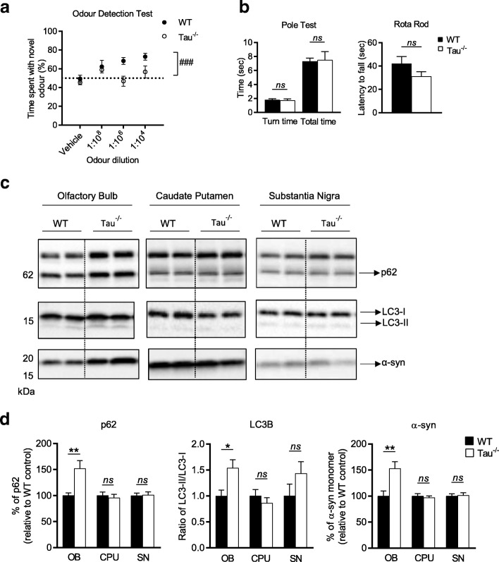
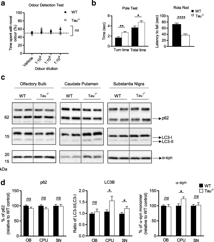
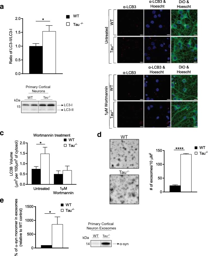
Parkinson's disease is diagnosed upon the presentation of motor symptoms, resulting from substantial degeneration of dopaminergic neurons in the midbrain. Prior to diagnosis, there is a lengthy prodromal stage in which non-motor symptoms, including olfactory deficits (hyposmia), develop. There is limited information about non-motor impairments and there is a need for directed research into these early pathogenic cellular pathways that precede extensive dopaminergic death in the midbrain. The protein tau has been identified as a genetic risk factor in the development of sporadic PD. Tau knockout mice have been reported as an age-dependent model of PD, and this study has demonstrated that they develop motor deficits at 15-months-old. We have shown that at 7-month-old tau knockout mice present with an overt hyposmic phenotype. This olfactory deficit correlates with an accumulation of α-synuclein, as well as autophagic impairment, in the olfactory bulb. This pathological feature becomes apparent in the striatum and substantia nigra of 15-month-old tau knockout mice, suggesting the potential for a spread of disease. Initial primary cell culture experiments have demonstrated that ablation of tau results in the release of α-synuclein enriched exosomes, providing a potential mechanism for disease spread. These alterations in α-synuclein level as well as a marked autophagy impairment in the tau knockout primary cells recapitulate results seen in the animal model. These data implicate a pathological role for tau in early Parkinson's disease.

Keywords: Autophagy; Neurodegeneration; Olfaction; Parkinson’s disease; Tau.


Conflict of interest statement

Ethics approval

This study was carried out in strict accordance with the Australian National Health and Medical Research Council published code of practice for animal research. The protocols were approved by The Florey Animal Ethics Committee (AEC number: 12–094 and 15–092). All efforts were made to minimize animal suffering.

Consent for publication

Not applicable.

Competing interests

The authors declare that they have no competing interests.

Publisher’s Note

Springer Nature remains neutral with regard to jurisdictional claims in published maps and institutional affiliations.

Figures

Fig. 1
Fig. 1
Olfactory deficit, motor performance and pathological features in 7-month-old tau−/− mice. a Odour detection test performed on 7-month-old tau−/− (n = 10–11) and littermate WT controls (n = 10–12). b Motor evaluation of 7-month-old tau−/− (n = 10) and littermate WT control (n = 11), including Rota Rod and Pole Test performance. c Representative western blots of cell lysate from olfactory bulb, caudate putamen and substantia nigra from 7-month-old tau−/− (n = 6) and littermate WT controls (n = 6) immunoblotted for p62, LC3B and α-syn. d Quantification of western blot densitometry presented as % of p62 relative to WT control, ratio of LC3-II/I relative to WT control and % of α-syn relative to WT control. Cell lysate for western blots normalised to automated total protein measurement via ChemiDoc stain-free detection software. ODT analysed by two-way repeated measures ANOVA (one factor repetition) with Fisher LSD post-hoc comparisons. # represents significant main effect of genotype, ### p < 0.001. Motor tests analysed by unpaired two-sided t test. Western blot analysed by unpaired two-sided t test from 3 independent repeats, * p < 0.05, ** p < 0.01, *** p < 0.001, **** p < 0.0001, ns: not significant. Full western blot images presented in Additional file 1: Figure S3
Fig. 2
Fig. 2
Olfactory deficit, motor performance and pathological features in 15-month-old tau−/− mice. a Odour detection test performed on 15-month-old tau−/− (n = 13) and littermate WT controls (n = 8). b Motor evaluation of 15-month-old tau−/− (n = 11) and littermate WT control (n = 8), including Rota Rod and Pole Test performance. c Representative western blots of cell lysate from olfactory bulb, caudate putamen and substantia nigra from 15-month-old tau−/− (n = 6) and littermate WT controls (n = 6) immunoblotted for p62, LC3B and α-syn. d Quantification of western blot densitometry presented as % of p62 relative to WT control, ratio of LC3-II/I relative to WT control and % of α-syn relative to WT control. Cell lysate for western blots normalised to automated total protein measurement via ChemiDoc stain-free detection software. ODT analysed by two-way repeated measures ANOVA (one factor repetition) with Fisher LSD post-hoc comparisons. Motor tests analysed by unpaired two-sided t test. Western blot analysed by unpaired two-sided t test from 3 independent repeats, * p < 0.05, ** p < 0.01, *** p < 0.001, **** p < 0.0001, ns: not significant. Full western blot images presented in Additional file 1: Figure S4
Fig. 3
Fig. 3
Tau−/− primary cortical neurons display autophagic impairment and have an increase in the release of α-synuclein enriched exosomes. a Quantification of western blot densitometry presented as ratio of LC3-II/I of tau−/− primary cortical neurons (n = 5) and WT primary cortical neurons (n = 5). b Representative images of tau−/− and WT primary cortical neurons immunostained for LC3B and post-stained with DiO and Hoechst 33,342 after incubation with either culture medium (untreated) or 1 μM Wortmannin for 8 h, scale bars: 10 μm. c Quantification of the number of LCB3 positive autophagosomes per 100 μm3 of DiO stained cytosol, in untreated and Wortmannin treated WT (n = 5) and tau−/− (n = 5) primary cortical neurons. d Representative electron micrograph images of tau−/− (n = 3) and WT (n = 3) exosome enriched cell culture media (scale bars: 200 μm) with quantitation of number of exosomes per 10 μM2. e Quantification of western blot densitometry from exosomes isolated from tau−/− (n = 2) and WT (n = 2) primary cortical neurons, presented as % of α-syn relative to WT control and representative western blot. Cell lysate for western blots normalised to automated total protein measurement via ChemiDoc stain-free detection software. Immunocytochemistry data analysed by one-way ANOVA. Western blot analysed by unpaired two-sided t test from 3 independent repeats, * p < 0.05, ** p < 0.01, *** p < 0.001, **** p < 0.0001, ns: not significant. Full western blot images presented in Additional file 1: Figure S5

Similar articles

Cited by

References

    1. Ahmed T, Van der Jeugd A, Blum D, Galas MC, D'Hooge R, Buee L, Balschun D. Cognition and hippocampal synaptic plasticity in mice with a homozygous tau deletion. Neurobiol Aging. 2014;35:2474–2478. doi: 10.1016/j.neurobiolaging.2014.05.005. - DOI - PubMed
    1. Alegre-Abarrategui J, Wade-Martins R. Parkinson disease, LRRK2 and the endocytic-autophagic pathway. Autophagy. 2009;5:1208–1210. doi: 10.4161/auto.5.8.9894. - DOI - PubMed
    1. Alonso AC, Zaidi T, Grundke-Iqbal I, Iqbal K. Role of abnormally phosphorylated tau in the breakdown of microtubules in Alzheimer disease. Proc Natl Acad Sci U S A. 1994;91:5562–5566. doi: 10.1073/pnas.91.12.5562. - DOI - PMC - PubMed
    1. Alvarez-Erviti L, Seow Y, Schapira AH, Gardiner C, Sargent IL, Wood MJ, Cooper JM. Lysosomal dysfunction increases exosome-mediated alpha-synuclein release and transmission. Neurobiol Dis. 2011;42:360–367. doi: 10.1016/j.nbd.2011.01.029. - DOI - PMC - PubMed
    1. Attems J, Jellinger KA. Olfactory tau pathology in Alzheimer disease and mild cognitive impairment. Clin Neuropathol. 2006;25:265–271. - PubMed

Publication types

MeSH terms