. 2018 Jul 5;3(4):e00303-18.
doi: 10.1128/mSphere.00303-18.

Transporters MRP1 and MRP2 Regulate Opposing Inflammatory Signals To Control Transepithelial Neutrophil Migration during Streptococcus pneumoniae Lung Infection

Affiliations

Transporters MRP1 and MRP2 Regulate Opposing Inflammatory Signals To Control Transepithelial Neutrophil Migration during Streptococcus pneumoniae Lung Infection

Andrew Zukauskas et al. mSphere. .

Abstract

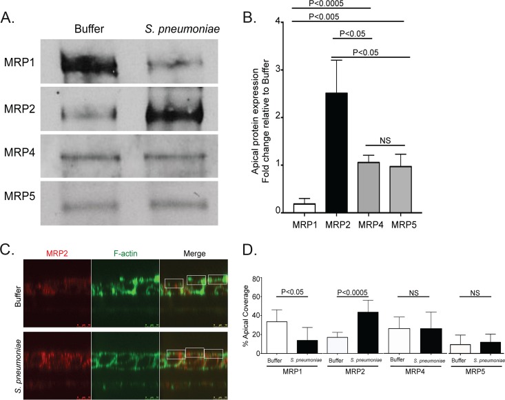
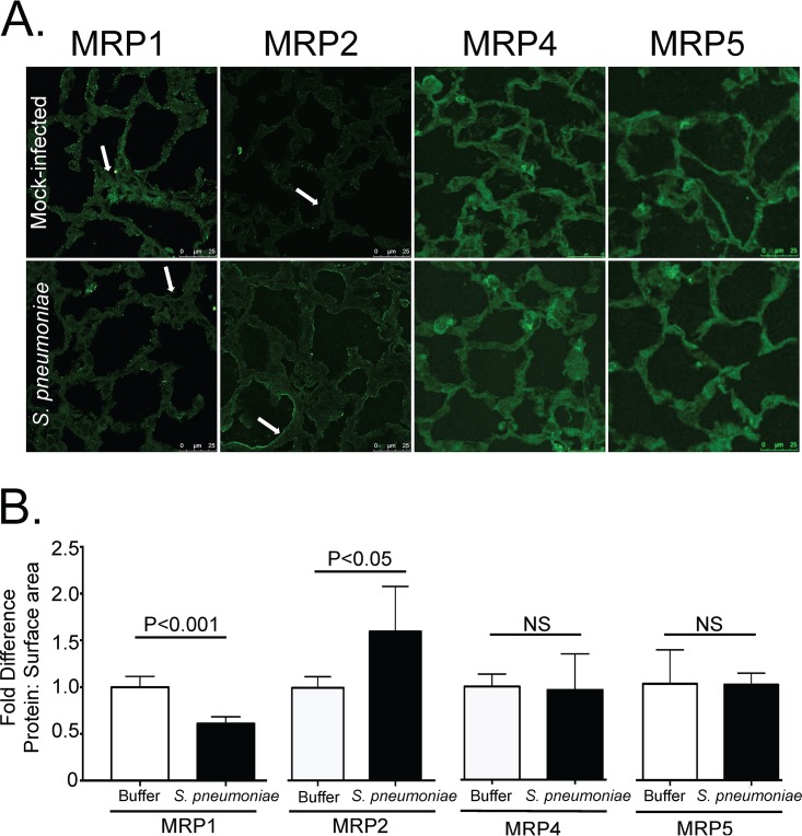
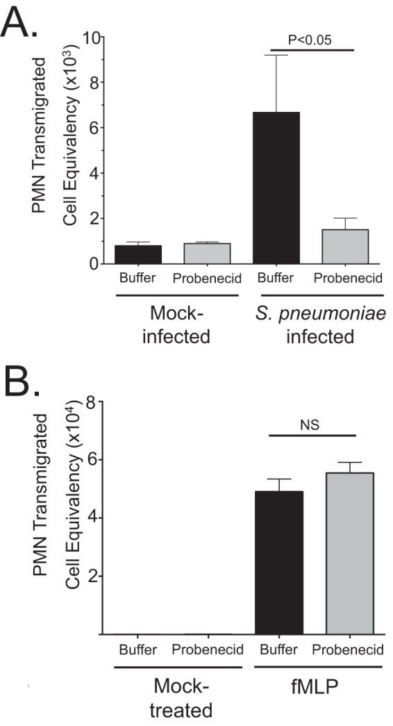
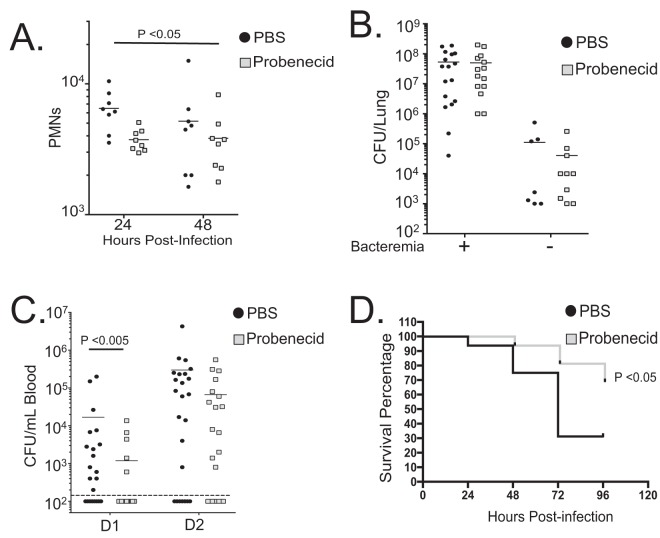
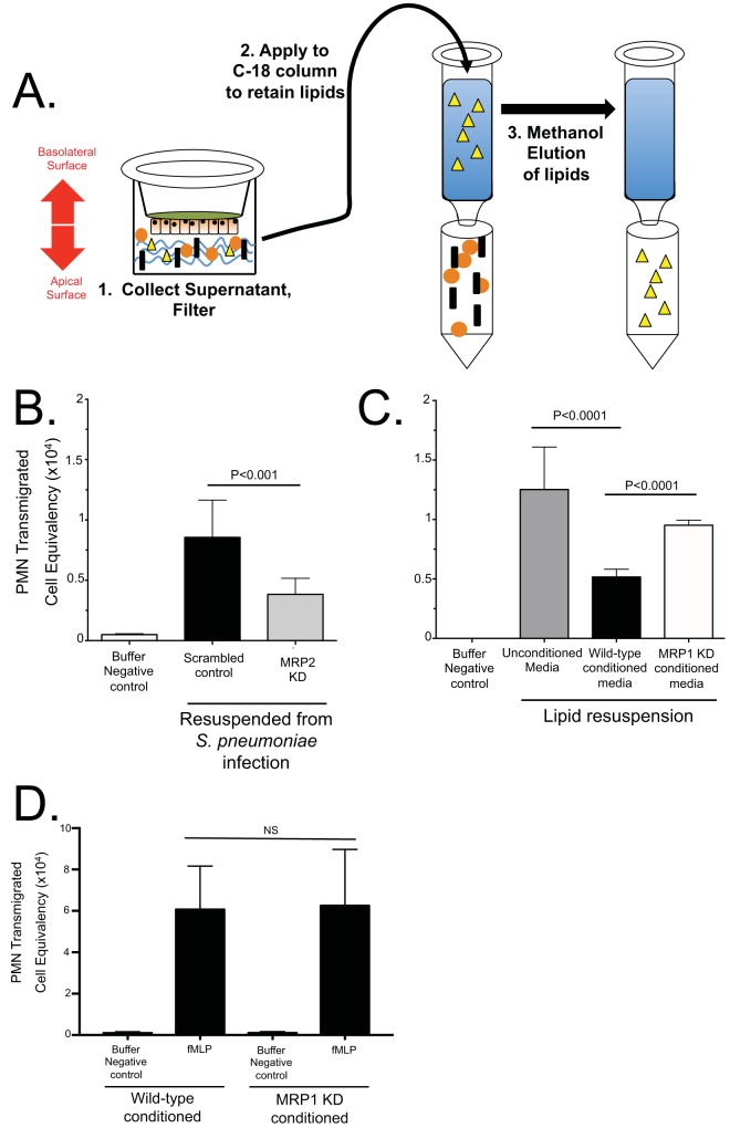
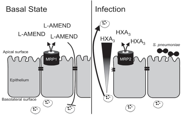
Streptococcus pneumoniae remains a source of morbidity and mortality in both developed and underdeveloped nations of the world. Disease can manifest as pneumonia, bacteremia, and meningitis, depending on the localization of infection. Interestingly, there is a correlation in experimental murine infections between the development of bacteremia and influx of neutrophils into the pulmonary lumen. Reduction of this neutrophil influx has been shown to improve survivability during infection. In this study, we use in vitro biotinylation and neutrophil transmigration and in vivo murine infection to identify a system in which two epithelium-localized ATP-binding cassette transporters, MRP1 and MRP2, have inverse activities dictating neutrophil transmigration into the lumen of infected mouse lungs. MRP1 effluxes an anti-inflammatory molecule that maintains homeostasis in uninfected contexts, thus reducing neutrophil infiltration. During inflammatory events, however, MRP1 decreases and MRP2 both increases and effluxes the proinflammatory eicosanoid hepoxilin A3. If we then decrease MRP2 activity during experimental murine infection with S. pneumoniae, we reduce both neutrophil infiltration and bacteremia, showing that MRP2 coordinates this activity in the lung. We conclude that MRP1 assists in depression of polymorphonuclear cell (PMN) migration by effluxing a molecule that inhibits the proinflammatory effects of MRP2 activity.IMPORTANCEStreptococcus pneumoniae is a Gram-positive bacterium that normally inhabits the human nasopharynx asymptomatically. However, it is also a major cause of pneumonia, bacteremia, and meningitis. The transition from pneumonia to bacteremia is critical, as patients that develop septicemia have ~20% mortality rates. Previous studies have shown that while neutrophils, a major bacterium-induced leukocyte, aid in S. pneumoniae elimination, they also contribute to pathology and may mediate the lung-to-blood passage of the bacteria. Herein, we show that epithelium-derived MRP1 and MRP2 efflux immunomodulatory agents that assist in controlling passage of neutrophils during infection and that limiting neutrophil infiltration produced less bacteremia and better survival during murine infection. The importance of our work is twofold: ours is the first to identify an MRP1/MRP2 axis of neutrophil control in the lung. The second is to provide possible therapeutic targets to reduce excess inflammation, thus reducing the chances of developing bacteremia during pneumococcal pneumonia.

Keywords: MRP1; MRP2; PMN; Streptococcus pneumoniae; hepoxilin A3; neutrophil; pneumococcus.


Figures

FIG 1
FIG 1
MRP1 protein on the apical surface reduces during infection with Streptococcus pneumoniae, while MRP2 increases. The apical surface examination of mock-infected or S. pneumoniae serotype 4 (TIGR4)-infected polarized H292 cells. The cells were treated with HBSS (Buffer) or infected (S. pneumoniae), washed, and allowed to rest at 37°C for 1 h postinfection. (A) Apical surfaces were then labeled with biotin and lysed. Samples were normalized to protein content against a BSA standard, exposed to streptavidin beads, and subjected to sodium dodecyl sulfate-polyacrylamide gel electrophoresis (SDS-PAGE). The blots were probed with primary antibodies for the selected proteins. A representative Western blot of apical biotinylation probing MRP expression is shown. (B) Densitometry of pooled Western blot samples from multiple experiments. Statistics calculated using two-tailed Student’s t test compared with MRP4 or MRP5 (n = 3). (C) Buffer-treated or infected cells were fixed and stained for MRP2 and F-actin. Immunofluorescence cross-section Z-stack images of F-actin were utilized to identify cellular borders and apical surface (green). The corresponding region of MRP2-stained Z-stack (red) was marked for the particular region of interest (white boxes) and calculated for the total apical coverage using ImageJ. (D) The calculated percentage of the area taken up by the indicated MRPs in panel C via calculations completed with ImageJ (see Materials and Methods). P values were calculated using two-tailed Student’s t test comparing the percentages for the given protein in uninfected and infected cells (n = 8). Values that are not significantly different (NS) are indicated. Quantification samples for biotinylation and immunofluorescence were taken from at least two separate infections with similar results.
FIG 2
FIG 2
S. pneumoniae infection reduces MRP1 and increases MRP2 upon pulmonary infection in mice. Mice were infected via an intratracheal route with S. pneumoniae and sacrificed 2 days postinfection. The lungs were excised, reinflated, sectioned, and stained for MRP1, MRP2, MRP4, or MRP5. (A) Representative images from three different experiments. Arrows indicate points of interest. In particular, areas of similar density in MRP1-stained lungs appear to have reduced expression in infected lungs compared to uninfected lungs (arrow). MRP2 staining appears to increase drastically on the cell periphery during infection compared to mock-infected lungs (arrow). No such increases or decreases are observed for MRP4 and MRP5. (B) Quantification of staining. Antibody staining was quantified and normalized to surface area (measured by F-actin [not shown]). Fold differences of signal to surface area for each given antibody comparing infected (S. pneumoniae) to uninfected (Buffer) animals are shown. Values are expressed as fold increase or decrease compared to the values for uninfected samples.
FIG 3
FIG 3
MRP2 inhibition via probenecid reduces PMN migration across polarized pulmonary epithelial cells. (A and B) Polarized NCI-H292 cells on filter membranes were incubated with 100 µM MRP2 inhibitor probenecid or mock treated for 1 h with PBS before apical infection with S. pneumoniae at an MOI of 10 (A) or the HxA3-independent bacterial product formyl-methionyl-leucyl-phenylalanine (fMLP) (B). For both panels A and B, mock-infected or mock-treated cells were exposed to HBSS for the treatment time period, and basolateral-to-apical migration of PMNs was quantified with a myeloperoxidase assay against a standard number of PMNs (see Materials and Methods). The results of one representative experiment from at least three experiments performed are shown. Statistical significance was calculated using two-tailed Student’s t test.
FIG 4
FIG 4
MRP2 inhibition mitigates pulmonary burden and bacteremia following lung challenge with S. pneumoniae. Using an in vivo model of PMN migration, we examined the results of MRP2 inhibition. C57BL/6 mice were treated with either PBS or probenecid 3 h before and 3 h after intratracheal application of 2.5 × 10⁵ S. pneumoniae. Four sets of mice were infected in this way. (A) Comparing the number of PMNs found in the lumen after isolating BAL fluid samples after 24 and 48 h postinfection. PMNs, identified as Ly6g-positive cells, were quantified by flow cytometry (n = 8). (B) Overall lung burden from mice sacrificed on day 2. Mice were sacrificed, lungs were excised and homogenized, and total bacterial burden was calculated using serial dilutions (n = 24 for each condition). The presence (+) or absence (−) of bacteremia is indicated as follows: +, mice had detected levels of bacteria in blood 48 h postinfection; −, no bacteria were detected in blood during tail vein bleeds. (C) Bacteremia, as measured by tail vein bleeds, from the cohorts in panel B (see Materials and Methods). Detected events of colony formation on day 1 (D1) and day 2 (D2) (n = 24) are shown. The broken line represents the limit of detection, and as such, values for mice without visible bacteremia were represented as 100 CFU/ml, just below this level of detection. In panels B and C, statistical significance was calculated using the Mann-Whitney test. (D) Survival experiment with the fourth set of mice. There were 16 mice in a group for each condition. Statistical significance was calculated using Mantel-Cox test and Gehan-Breslow-Wilcoxon test. Probenecid treatment consistently increased survival by approximately 30 to 40% during survival experiments and often delayed symptoms, such as lethargy. Mouse experiments were repeated, and similar results were observed in at least two different experiments.
FIG 5
FIG 5
MRP2 and MRP1 promote the secretion of proinflammatory lipids and anti-inflammatory molecules, respectively. (A) Proinflammatory lipids were isolated from apical supernatants of pneumococcus-infected H292 monolayers. HBSS subjected to the apical surface of pneumococcus-infected H292 cells were enriched on C18 columns, which retain the proinflammatory lipids, and eluted in methanol for storage (see Materials and Methods). Yellow triangles represent proinflammatory lipids. (B) Lipids from MRP2-competent (Scrambled control) cells and MRP2-deficient (MRP2 knockdown [MRP2 KD]) cells were enriched and eluted with methanol. This methanol/lipid solution was evaporated under a constant stream of nitrogen and resuspended in HBSS to be used as PMN chemoattractant during a PMN migration assay through a monolayer of naive wild-type H292 cells. (C) Proinflammatory lipids isolated from wild-type cells infected with Streptococcus pneumoniae from panel A were resuspended with either unconditioned media, conditioned media from MRP1-competent scrambled-control cells (Wild-type conditioned media), or conditioned media from MRP1 knockdown cells (MRP1 KD conditioned media) and applied to the apical chamber of naive cells to act as a chemoattractant during a PMN migration assay. HBSS without any lipid acted as a negative control (Buffer Negative control). (D) MRP1-conditioned media failed to inhibit HxA3-independent PMN-migration produced using formyl-methionyl-leucyl-phenylalanine (fMLP). The values were not significantly different (NS) by two-tailed Student’s t test.
FIG 6
FIG 6
Epithelial MRPs assist in controlling pro- and anti-inflammatory states. As shown by this work, lung epithelium expresses large amounts of MRP1 and small amounts of MRP2 at basal (uninfected) states. MRP1 effluxes molecules with anti-inflammatory activity, here termed L-AMEND, which acts to suppress PMN migration. During infection with Streptococcus pneumoniae, MRP1 is reduced and MRP2 increases on the apical surface of the epithelium. MRP2 mediates release of the proinflammatory molecule hepoxilin A3 (HXA3), which creates a chemokine gradient to draw PMNs to the site of infection. By both reducing MRP1 and increasing MRP2, the epithelium works to maximize the transepithelial PMN migration; however, this PMN migration also disrupts epithelial tight junctions and can lead to infiltration of the intruding S. pneumoniae.

Similar articles

Cited by

References

    1. Centers for Disease Control and Prevention 2015. Epidemiology and prevention of vaccine-preventable diseases, 13th ed. Communication and Education Branch, National Center for Immunization and Respiratory Diseases, Centers for Disease Control and Prevention, Atlanta, GA.
    1. Centers for Disease Control and Prevention 2012. Manual for the surveillance of vaccine-preventable diseases. National Center for Immunization and Respiratory Disease, Centers for Disease Control and Prevention, Atlanta, GA.
    1. Baird BR, Cheronis JC, Sandhaus RA, Berger EM, White CW, Repine JE. 1986. O2 metabolites and neutrophil elastase synergistically cause edematous injury in isolated rat lungs. J Appl Physiol 61:2224–2229. doi:10.1152/jappl.1986.61.6.2224. - DOI - PubMed
    1. Flick MR, Perel A, Staub NC. 1981. Leukocytes are required for increased lung microvascular permeability after microembolization in sheep. Circ Res 48:344–351. doi:10.1161/01.RES.48.3.344. - DOI - PubMed
    1. Menéndez R, Cavalcanti M, Reyes S, Mensa J, Martinez R, Marcos MA, Filella X, Niederman M, Torres A. 2008. Markers of treatment failure in hospitalised community acquired pneumonia. Thorax 63:447–452. doi:10.1136/thx.2007.086785. - DOI - PubMed

Publication types

Substances