. 2018 Aug 29;92(18):e00526-18.
doi: 10.1128/JVI.00526-18. Print 2018 Sep 15.

Suppressor of Cytokine Signaling 1 (SOCS1) and SOCS3 Are Stimulated within the Eye during Experimental Murine Cytomegalovirus Retinitis in Mice with Retrovirus-Induced Immunosuppression

Affiliations

Suppressor of Cytokine Signaling 1 (SOCS1) and SOCS3 Are Stimulated within the Eye during Experimental Murine Cytomegalovirus Retinitis in Mice with Retrovirus-Induced Immunosuppression

Hsin Chien et al. J Virol. .

Abstract

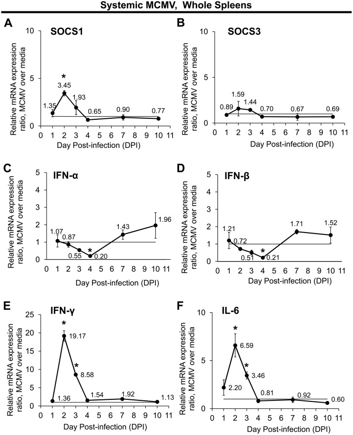
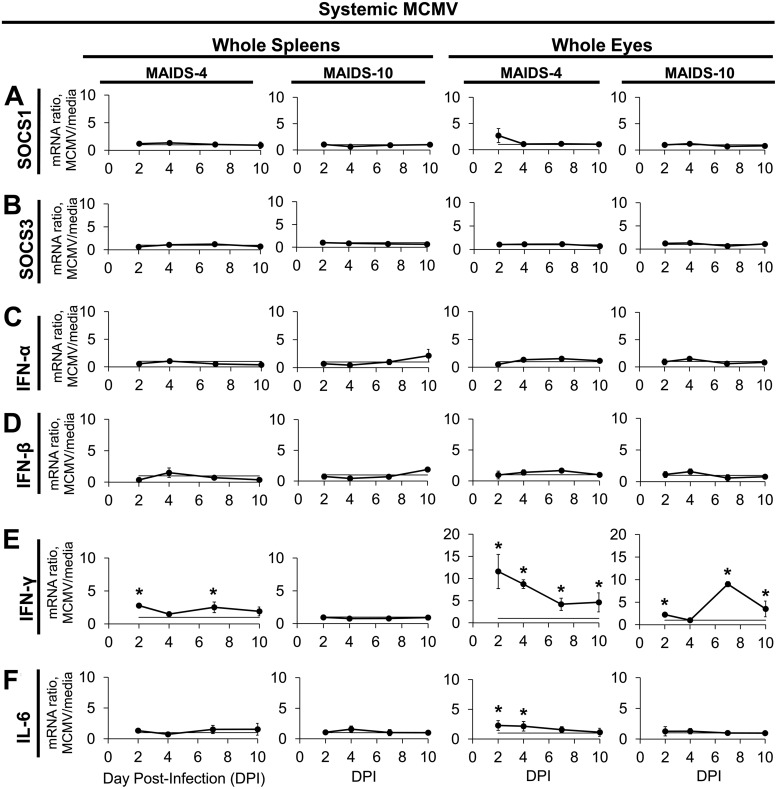
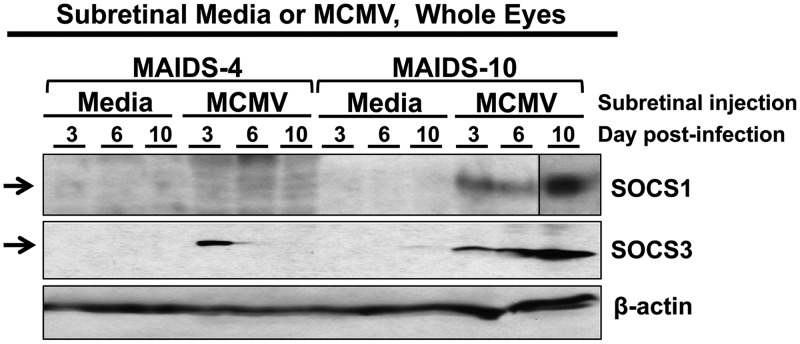
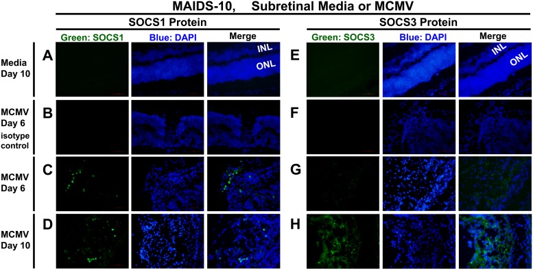
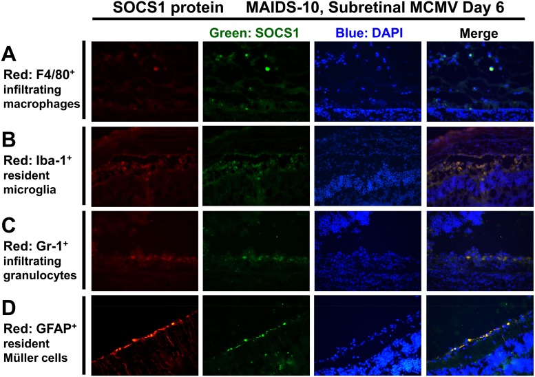
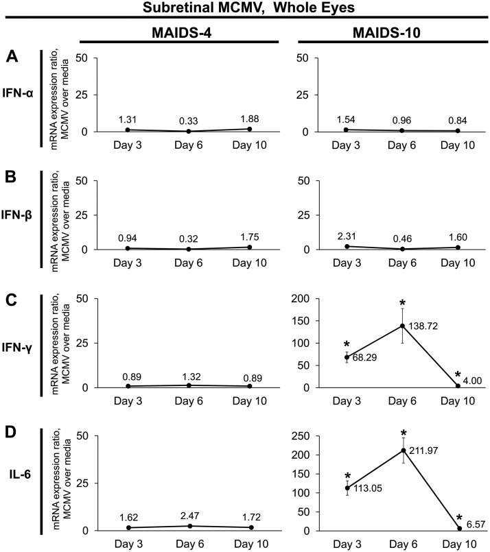
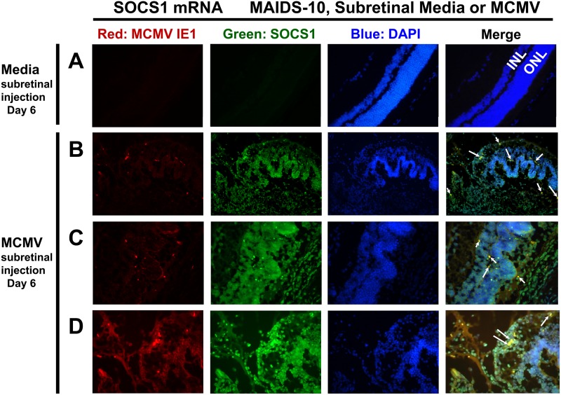
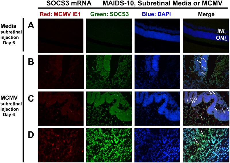
AIDS-related human cytomegalovirus retinitis remains the leading cause of blindness among untreated HIV/AIDS patients worldwide. To study mechanisms of this disease, we used a clinically relevant animal model of murine cytomegalovirus (MCMV) retinitis with retrovirus-induced murine AIDS (MAIDS) that mimics the progression of AIDS in humans. We found in this model that MCMV infection significantly stimulates ocular suppressor of cytokine signaling 1 (SOCS1) and SOCS3, host proteins which hinder immune-related signaling by cytokines, including antiviral type I and type II interferons. The present study demonstrates that in the absence of retinal disease, systemic MCMV infection of mice without MAIDS, but not in mice with MAIDS, leads to mild stimulation of splenic SOCS1 mRNA. In sharp contrast, when MCMV is directly inoculated into the eyes of retinitis-susceptible MAIDS mice, high levels of intraocular SOCS1 and SOCS3 mRNA and protein are produced which are associated with significant intraocular upregulation of gamma interferon (IFN-γ) and interleukin-6 (IL-6) mRNA expression. We also show that infiltrating macrophages, granulocytes, and resident retinal cells are sources of intraocular SOCS1 and SOCS3 protein production during development of MAIDS-related MCMV retinitis, and SOCS1 and SOCS3 mRNA transcripts are detected in retinal areas histologically characteristic of MCMV retinitis. Furthermore, SOCS1 and SOCS3 are found in both MCMV-infected cells and uninfected cells, suggesting that these SOCS proteins are stimulated via a bystander mechanism during MCMV retinitis. Taken together, our findings suggest a role for MCMV-related stimulation of SOCS1 and SOCS3 in the progression of retinal disease during ocular, but not systemic, MCMV infection.IMPORTANCE Cytomegalovirus infection frequently causes blindness in untreated HIV/AIDS patients. This virus manipulates host cells to dysregulate immune functions and drive disease. Here, we use an animal model of this disease to demonstrate that cytomegalovirus infection within eyes during retinitis causes massive upregulation of immunosuppressive host proteins called SOCS. As viral overexpression of SOCS proteins exacerbates infection with other viruses, they may also enhance cytomegalovirus infection. Alternatively, the immunosuppressive effect of SOCS proteins may be protective against immunopathology during cytomegalovirus retinitis, and in such a case SOCS mimetics or overexpression treatment strategies might be used to combat this disease. The results of this work therefore provide crucial basic knowledge that contributes to our understanding of the mechanisms of AIDS-related cytomegalovirus retinitis and, together with future studies, may contribute to the development of novel therapeutic targets that could improve the treatment or management of this sight-threatening disease.

Keywords: AIDS-related cytomegalovirus retinitis; MAIDS; SOCS1; SOCS3; murine cytomegalovirus (MCMV); retinitis; suppressor of cytokine signaling (SOCS).


Figures

FIG 1
FIG 1
Systemic MCMV infection of immunologically normal mice without MAIDS differentially affects temporal mRNA expression of SOCS1, SOCS3, and SOCS-inducing cytokines in whole splenic cells. Groups of C57BL/6 mice without MAIDS were injected intraperitoneally with 104 PFU MCMV or an equal volume of medium (controls). Whole spleens collected at 1, 2, 3, 4, 7, and 10 days postinfection (dpi) were assessed for SOCS1 (A), SOCS3 (B), IFN-α (C), IFN-β (D), IFN-γ (E), and IL-6 (F) mRNA expression by quantitative RT-PCR assay. Mean mRNA expression ratios from MCMV samples are shown relative to their respective medium controls per day by the 2−ΔΔCT method. Values represent means from n = 8 mice per group from two independent experiments, and error bars indicate standard errors of the means (SEM). Statistical significance between groups per day (*, P < 0.05) was determined by Student's t test.
FIG 2
FIG 2
Within the spleens and eyes of MAIDS-4 or MAIDS-10 mice, systemic MCMV infection stimulates IFN-γ and IL-6 mRNA expression but does not affect SOCS1, SOCS3, IFN-α, or IFN-β mRNA transcripts. Groups of C57BL/6 mice with MAIDS-4 or MAIDS-10 were injected intraperitoneally with 104 PFU MCMV or an equal volume of medium (controls). Whole spleens and whole eyes collected at 2, 4, 7, and 10 dpi were assessed for SOCS1 (A), SOCS3 (B), IFN-α (C), IFN-β (D), IFN-γ (E), and IL-6 (F) mRNA expression by real-time quantitative RT-PCR assay. Mean mRNA expression ratios (fold change) from MCMV samples are shown relative to their respective medium controls per day by the 2−ΔΔCT method. Values represent means ± standard deviations (SD) from n = 5 mice per group from two independent experiments. Statistical significance between groups per day (*, P < 0.05) was determined by Student's t test.
FIG 3
FIG 3
Ocular SOCS1 and SOCS3 proteins are stimulated in the MCMV-infected eyes of retinitis-susceptible mice with MAIDS-10. Groups of C57BL/6 mice with MAIDS-4 (retinitis resistant) or MAIDS-10 (retinitis susceptible) were injected subretinally with 104 PFU MCMV (left eyes) or an equal volume of medium (right eyes; controls). Whole eyes (n = 5 per group, pooled) were collected at 3, 6, and 10 days postinfection, and ocular protein was extracted and analyzed by Western blotting for SOCS1 and SOCS3 proteins. SOCS1 contains composite images from Western blots with identical parameters.
FIG 4
FIG 4
Ocular SOCS1 and SOCS3 proteins are expressed in the retinas of MCMV-infected MAIDS-10 mice during MCMV retinitis. Whole eyes were collected from MAIDS-10 mice at days 6 and 10 after subretinal injection with 104 PFU MCMV (left eyes) (B to D and F to H) or an equal volume of medium (right eyes; control) (A and E). Formalin-fixed cryosections were analyzed by fluorescence microscopy for SOCS1 (A, C, and D) or SOCS3 (E, G, and H) proteins (green) or IgG isotype control antibody (B and F). Nuclei were counterstained with DAPI (blue). The inner nuclear layer (INL) and outer nuclear layer (ONL) of the retina are indicated for medium controls, where, in the absence of MCMV retinitis, distinct retinal layers are still intact. Original magnification, ×400.
FIG 5
FIG 5
During MAIDS-related MCMV retinitis, SOCS1 protein is detectable in resident cells and infiltrating immune cells of the retinal tissue. Whole eyes were collected from MAIDS-10 mice 6 days after subretinal injection with 104 PFU MCMV. Formalin-fixed cryosections were immunofluorescently double stained as described in Materials and Methods for detection of SOCS1 protein (green) plus one of the following cell markers (red): F4/80 for infiltrating macrophages (A), Iba-1 for microglia (B), Gr-1 (Ly-6G) for granulocytes (neutrophils) (C), or GFAP for activated Müller cells (D). Nuclei were counterstained with DAPI (blue). Original magnification, ×400.
FIG 6
FIG 6
During MAIDS-related MCMV retinitis, SOCS3 protein is present in resident cells and infiltrating immune cells of the retina. As described for Fig. 5, whole eyes were collected from MAIDS-10 mice on day 6 following subretinal injection with 104 PFU MCMV. Formalin-fixed cryosections were immunofluorescently double stained for detection of SOCS3 protein (green) plus cell markers F4/80 (A), Iba-1 (B), Gr-1 (Ly-6G) (C), or GFAP (D) in the red channel. Nuclei were counterstained with DAPI (blue). Original magnification, ×400.
FIG 7
FIG 7
SOCS-inducing cytokines IFN-γ and IL-6, but not IFN-α or IFN-β, are transcriptionally upregulated concurrently with SOCS1 and SOCS3 during MAIDS-related MCMV retinitis. Groups of C57BL/6 mice with MAIDS-4 (retinitis resistant) or MAIDS-10 (retinitis susceptible) were injected subretinally with 104 PFU MCMV (left eyes) or an equal volume of medium (right eyes, controls). Whole eyes were collected at 3, 6, and 10 days postinfection and analyzed for IFN-α (A), IFN-β (B), IFN-γ (C), and IL-6 (D) mRNA expression by real-time quantitative RT-PCR assay. Mean mRNA expression ratios (fold change) from MCMV-infected eyes are shown relative to their medium-injected control eyes by the 2−ΔΔCT method. Values represent means ± SD from n = 5 mice per group. (Note that the y axis scaling differs between gene targets.) Statistical significance between treatment with MCMV and subretinal medium per day (*, P < 0.05) was determined by Student's t test.
FIG 8
FIG 8
SOCS1 mRNA transcripts are detectable during MAIDS-related MCMV retinitis not only in retinal cells that are directly infected with MCMV but also in uninfected bystander retinal cells. Eyes from MAIDS-10 mice were collected 6 days following subretinal injection with medium (control) (A) or 104 PFU MCMV (B to D). Cryosections were double stained by fluorescent in situ hybridization (FISH) per Materials and Methods for mRNA transcripts of SOCS1 (green) and MCMV IE1 (red). Nuclei were counterstained with DAPI (blue). The inner nuclear layer (INL) and outer nuclear layer (ONL) of the retina are indicated for medium controls. Same-cell colocalization of SOCS1 and MCMV IE1 mRNA expression is indicated by white arrows. Ocular sections containing histopathologically characteristic areas of MCMV retinitis are shown: transition zones between retinal folding and retinal necrosis (B), retinal folding (C), and retinal necrosis (D). Original magnification, 200× (A and B) or 400× (C and D).
FIG 9
FIG 9
SOCS3 mRNA transcripts are expressed during MAIDS-related MCMV retinitis in retinal cells directly infected with MCMV as well as in uninfected bystander retinal cells. As described for Fig. 8, eyes from MAIDS-10 mice were collected 6 days following subretinal injection with medium (control) (A) or 104 PFU MCMV (B to D). Cryosections were double stained by FISH for mRNA transcripts of SOCS3 (green) and MCMV IE1 (red). Nuclei were counterstained with DAPI (blue). The inner nuclear layer (INL) and outer nuclear layer (ONL) of the retina are indicated for medium controls. Same-cell colocalization of SOCS3 and MCMV IE1 mRNA transcripts is indicated by white arrows. Sections containing histopathologically characteristic areas of MCMV retinitis are shown: transition zones between retinal folding and retinal necrosis (B), retinal folding (C), and retinal necrosis (D). Original magnification, 200× (A) or 400× (B to D).

Similar articles

Cited by

References

    1. Staras SA, Dollard SC, Radford KW, Flanders WD, Pass RF, Cannon MJ. 2006. Seroprevalence of cytomegalovirus infection in the United States, 1988-1994. Clin Infect Dis 43:1143–1151. doi:10.1086/508173. - DOI - PubMed
    1. Mocarski ES Jr, Shenk T, Pass RF. 2007. Cytomegalovirus, p 2702–2772. In Knipe DM, Howley PM (ed), Fields virology, vol 2 Lippincott Williams & Wilkins, Philadelphia, PA.
    1. Hahn G, Jores R, Mocarski ES. 1998. Cytomegalovirus remains latent in a common precursor of dendritic and myeloid cells. Proc Natl Acad Sci U S A 95:3937–3942. doi:10.1073/pnas.95.7.3937. - DOI - PMC - PubMed
    1. Mocarski ES Jr, Shenk T, Griffiths PD, Pass RF. 2013. Cytomegaloviruses, p 1960–2014. In Knipe DM, Howley PM, Cohen JI, Griffin DE, Lamb RA, Martin MA, Racaniello VR, Roizman B (ed), Fields virology, 6th ed, vol 2 Lippincott Williams & Wilkins, Philadelphia, PA.
    1. Dix RD, Cousins SW. 2004. AIDS-related cytomegalovirus retinitis: lessons from the laboratory. Curr Eye Res 29:91–101. doi:10.1080/02713680490504641. - DOI - PubMed

Publication types

MeSH terms

Substances

LinkOut - more resources