Review
. 2018 Nov;103(11):1760-1771.
doi: 10.3324/haematol.2018.195867. Epub 2018 Jul 5.

Preclinical human models and emerging therapeutics for advanced systemic mastocytosis

Affiliations
Review

Preclinical human models and emerging therapeutics for advanced systemic mastocytosis

Michel Arock et al. Haematologica. 2018 Nov.

Abstract

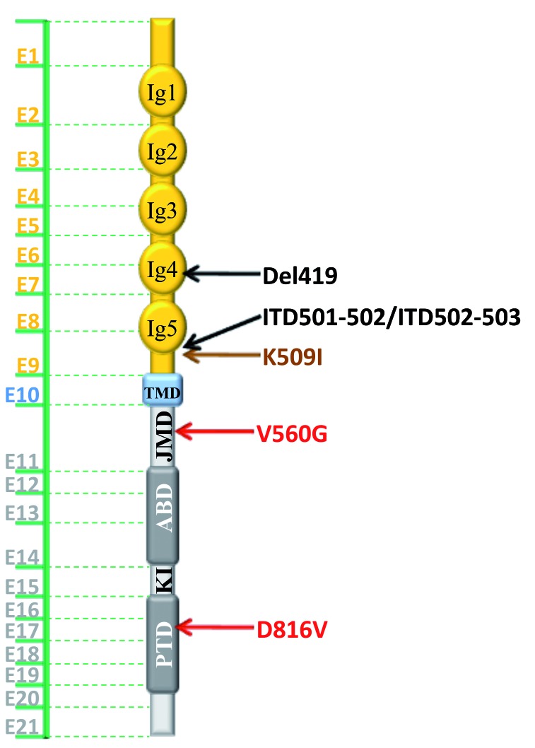
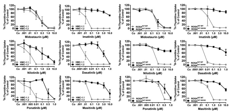
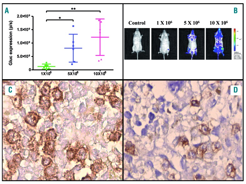
Mastocytosis is a term used to denote a group of rare diseases characterized by an abnormal accumulation of neoplastic mast cells in various tissues and organs. In most patients with systemic mastocytosis, the neoplastic cells carry activating mutations in KIT Progress in mastocytosis research has long been hindered by the lack of suitable in vitro models, such as permanent human mast cell lines. In fact, only a few human mast cell lines are available to date: HMC-1, LAD1/2, LUVA, ROSA and MCPV-1. The HMC-1 and LAD1/2 cell lines were derived from patients with mast cell leukemia. By contrast, the more recently established LUVA, ROSA and MCPV-1 cell lines were derived from CD34+ cells of non-mastocytosis donors. While some of these cell lines (LAD1/2, LUVA, ROSAKIT WT and MCPV-1) do not harbor KIT mutations, HMC-1 and ROSAKIT D816V cells exhibit activating KIT mutations found in mastocytosis and have thus been used to study disease pathogenesis. In addition, these cell lines are increasingly employed to validate new therapeutic targets and to screen for effects of new targeted drugs. Recently, the ROSAKIT D816V subclone has been successfully used to generate a unique in vivo model of advanced mastocytosis by injection into immunocompromised mice. Such a model may allow in vivo validation of data obtained in vitro with targeted drugs directed against mastocytosis. In this review, we discuss the major characteristics of all available human mast cell lines, with particular emphasis on the use of HMC-1 and ROSAKIT D816V cells in preclinical therapeutic research in mastocytosis.


Figures

Figure 1.
Figure 1.
Normal structure of the KIT receptor and KIT mutations described in human mast cell leukemia-like cell lines and in patients with mastocytosis. In humans, KIT, located on chromosome 4q12, contains 21 exons transcribed/translated into a transmembrane receptor with tyrosine kinase activity (145 kDa and 976 amino acids). KIT is presented in its monomeric form, but dimerizes as a result of stem cell factor (SCF) ligation. The extracellular domain, in yellow, comprises five immunoglobulin (Ig)-like subunits where the ligand binding site (SCF for KIT) and the dimerization site are located. The cytoplasmic region contains a transmembrane domain (TMD) made by a single helix, in blue. The intracellular portion of KIT, in gray, contains an auto-inhibitory juxtamembrane domain (JMD) and a kinase domain (in dark gray) which is split into two parts: an ATP-binding domain (ABD) and a phosphotransferase domain (PTD) linked by a large kinase insert (KI) domain of ~ 60–100 residues. For the sake of clarity and readability, only the most frequent mutations found in patients and/or in human MC lines are represented. In red, mutants found in adult patients and in cell lines (KIT D816V found in >80% of adult patients with systemic mastocytosis and 30% of children with cutaneous mastocytosis, as well as in HMC-1.2 and ROSAKIT D816V MCL-like cell lines, and KIT V560G found in the MCL-like cell lines HMC-1.1 and HMC-1.2, but only in a very few adult patients). In black, three of the KIT defects most frequently found in pediatric patients: KIT Del419 (NM_000222.2(KIT):c.1255_1257del, p.Asp419del), KIT ITD501-502 (NM_000222.2(KIT):c.1500_1505dup, p.Ser501_Ala502dup) and KIT ITD502-503 (NM_000222.2(KIT):c.1503_1508dup, p.Ala502_Tyr503dup) and in brown, the KIT K509I mutant found in several familial cases of the disease. For a complete overview of the various KIT mutations found in pediatric and adult mastocytosis patients, see Valent et al. Del, deletion; ITD, internal tandem duplication.
Figure 2.
Figure 2.
Dose-dependent inhibition of the proliferation of wild-type and mutant KIT human mast cell lines by various tyrosine kinase inhibitors in vitro. Human mast cell lines expressing KIT D816V (HMC-1.2 or ROSAKIT D816V; black lines) or lacking KIT D816V (HMC-1.1 or ROSAKIT WT; gray lines) were incubated in control medium (Co) or in medium containing various concentrations of tyrosine kinase inhibitors (as indicated) at 37°C for 48 h. Thereafter, 3H-thymidine uptake was measured. Results are expressed as percent of 3H-thymidine uptake compared to the control and represent the mean ± standard deviation of three different experiments.
Figure 3.
Figure 3.
Dose-dependent inhibition of the proliferation of KIT D816V+ human mast cell lines by STAT5 inhibitors in vitro. ROSAKIT D816V and HMC-1.2 cells were cultured in 96-well plates for 72 h in control medium containing 0.1% dimethylsulfoxide (DMSO) or with increasing concentrations (between 1.0 and 50.0 μM) of SF-1066, a very weak STA5 inhibitor (Ki >25 μM on STAT5), and of more specific and potent STAT5 inhibitors BP-1-102 and BP-1-108 (Ki >10 μM on STAT5). Viability was calculated in each condition by the MTT method. Results are expressed as percent of control and represent the mean ± standard deviation of triplicate experiments. The half maximal inhibitory concentration (IC50) at day 3 of each compound for each cell line was calculated using Prism GraphPad 4.0 software after plotting log concentration versus response. As expected, while the IC50 for SF-1066 was >50 μM, IC50 values for the more STAT5-specific compounds were lower: 11 μM for BP-1-102 on both KIT D816V+ neoplastic human MC lines and 34 and 22 μM for BP-1-108 for, respectively, HMC-1.2 and ROSAKIT D816V cells. Although these values are still irrelevant at the pharmacological level, they open hopes that drug optimization might lead in a near future to the design of more potent small molecules inhibiting STAT5 activity.
Figure 4.
Figure 4.
Engraftment of ROSAKIT D816V-Gluc cells in NSG mice. Increasing numbers of ROSAKIT D816V-Gluc cells (1×10⁶, 5×10⁶ or 10×106) were injected into the tail vein of NOD-SCID IL-2Rγ-/- (NSG) mice (3 groups; 6 mice per group) 24 h after irradiation at 2.5 Gy from a cesium-137 source. After 10 weeks, engraftment was assessed using (A) quantitative measurements of Gluc activity in plasma and (B) in vivo bioluminescence imaging (IVIS) on engrafted mice. Ten weeks after engraftment, mice were sacrificed and (C) bone marrow and (D) spleen sections from the three groups of mice were stained by immunohistochemistry with an anti-human tryptase antibody. Staining was visualized by a Histomouse Kit, showing human MC with a brownish stain, which massively invaded the bone marrow and to a lesser extent the spleen. (C) and (D) are from one representative mouse from the group injected with 10×106 ROSAKIT D816V-Gluc cells. Original magnification, ×100.

Similar articles

Cited by

References

    1. DeBruin EJ, Gold M, Lo BC, et al. Mast cells in human health and disease. Methods Mol Biol. 2015;1220:93–119. - PubMed
    1. Galli SJ, Tsai M. IgE and mast cells in allergic disease. Nat Med. 2012;18(5):693–704. - PMC - PubMed
    1. Kirshenbaum AS, Metcalfe DD. Growth of human mast cells from bone marrow and peripheral blood-derived CD34+ pluripotent progenitor cells. Methods Mol Biol. 2006;315:105–112. - PubMed
    1. Galli SJ, Tsai M, Wershil BK, Tam SY, Costa JJ. Regulation of mouse and human mast cell development, survival and function by stem cell factor, the ligand for the c-kit receptor. Int Arch Allergy Immunol. 1995;107(1–3):51–53. - PubMed
    1. Brown JM, Wilson TM, Metcalfe DD. The mast cell and allergic diseases: role in pathogenesis and implications for therapy. Clin Exp Allergy. 2008;38(1):4–18. - PubMed

Publication types