. 2018 Oct;103(10):1720-1729.
doi: 10.3324/haematol.2017.187385. Epub 2018 Jul 5.

CD16+NK-92 and anti-CD123 monoclonal antibody prolongs survival in primary human acute myeloid leukemia xenografted mice

Affiliations

CD16+NK-92 and anti-CD123 monoclonal antibody prolongs survival in primary human acute myeloid leukemia xenografted mice

Brent A Williams et al. Haematologica. 2018 Oct.

Abstract

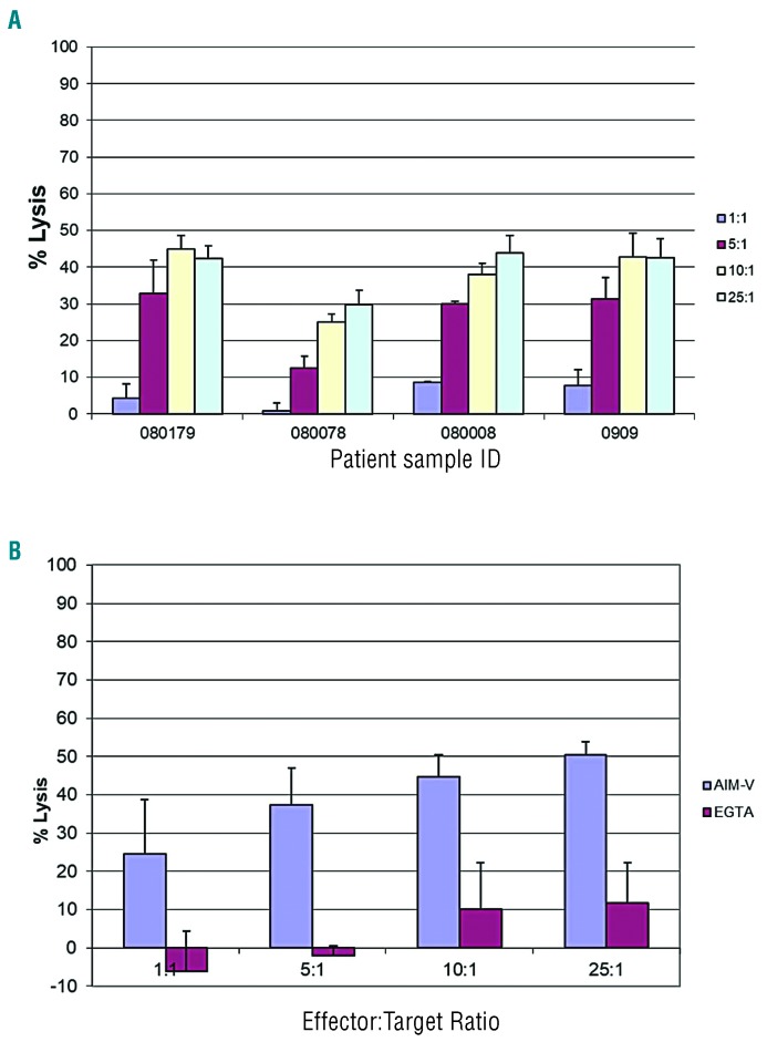
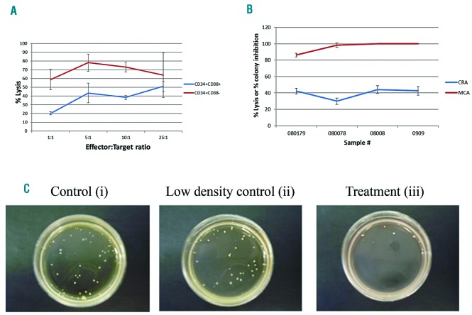
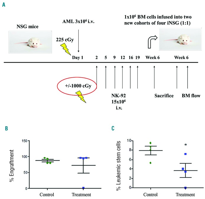
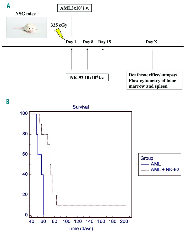
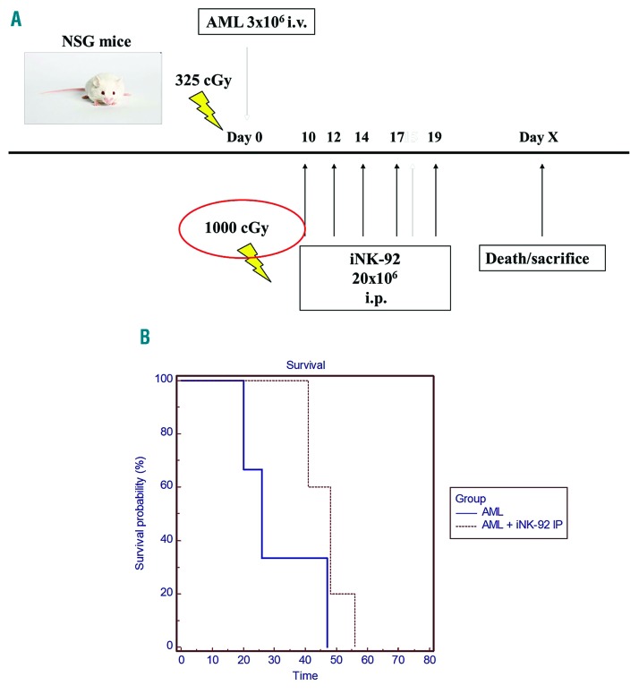
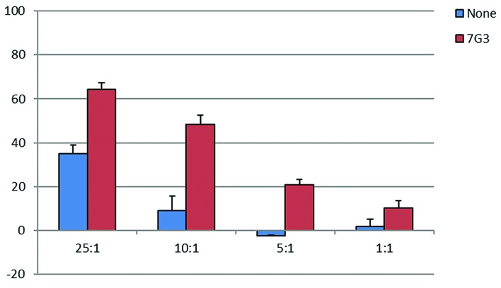
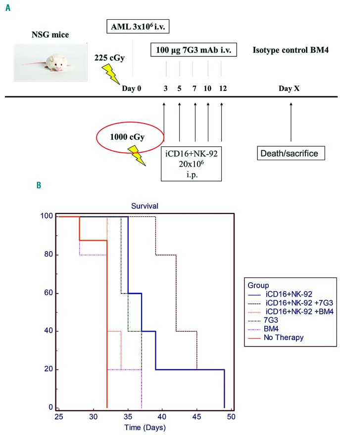
Patients with acute myeloid leukemia (AML) often relapse after initial therapy because of persistence of leukemic stem cells that frequently express the IL-3 receptor alpha chain CD123. Natural killer (NK) cell-based therapeutic strategies for AML show promise and we explore the NK cell lines, NK-92 and CD16+ NK-92, as a treatment for AML. NK-92 has been tested in phase I clinical trials with minimal toxicity; irradiation prior to infusion prevents risk of engraftment. The CD16 negative NK-92 parental line was genetically modified to express the high affinity Fc gamma receptor, enabling antibody-dependent cell-mediated cytotoxicity, which we utilized in combination with an anti-CD123 antibody to target leukemic stem cells. NK-92 was preferentially cytotoxic against leukemic stem and progenitor cells compared with bulk leukemia in in vitro assays, while CD16+ NK-92 in combination with an anti-CD123 mAb mediated antibody-dependent cell-mediated cytotoxicity against CD123+ leukemic targets. Furthermore, NK-92 infusions (with or without prior irradiation) improved survival in a primary AML xenograft model. Mice xenografted with primary human AML cells had a superior survival when treated with irradiated CD16+NK-92 cells and an anti-CD123 monoclonal antibody (7G3) versus treatment with irradiated CD16+NK-92 cells combined with an isotype control antibody. In this proof-of-principle study, we show for the first time that a CD16+NK-92 cell line combined with an antibody that targets a leukemic stem cell antigen can lead to improved survival in a relevant pre-clinical model of AML.


Figures

Figure 1.
Figure 1.
Chromium release assay of NK-92 against primary acute myeloid leukemia (AML) samples. (A) Four freshly thawed primary AML blast samples were labeled with 100 μCi of Na251CrO4 prior to treatment with NK-92 at four E:T ratios. (B) AML blast sample 080078 was tested in a separate experiment at four E:T ratios with and without calcium chelator EGTA 4 (mM) and MgCl2 (3 mM). Data are presented as the mean percent lysis of triplicate samples (+/−Standard Deviation) from a representative experiment carried out three times.
Figure 2.
Figure 2.
NK-92 cytotoxicity against sorted leukemic stem cells and clonogenic leukemic cells relative to bulk leukemia cells. (A) Primary acute myeloid leukemia (AML) samples were sorted into CD34+CD38 and CD34+CD38+ fractions for subsequent testing in a chromium release assay with NK-92 at a 25:1 E:T ratio. Data are presented as the mean percent (%) lysis of triplicate samples (+/−Standard Deviation) representative of two separate experiments. (B) Four primary AML samples were incubated with or without NK-92 at a 25:1 E:T ratio for 4 hours in 96-well U bottom plates and utilized in either a chromium release assay (CRA) or a methylcellulose cytotoxicity assay (MCA) conducted on the same day. The % lysis values and % colony inhibition values are plotted together (B). An example of the methylcellulose cytotoxicity assay (C) shows a representative assay for one sample (080179) with a control (AML only) (i), low density control (AML + NK-92 infused into methylcellulose only) (ii), and treatment group (AML + NK-92 co-incubated together in a 96-well plate well and then infused in methylcellulose) (iii).
Figure 3.
Figure 3.
Effect of iNK-92 on secondary bone marrow (BM) engraftment of acute myeloid leukemia (AML) cells and leukemic stem cells (LSCs). (A) 3×10⁶ AML cells were also infused intravenously (i.v.) into two cohorts of 4 mice and treated with and without iNK-92 from day 2 and given 15×106 cells twice weekly to a total dose of 75×106 (A). BM (1×10⁶ cells) from each of 4 primary recipients in control and treatment was serially transplanted 1:1 into 4 new NOD/SCID gammanull (NSG) mice. These mice were sacrificed at six weeks and BM assayed for overall leukemic engraftment as determined by presence of % human CD45+ cells (B) and LSC engraftment as determined by % human CD34+CD38CD123+ cells (C) (*P=0.05).
Figure 4.
Figure 4.
NK-92 therapy of primary acute myeloid leukemia (AML) xenografted NOD/SCID gammanull (NSG) mice. 3×10⁶ primary AML cells were injected intravenously (i.v.) via tail vein into irradiated NOD/SCID gamma null mice to establish disease in control (n=5) and therapy mice (n=10). 10×106 NK-92 were infused via tail vein weekly for three weeks starting on the day of AML inoculation in treatment group (A). Mice were monitored for signs of leukemia and sacrificed at humane end points. Kaplan-Meier survival curves were generated to compare survival in control and treatment groups (P<0.01) (B).
Figure 5.
Figure 5.
iNK-92 therapy of primary acute myeloid leukemia (AML) xenografted NOD/SCID gammanull (NSG) mice. 3×10⁶ primary AML cells were injected intravenously (i.v.) via tail vein into irradiated NGS to establish disease in control (n=5) and therapy (n=5) mice. iNK-92 given intraperitoneally (i.p.) 20×206 weekly for six weeks were used to treat AML xenografted mice starting ten days after inoculation (A). Mice were monitored for signs of leukemia and sacrificed at humane end points (B). Kaplan-Meier survival curves were generated to compare survival in control and treatment groups (P=0.0566).
Figure 6.
Figure 6.
CD16+NK-92 in vitro ADCC assay against primary acute myeloid leukemia (AML) and OCI/AML5. OCI/AML5 cells were labeled with 100 μCi of Na251CrO4 for 2 hours +/− 10 μg/mL of 7G3 (anti-CD123 mAb) prior to treatment with CD16+NK-92 in 96-well plates in a standard chromium release assay. Data are presented as the mean percent lysis of triplicate samples (+/−Standard Deviation) from a representative experiment carried out twice.
Figure 7.
Figure 7.
iCD16+NK-92 +/− 7G3 or isotype control treatment of primary acute myeloid leukemia (AML) xenografted mice. NOD/SCID gammanull (NSG) mice were inoculated intravenously (i.v.) with 3×10⁶ passage human AML spleen-derived cells (day 0) and treated with iCD16+NK-92 +/− 7G3 or BM4 × 5 doses [intraperitoneal injection (i.p.)] (3×/week) starting on day 3 (A). Controls included no therapy and antibodies alone (n=5 for all groups). Survival was determined using Kaplan-Meier survival analysis with a log rank test (B).

References

    1. Hurwitz CA, Mounce KG, Grier HE. Treatment of patients with acute myelogenous leukemia: review of clinical trials of the past decade. J Pediatr Hematol Oncol. 1995;17(3):185–197. - PubMed
    1. Lowenberg B, Downing JR, Burnett A. Acute myeloid leukemia. N Engl J Med. 1999;341(14):1051–1062. - PubMed
    1. Ribeiro RC, Razzouk BI, Pounds S, Hijiya N, Pui CH, Rubnitz JE. Successive clinical trials for childhood acute myeloid leukemia at St Jude Children’s Research Hospital, from 1980 to 2000. Leukemia. 2005; 19(12):2125–2129. - PubMed
    1. Lapidot T, Sirard C, Vormoor J, et al. A cell initiating human acute myeloid leukaemia after transplantation into SCID mice. Nature. 1994;367(6464):645–648. - PubMed
    1. Bonnet D, Dick JE. Human acute myeloid leukemia is organized as a hierarchy that originates from a primitive hematopoietic cell. Nat Med. 1997;3(7):730–737. - PubMed

Publication types

MeSH terms