. 2018 Aug 24;293(34):13044-13058.
doi: 10.1074/jbc.RA118.003845. Epub 2018 Jul 5.

Legionella pneumophila effector Lem4 is a membrane-associated protein tyrosine phosphatase

Affiliations

Legionella pneumophila effector Lem4 is a membrane-associated protein tyrosine phosphatase

Ksenia Beyrakhova et al. J Biol Chem. .

Abstract

Legionella pneumophila is a Gram-negative pathogenic bacterium that causes severe pneumonia in humans. It establishes a replicative niche called Legionella-containing vacuole (LCV) that allows bacteria to survive and replicate inside pulmonary macrophages. To hijack host cell defense systems, L. pneumophila injects over 300 effector proteins into the host cell cytosol. The Lem4 effector (lpg1101) consists of two domains: an N-terminal haloacid dehalogenase (HAD) domain with unknown function and a C-terminal phosphatidylinositol 4-phosphate-binding domain that anchors Lem4 to the membrane of early LCVs. Herein, we demonstrate that the HAD domain (Lem4-N) is structurally similar to mouse MDP-1 phosphatase and displays phosphotyrosine phosphatase activity. Substrate specificity of Lem4 was probed using a tyrosine phosphatase substrate set, which contained a selection of 360 phosphopeptides derived from human phosphorylation sites. This assay allowed us to identify a consensus pTyr-containing motif. Based on the localization of Lem4 to lysosomes and to some extent to plasma membrane when expressed in human cells, we hypothesize that this protein is involved in protein-protein interactions with an LCV or plasma membrane-associated tyrosine-phosphorylated host target.

Keywords: binding domain; pathogenesis; phosphatase; phosphatidylinositol; structural biology; substrate specificity.


Conflict of interest statement

The authors declare that they have no conflicts of interest with the contents of this article

Figures

Figure 1.
Figure 1.
HAD-like domain of Lem4 is homologous to MDP-1. A, schematic representation of domain organization of Lem4. B, overall fold of Lem4-N in rainbow colors from blue at the N terminus to red at the C terminus. The secondary structure elements are labeled. Mg2+ ion bound in the active center is shown as a green sphere. C, superposition of Lem4-N (cyan) and MDP-1 (orange). Mg2+ ions are colored cyan in Lem4-N and orange in MDP-1. Tungstate ion bound in the active site of MDP-1 is shown as orange sticks. The cap insertions are marked by oval dashed line.
Figure 2.
Figure 2.
Lem4-N activity against a set of potential small molecule substrates. The release of phosphate was measured using Malachite Green assay. Lem4(6–218) was added to 40 μl of solution containing 0.1 mm substrate, 50 mm sodium acetate, pH 5.5, 0.1 m NaCl, 5 mm MgCl2 to the final concentration of 10 μg/ml. Reactions were carried out for 30 min at 30 °C. The mean values of the two replicates are shown; the bars indicate the range of the data.
Figure 3.
Figure 3.
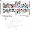
Consensus motif recognized by Lem4-N. A, graphical representation of the consensus motif was generated with pLogo web server using 53 peptides with dephosphorylation level >60% as a foreground set. All the 355 peptides from the substrate array served as a background set. Residue heights are scaled relative to their statistical significance (64). Over- and under-represented residues are positioned above and below the x axis, respectively. Phosphorylated Tyr is “fixed” in a zero position and highlighted in gray. The horizontal red lines correspond to statistically significant values (p = 0.05) as described previously (64). B, activity assay of Lem4-N with different substrates: pTyr, general phosphatase substrate ENDpYINASL and two peptides identified as good substrates in a peptide array. For all substrates in 0.2 mm concentration, Lem4-N was added to a final concentration of 50 nm; final reaction volume was 1 ml, and reactions were performed in triplicates. Reaction products (free tyrosine or dephosphorylated peptides) were detected by measuring A280 of the reaction mixture.
Figure 4.
Figure 4.
Conserved motifs in HAD enzymes. A, structure-based alignment of conserved motifs I–IV of different members of HAD superfamily (left column, protein name is followed by the PDB code of the corresponding structure used for alignment). Amino acid numbering is according to Lem4 sequence; highly conserved residues are marked with asterisks. Multiple structural alignment was performed with PDBeFold (http://www.ebi.ac.uk/msd-srv/ssm/) (65) and visualized with ESPript (http://espript.ibcp.fr) (66) (Please note that the JBC is not responsible for the long-term archiving and maintenance of this site or any other third party hosted site.). B, active site of Lem4-N with conserved motifs I–IV colored magenta, green, cyan, and yellow, respectively. Blue dashed lines indicate Mg2+ coordination sphere. Green dashed lines show hydrogen bonds between the bound phosphate and the protein residues. C, superposition based on the active-site residues of Lem4 (white carbons, wheat magnesium, and orange PO4) and MDP-1 (green carbons, dark green magnesium, and blue WO4) showing close structural similarity. The residues from the active site center are shown in a stick representation and labeled according to the Lem4 numbering. Mg2+ ions and PO4/WO4 superpose from both structures very well.
Figure 5.
Figure 5.
Active center of Lem4 with and without substrate showing the molecules located in the active site. Difference electron density maps were calculated with phases obtained after the refinement of Lem4-N or Lem4-N(D25N) that excluded the molecules in the active site. The maps are contoured at 4.5σ level and colored green. Magnesium ion is represented by a cyan sphere; waters are represented by smaller red spheres, and phosphate and acetate are in orange. A, active center of the substrate-trapping mutant Lem4-N(D25N) in the presence of phosphorylated peptide. The Mg2+, phosphate, and water molecules were modeled in the active center. B, active center of native Lem4-N with Mg2+ and water molecules modeled. C, active center of Lem4-N(D25N) in the absence of phosphorylated substrate. The Mg2+, waters, and acetate were modeled.
Figure 6.
Figure 6.
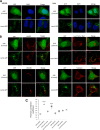
Lem4 co-localizes with lysosomes in transfected mammalian cells. A, HEK293 (left panel) or HeLa cells (right panel) were transfected with plasmids encoding Lem4-GFP or GFP alone (green), fixed with 4% (w/v) of paraformaldehyde (PFA) 24 h post-transfection, and imaged by confocal microscopy. B, HEK293 cells transfected with plasmids encoding Lem4-GFP or GFP alone and labeled for different compartments. Top left panel, cells were incubated with the medium containing anti-LAMP1; top right panel, cells were incubated with the medium containing 50 nm LysoTracker Red probe; bottom left panel, cells were incubated with anti-GM130; bottom right panel, cells incubated with anti-calnexin antibody, followed by Alexa Fluor-546 (red) secondary antibody. Cells were fixed and imaged by confocal microscopy. Scale bars, 5 μm. C, quantitation of co-localization by Pearson coefficient. The Pearson correlation coefficient was determined with Coloc 2 plugin of the ImageJ software and was calculated with at least 30 cells (mean and S.D. of the mean from each experiment, ***, p < 0.001).
Figure 7.
Figure 7.
C-terminal PI(4)P-binding domain of Lem4 is responsible for its localization in mammalian cells. A, HEK293 cells transfected with a plasmid encoding Lem4(1–200)-GFP or Lem4(200–322)-GFP (green) and stained with anti-LAMP1 antibody (followed by Alexa Fluor-546 (red) secondary antibody) (left panel) or LysoTracker (red) (right panel), and then cells were fixed and imaged by confocal microscopy. Scale bars, 5 μm. B, quantitation of co-localization of truncated Lem4 with lysosome marker by Pearson coefficient. The Pearson correlation coefficient was determined with coloc 2 plugin of the ImageJ software and was calculated with at least 30 cells (mean and S.D., of the mean from each experiment, ***, p < 0.001). C, macrophage cell line Raw264.7 was transfected with plasmids encoding GFP alone, Lem4-GFP, Lem4(1–200)-GFP, or Lem4(200–322)-GFP, fixed with 4% (w/v) of PFA 24 h post-transfection, and imaged by confocal microscopy. Scale bars, 5 μm.
Figure 8.
Figure 8.
Effect of Lem4 on tyrosine phosphorylation of signal proteins in HEK293T cell line, as detected by tyrosine phosphorylation ProArray (Full Moon BioSystems). Phosphorylation levels were increased by at least 30% at 30 sites and decreased by at least 30% at four sites in the cells overexpressing Lem4-GFP. Red dashed line indicates the level of phosphorylation in the control GFP-expressing cell line.

References

    1. Fields B. S., Benson R. F., and Besser R. E. (2002) Legionella and Legionnaires' disease: 25 years of investigation. Clin. Microbiol. Rev. 15, 506–526 10.1128/CMR.15.3.506-526.2002 - DOI - PMC - PubMed
    1. Newton H. J., Ang D. K., van Driel I. R., and Hartland E. L. (2010) Molecular pathogenesis of infections caused by Legionella pneumophila. Clin. Microbiol. Rev. 23, 274–298 10.1128/CMR.00052-09 - DOI - PMC - PubMed
    1. Fields B. S. (1996) The molecular ecology of legionellae. Trends Microbiol. 4, 286–290 10.1016/0966-842X(96)10041-X - DOI - PubMed
    1. Declerck P. (2010) Biofilms: the environmental playground of Legionella pneumophila. Environ. Microbiol. 12, 557–566 10.1111/j.1462-2920.2009.02025.x - DOI - PubMed
    1. Horwitz M. A. (1983) The Legionnaires' disease bacterium (Legionella pneumophila) inhibits phagosome–lysosome fusion in human monocytes. J. Exp. Med. 158, 2108–2126 10.1084/jem.158.6.2108 - DOI - PMC - PubMed

Publication types

Grants and funding

LinkOut - more resources