. 2018 Aug 31;293(35):13415-13426.
doi: 10.1074/jbc.RA118.002751. Epub 2018 Jul 5.

Interaction via the N terminus of the type IV secretion system (T4SS) protein VirB6 with VirB10 is required for VirB2 and VirB5 incorporation into T-pili and for T4SS function

Affiliations

Interaction via the N terminus of the type IV secretion system (T4SS) protein VirB6 with VirB10 is required for VirB2 and VirB5 incorporation into T-pili and for T4SS function

Charline Mary et al. J Biol Chem. .

Abstract

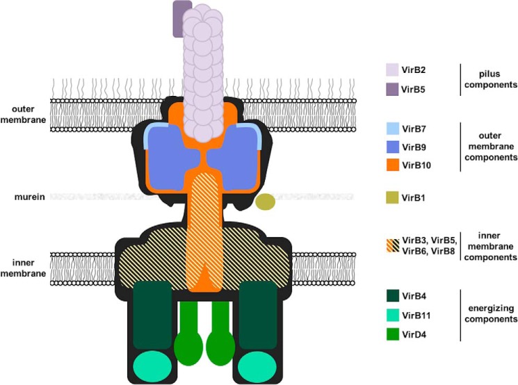
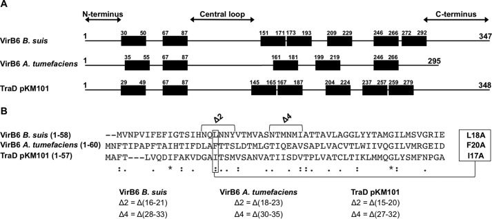
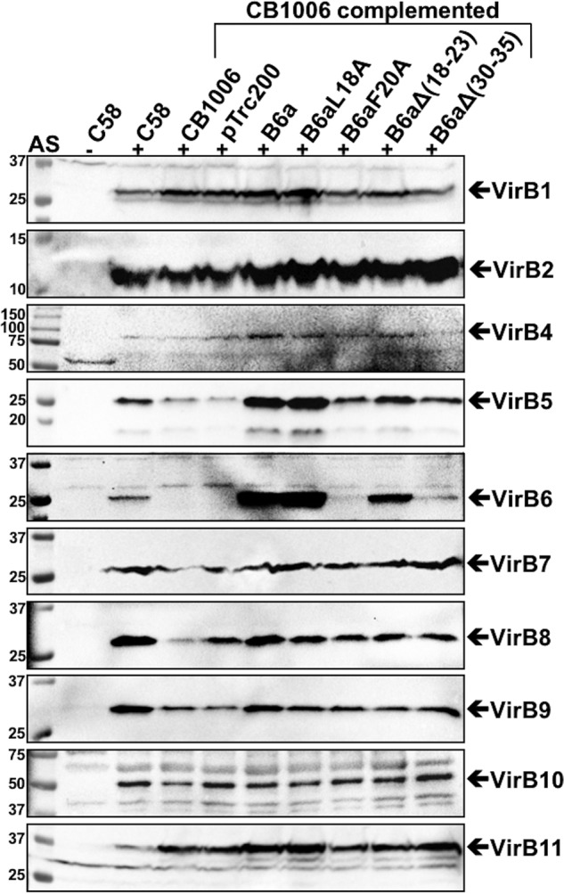
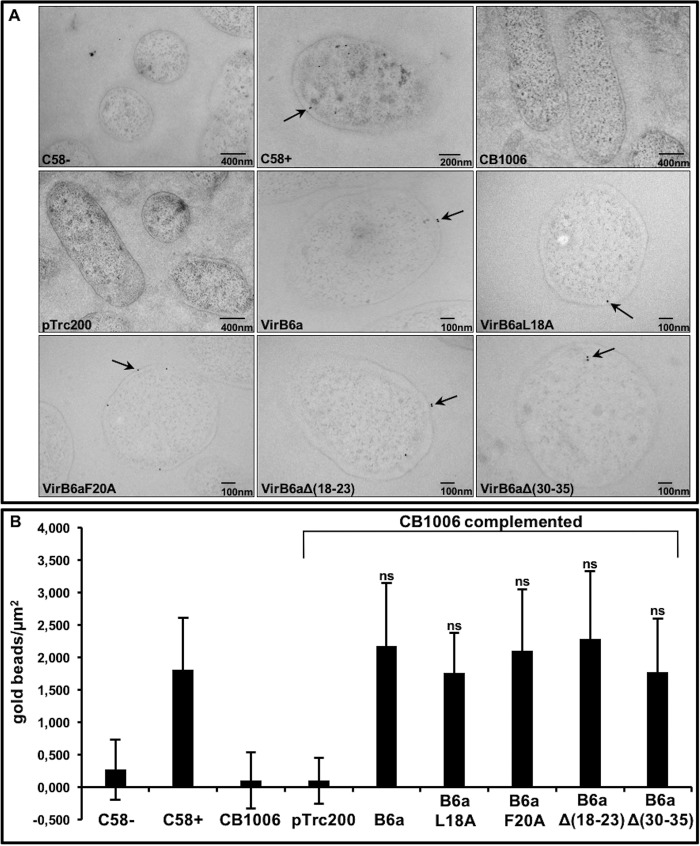
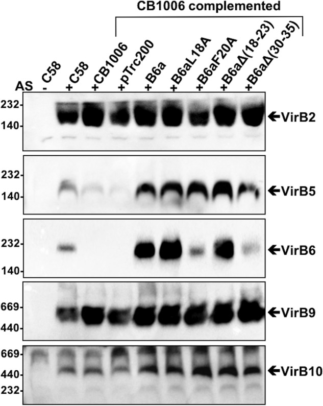
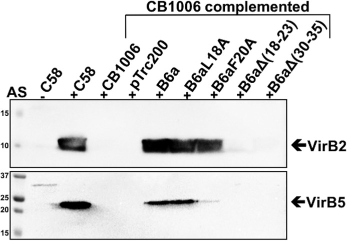
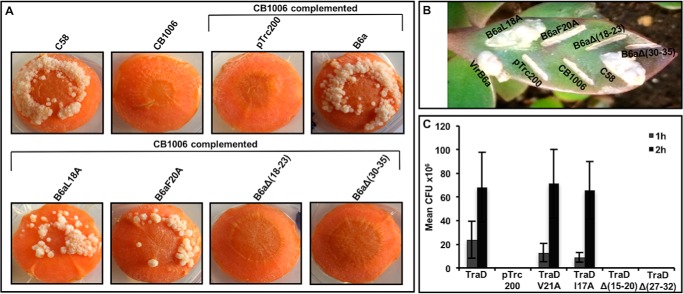
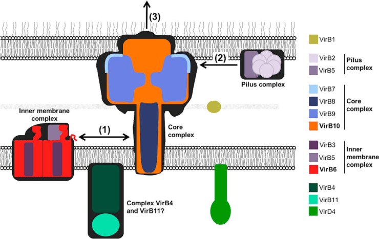
Many bacterial pathogens employ multicomponent protein complexes such as type IV secretion systems (T4SSs) to transfer virulence factors into host cells. Here we studied the interaction between two essential T4SS components: the very hydrophobic inner membrane protein VirB6, which may be a component of the translocation channel, and VirB10, which links the inner and outer bacterial membranes. To map the interaction site between these two T4SS components, we conducted alanine scanning and deleted six-amino acid stretches from the N-terminal periplasmic domain of VirB6 from Brucella suis Using the bacterial two-hybrid system to analyze the effects of these alterations on the VirB6-VirB10 interaction, we identified the amino acid regions 16-21 and 28-33 and Leu-18 in VirB6 as being required for this interaction. SDS-PAGE coupled with Western blotting of cell lysates and native PAGE of detergent-extracted membrane proteins revealed that the corresponding VirB6 residues in Agrobacterium tumefaciens (Phe-20 and amino acids 18-23 and 30-35) modulate the stability of both VirB6 and VirB5. However, the results from immuno-EM and super-resolution microscopy suggested that these regions and residues are not required for membrane association or for polar localization of VirB6. The six-amino acid deletions in the N terminus of VirB6 abolished pilus formation and virulence of A. tumefaciens, and the corresponding deletions in the VirB6 homolog TraD from the plasmid pKM101-T4SS abrogated plasmid transfer. Our results indicate that specific residues of the VirB6 N-terminal domain are required for VirB6 stabilization, its interaction with VirB10, and the incorporation of VirB2 and VirB5 into T-pili.

Keywords: Agrobacterium tumefaciens; T-pilus; bacteria; bacterial pathogenesis; membrane protein; protein secretion; protein–protein interaction; type IV secretion system; virulence; virulence factor.


Conflict of interest statement

The authors declare that they have no conflicts of interest with the contents of this article

Figures

Figure 1.
Figure 1.
Model of the type IV secretion system with the known or suspected localization of its 12 components.
Figure 2.
Figure 2.
Results of bacterial two-hybrid analysis to assess the interactions of VirB10b with variants of VirB6b(1–168). A, bacterial two-hybrid assay measuring β-gal activity in the E. coli indicator strain BTH101 resulting from the interactions of CyaA fusion proteins to VirB10b and VirB6b(1–168) carrying substitutions of the indicated amino acids by alanine or deletions of blocks of six amino acids. Positive control (+), fusions to VirB10b and VirB6b(1–168) WT and negative control; (−), fusion to VirB10b and a strain not expressing an interaction partner. B, analysis of protein levels after SDS-PAGE and Western blotting using anti-CyaA and anti-VirB10b antibodies. Molecular masses of reference proteins are shown on the left (in kilodaltons). Values and standard deviations were calculated from five independent experiments. The p values were obtained by t test, and significant differences were observed between several variants and the WT (****, p < 0.0001).
Figure 3.
Figure 3.
Sequence alignment between VirB6b, VirB6a, and TraD. A, schematic of the overall topology of VirB6 homologs realized according to the consensus prediction of the membrane protein topology program TOPCONS (38). B, CLUSTAL multiple sequence alignment by MUSCLE (3.8), indicating which residues and deletions in VirB6a and TraD correspond to the ones identified with the BTH assay in VirB6b. Sequence similarity is indicated as follows: asterisks, identical (single, fully conserved residue); colons, conserved substitutions (strongly similar properties); periods, semiconserved substitutions (weakly similar properties).
Figure 4.
Figure 4.
VirB proteins levels in strains C58 and CB1006 after complementation with VirB6a and its variants. Shown are results of SDS-PAGE and Western blot analysis with VirB protein–specific antisera of A. tumefaciens C58 cultivated without (−AS) or with virulence gene induction (+AS) and virB6 deletion (CB1006) transformed with empty vector pTrc200- or pTrc200-expressing VirB6a or its variants VirB6aL18A, VirB6aF20A, VirB6aΔ(18–23), and VirB6aΔ(30–35). The signals of the expected molecular masses of VirB proteins are indicated by arrows. Molecular masses of reference proteins are shown on the left (in kilodaltons). Representative results of five repetitions are shown.
Figure 5.
Figure 5.
Immunogold labeling using VirB6-specific antisera to localize Virb6-WT and its variants in A. tumefaciens. A, results of immuno-EM analysis with VirB6-specific antisera and colloidal gold–labeled protein A of A. tumefaciens C58 cultivated without (−AS) or with virulence gene induction (+AS) and virB6 deletion (CB1006) transformed with empty vector pTrc200- or pTrc200-expressing VirB6a or its variants VirB6aL18A, VirB6aF20A, VirB6aΔ(18–23), and VirB6aΔ(30–35). Gold beads are indicated by arrows. B, graphic representation of the number of gold beads per square micrometer. Values and standard deviations were calculated from three independent experiments. The p values were obtained by t test, and no significant differences were observed between strain C58 and complemented CB1006. ns, p ≥ 0.05.
Figure 6.
Figure 6.
Structural illumination microscopy of sfGFP-fused VirB6a and its variants. A, C-terminal fusions of VirB6 and of variants VirB6aL18A, VirB6aF20A, VirB6aΔ(18–23), and VirB6aΔ(30–35) to sfGFP were expressed in strain CB1006 and imaged by SIM. B, quantification of the fluorescence using the CTCF method, showing preferentially polar localization for each sample (****, p < 0.0001). Values and standard deviations were calculated from three independent experiments. The p values were obtained by t test, for each construction, between the overall bacteria and pole 1 or pole 2. ns, p > 0.05; *, p < 0.05; **, p < 0.01; ***, p < 0.001, ****, p < 0.0001.
Figure 7.
Figure 7.
Native gel electrophoresis to separate detergent-extracted VirB protein complexes. Membranes were extracted with the detergent DDM from A. tumefaciens C58 cultivated without (−AS) or with virulence gene induction (+AS) and virB6 deletion (CB1006) transformed with empty vector pTrc200- or pTrc200-expressing VirB6a or its variants VirB6aL18A, VirB6aF20A, VirB6aΔ(18–23), and VirB6aΔ(30–35). DDM-solubilized membrane proteins were separated by native PAGE followed by Western blotting with specific antisera and detected in different molecular mass ranges, which are indicated by arrows. Molecular masses of reference proteins are shown on the left (in kilodaltons). Representative results of four repetitions are shown.
Figure 8.
Figure 8.
Analysis of the formation of T-pili. T-pili were isolated from A. tumefaciens C58 cultivated without (−AS) or with virulence gene induction (+AS) and virB6 deletion (CB1006) transformed with empty vector pTrc200- or pTrc200-expressing VirB6a or its variants VirB6aL18A, VirB6aF20A, VirB6aΔ(18–23), and VirB6aΔ(30–35). High-molecular-mass extracellular structures were isolated from the surface of A. tumefaciens by shearing, followed by ultracentrifugation. The T-pilus components VirB2 and VirB5 were detected by SDS-PAGE and Western blotting with specific antisera. Molecular masses of reference proteins are shown on the left (in kilodaltons). Representative results of four repetitions are shown.
Figure 9.
Figure 9.
Virulence and conjugation assays. A and B, carrot disc (A) and K. daigremontiana (B) assays showing the formation of tumors after infection with A. tumefaciens C58, virB6 deletion (CB1006), and CB1006 transformed with empty vector pTrc200- or pTrc200-expressing VirB6a or its variants VirB6aL18A, VirB6aF20A, VirB6aΔ(18–23), and VirB6aΔ(30–35). Representative results of three repetitions are shown. C, conjugation assays between the pKM101-carrying donor strain FM433 and the plasmid-free recipient WL400 were conducted for 1 h or 2 h. Donor cells were pKM101 WT and a traD insertion mutant transformed with pTrc200 (empty vector) or complemented with pTrc200-expressing TraD or its variants TraDV21A, TraDI17A, TraDΔ(15–20), and TraD Δ(27–32). Conjugation assays showing the effects of changes on the capacity of TraD and its variants to complement (CFU, cfu of the recipient strain). Values and standard deviations were calculated from three independent experiments.
Figure 10.
Figure 10.
Model for the role of the VirB6–VirB10 interaction for T4SS assembly. The interaction between VirB6 and VirB10 (1) enables the recruitment of the components of the T-pilus VirB2 and VirB5 (2) to the T4SS and is an essential step for pilus biogenesis (3).

Similar articles

Cited by

References

    1. Fronzes R., Christie P. J., and Waksman G. (2009) The structural biology of type IV secretion systems. Nat. Rev. Microbiol. 7, 703–714 10.1038/nrmicro2218 - DOI - PMC - PubMed
    1. Trokter M., Felisberto-Rodrigues C., Christie P. J., and Waksman G. (2014) Recent advances in the structural and molecular biology of type IV secretion systems. Curr. Opin. Struct. Biol. 27C, 16–23 10.1016/j.sbi.2014.02.006 - DOI - PMC - PubMed
    1. Cascales E., and Christie P. J. (2003) The versatile bacterial type IV secretion systems. Nat. Rev. Microbiol. 1, 137–149 10.1038/nrmicro753 - DOI - PMC - PubMed
    1. Zupan J., Muth T. R., Draper O., and Zambryski P. (2000) The transfer of DNA from Agrobacterium tumefaciens into plants: a feast of fundamental insights. Plant J. 23, 11–28 10.1046/j.1365-313x.2000.00808.x - DOI - PubMed
    1. Christie P. J., and Gordon J. E. (2014) The Agrobacterium Ti plasmids. Microbiol Spectr. 2, 10.1128/microbiolspec.PLAS-0010-2013 10.1128/microbiolspec.PLAS-0010-2013 - DOI - DOI - PMC - PubMed

Publication types

MeSH terms

Substances

Grants and funding

LinkOut - more resources