Comparative Study
. 2018 Aug;38(8):1890-1900.
doi: 10.1161/ATVBAHA.118.310979.

Serum Amyloid A Is an Exchangeable Apolipoprotein

Affiliations
Comparative Study

Serum Amyloid A Is an Exchangeable Apolipoprotein

Patricia G Wilson et al. Arterioscler Thromb Vasc Biol. 2018 Aug.

Abstract

Objective- SAA (serum amyloid A) is a family of acute-phase reactants that have proinflammatory and proatherogenic activities. SAA is more lipophilic than apoA-I (apolipoprotein A-I), and during an acute-phase response, <10% of plasma SAA is found lipid-free. In most reports, SAA is found exclusively associated with high-density lipoprotein; however, we and others have reported SAA on apoB (apolipoprotein B)-containing lipoproteins in both mice and humans. The goal of this study was to determine whether SAA is an exchangeable apolipoprotein. Approach and Results- Delipidated human SAA was incubated with SAA-free human lipoproteins; then, samples were reisolated by fast protein liquid chromatography, and SAA analyzed by ELISA and immunoblot. Both in vitro and in vivo, we show that SAA associates with any lipoprotein and does not remain in a lipid-free form. Although SAA is preferentially found on high-density lipoprotein, it can exchange between lipoproteins. In the presence of CETP (cholesterol ester transfer protein), there is greater exchange of SAA between lipoproteins. Subjects with diabetes mellitus, but not those with metabolic syndrome, showed altered SAA lipoprotein distribution postprandially. Proteoglycan-mediated lipoprotein retention is thought to be an underlying mechanism for atherosclerosis development. SAA has a proteoglycan-binding domain. Lipoproteins containing SAA had increased proteoglycan binding compared with SAA-free lipoproteins. Conclusions- Thus, SAA is an exchangeable apolipoprotein and increases apoB-containing lipoproteins' proteoglycan binding. We and others have previously reported the presence of SAA on low-density lipoprotein in individuals with obesity, diabetes mellitus, and metabolic syndrome. We propose that the presence of SAA on apoB-containing lipoproteins may contribute to cardiovascular disease development in these populations.

Keywords: apolipoproteins; atherosclerosis; inflammation; metabolic syndrome; models, animal.


Figures
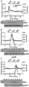
Fig. 1
Fig. 1. SAA does not remain lipid free
Lipid-free human SAA was incubated for 3 hours with SAA-free human VLDL (A), LDL (B), or HDL (C) (each at a ratio of 200 μg SAA to 1 mg lipoprotein protein) then subjected to FPLC. SAA in individual fractions was assessed by ELISA (upper panels) or immunoblotting (lower panels). FPLC fractions corresponding to each lane are indicated above the immunoblot. Shown is the mean±SEM of the cholesterol absorbance (left Y axis) and SAA found in each FPLC fraction (right Y axis). Data shown is mean±SEM from n=3 separate experiments for each.
Fig. 2
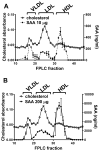
Fig. 2. SAA associates with all lipoproteins
Lipid-free human SAA was incubated for 3 hours with a mixture of human VLDL, LDL and HDL [at a ratio of 10 μg (panel A) or 200 μg (panel B) SAA to 1 mg protein for each lipoprotein] then subjected to FPLC. SAA in individual fractions was assessed by ELISA. Shown is the mean±SEM of the cholesterol absorbance (left Y axis) and SAA found in each FPLC fraction (right Y axis). Data shown is mean±SEM from n=3 separate experiments for each.
Fig. 3
Fig. 3. SAA moves between lipoproteins in vivo
ApoE−/− x SAA1.1/2.1-DKO mice were injected with VLDL+ SAA (panel B, n=15), LDL+SAA (panel C, n=15), or HDL+SAA (panel D, n=12) and blood was collected at the indicated times. Not every mouse was bled at each time-point (n=3–6/data point). SAA was measured in an aliquot of plasma at each time point and is presented as mean±SEM (panel A). Samples were subjected to FPLC and SAA in individual fractions was assessed by ELISA. Shown is the mean±SEM of the SAA in each fraction (VLDL: black squares; LDL: open squares; HDL, gray triangles).
Fig. 4
Fig. 4. CETP facilitates the transfer of SAA from HDL to VLDL
A. HDL containing SAA was incubated with increasing concentrations of CETP in the presence of SAA-free VLDL, then subjected to non-denaturing gradient gel electrophoresis, and immunoblotted for SAA. The migration of standards with known radii (nm) are indicated on the left. Gel shown is representative of 3 separate experiments. B,C,D. ApoE−/− x SAA1.1/2.1-DKO mice were injected with murine AP-HDL in the absence (n=15, open squares) or presence of CETP expression (n=12, black squares) and blood was collected at the indicated times. AdCETP was injected 72 hours prior to injection with AP-HDL to induce CETP expression. Not every mouse was bled at each time-point (n=3–6/data point). B. SAA was measured in an aliquot of plasma at each time point and is presented as mean±SEM. C,D. Samples were subjected to FPLC and SAA in individual fractions was assessed by ELISA. Shown is the mean±SEM of the SAA in each fraction (VLDL: black squares; LDL: open squares; HDL, gray triangles).
Fig. 5
Fig. 5. The presence of SAA on LDL or VLDL increases their proteoglycan binding
Human SAA (closed circles) or saline (open circles) was incubated with human LDL or VLDL for 3 hours (at a ratio of 200 μg SAA to 1 mg lipoprotein protein). Increasing concentrations of LDL or VLDL (0–500 μg/mL lipoprotein as indicated) were then mixed with fixed amounts of radiolabeled proteoglycans for 1h under physiological pH and temperature before electrophoresis in agarose gels. Proteoglycans that are bound by lipoproteins are retained near the origin while free proteoglycans migrate into the gel. A. Gel shown is representative of n=5. B,C: Binding curves shown are mean±SEM from 5 separate experiments. Kd and Vmax (mean±SD) for curve fit are shown on the figures.
Fig. 6
Fig. 6. SAA lipoprotein distribution is significantly altered post-prandially in obese subjects with diabetes
Blood was collected from obese humans with or without metabolic syndrome (Obese, N=4, closed triangles, MetS, N=7, closed squares) or type 2 diabetes (DM, n=5, open squares) in the fasted state (time 0), and hourly for 8 hours after consumption of a high fat shake. A. Plasma SAA. B. Plasma triglycerides. C, D, E: VLDL/remnants (closed circles), LDL (open triangles), and HDL (open circles) were isolated by sequential density gradient ultracentrifugation and SAA content determined by ELISA. C. Obese subjects; D. MetS subjects; E. DM subjects. Data shown are mean±SEM from each individual subject.*p<0.05 between group comparison.
Fig. 7
Fig. 7. Endogenous enrichment of SAA on LDL or VLDL/remnants from diabetic subjects is associated with increased proteoglycan binding
Proteoglycan binding of lipoproteins collected fasting (with lowest SAA content, open circles) and at time of peak SAA content (closed circles) were compared within each individual with diabetes as described in legend to Fig. 5. Binding curves shown are mean±SEM from n=4 diabetic subjects. Kd and Vmax (mean±SD) for curve fit are shown on the figures.

References

    1. Kisilevsky R, Manley PN. Acute-phase serum amyloid a: Perspectives on its physiological and pathological roles. Amyloid. 2012;19:5–14. - PubMed
    1. Johnson BD, Kip KE, Marroquin OC, Ridker PM, Kelsey SF, Shaw LJ, Pepine CJ, Sharaf B, Bairey Merz CN, Sopko G, Olson MB, Reis SE. Serum amyloid a as a predictor of coronary artery disease and cardiovascular outcome in women: The national heart, lung, and blood institute-sponsored women’s ischemia syndrome evaluation (wise) Circulation. 2004;109:726–732. - PubMed
    1. Kosuge M, Ebina T, Ishikawa T, Hibi K, Tsukahara K, Okuda J, Iwahashi N, Ozaki H, Yano H, Kusama I, Nakati T, Umemura S, Kimura K. Serum amyloid a is a better predictor of clinical outcomes than c-reactive protein in non-st-segment elevation acute coronary syndromes. Circ J. 2007;71:186–190. - PubMed
    1. Ridker PM, Hennekens CH, Buring JE, Rifai N. C-reactive protein and other markers of inflammation in the prediction of cardiovascular disease in women. N Engl J Med. 2000;342:836–843. - PubMed
    1. Dong Z, Wu T, Qin W, An C, Wang Z, Zhang M, Zhang Y, Zhang C, An F. Serum amyloid a directly accelerates the progression of atherosclerosis in apolipoprotein e-deficient mice. Mol Med. 2011;17:1357–1364. - PMC - PubMed

Publication types

MeSH terms

LinkOut - more resources