

CRISPR-Cas9-Mediated Epitope Tagging Provides Accurate and Versatile Assessment of Myocardin-Brief Report
- PMID: 29976770
- PMCID: PMC6204210
- DOI: 10.1161/ATVBAHA.118.311171
CRISPR-Cas9-Mediated Epitope Tagging Provides Accurate and Versatile Assessment of Myocardin-Brief Report
Abstract
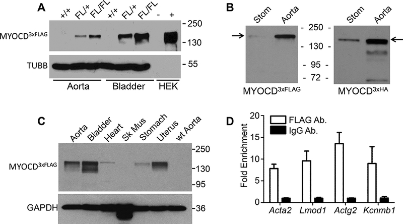
Objective- Unreliable antibodies often hinder the accurate detection of an endogenous protein, and this is particularly true for the cardiac and smooth muscle cofactor, MYOCD (myocardin). Accordingly, the mouse Myocd locus was targeted with 2 independent epitope tags for the unambiguous expression, localization, and activity of MYOCD protein. Approach and Results- 3cCRISPR (3-component clustered regularly interspaced short palindromic repeat) was used to engineer a carboxyl-terminal 3×FLAG or 3×HA epitope tag in mouse embryos. Western blotting with antibodies to each tag revealed a MYOCD protein product of ≈150 kDa, a size considerably larger than that reported in virtually all publications. MYOCD protein was most abundant in some adult smooth muscle-containing tissues with surprisingly low-level expression in the heart. Both alleles of Myocd are active in aorta because a 2-fold increase in protein was seen in mice homozygous versus heterozygous for FLAG-tagged Myocd. ChIP (chromatin immunoprecipitation)-quantitative polymerase chain reaction studies provide proof-of-principle data demonstrating the utility of this mouse line in conducting genome-wide ChIP-seq studies to ascertain the full complement of MYOCD-dependent target genes in vivo. Although FLAG-tagged MYOCD protein was undetectable in sections of adult mouse tissues, low-passaged vascular smooth muscle cells exhibited expected nuclear localization. Conclusions- This report validates new mouse models for analyzing MYOCD protein expression, localization, and binding activity in vivo and highlights the need for rigorous authentication of antibodies in biomedical research.
Keywords: allele; epitope; mice; muscle, smooth; myocardin.
Figures

References
-
- Partridge EC, Watkins TA, Mendenhall EM. Every transcription factor deserves its map: Scaling up epitope tagging of proteins to bypass antibody problems. Bioessays. 2016;38:801–811. - PubMed
-
- Jarvik JW, Telmer CA. Epitope tagging. Annu Rev Genet. 1998;32:601–618. - PubMed
-
- Chen YI, Maika SD, Stevens SW. Epitope tagging of proteins at the native chromosomal loci of genes in mice and in cultured vertebrate cells. J. Mol. Biol 2006;361:412–419. - PubMed
-
- Lee WJ, Kraus P, Lufkin T. Endogenous tagging of the murine transcription factor Sox5 with hemaglutinin for functional studies. Transgenic Res. 2012;21:293–301. - PubMed
Publication types
MeSH terms
Substances
Grants and funding
LinkOut - more resources
Full Text Sources
Other Literature Sources
Molecular Biology Databases
Research Materials
