. 2018 Aug;38(8):1901-1912.
doi: 10.1161/ATVBAHA.118.311150.

Neutrophil Extracellular Traps Induce Endothelial Cell Activation and Tissue Factor Production Through Interleukin-1α and Cathepsin G

Affiliations

Neutrophil Extracellular Traps Induce Endothelial Cell Activation and Tissue Factor Production Through Interleukin-1α and Cathepsin G

Eduardo J Folco et al. Arterioscler Thromb Vasc Biol. 2018 Aug.

Abstract

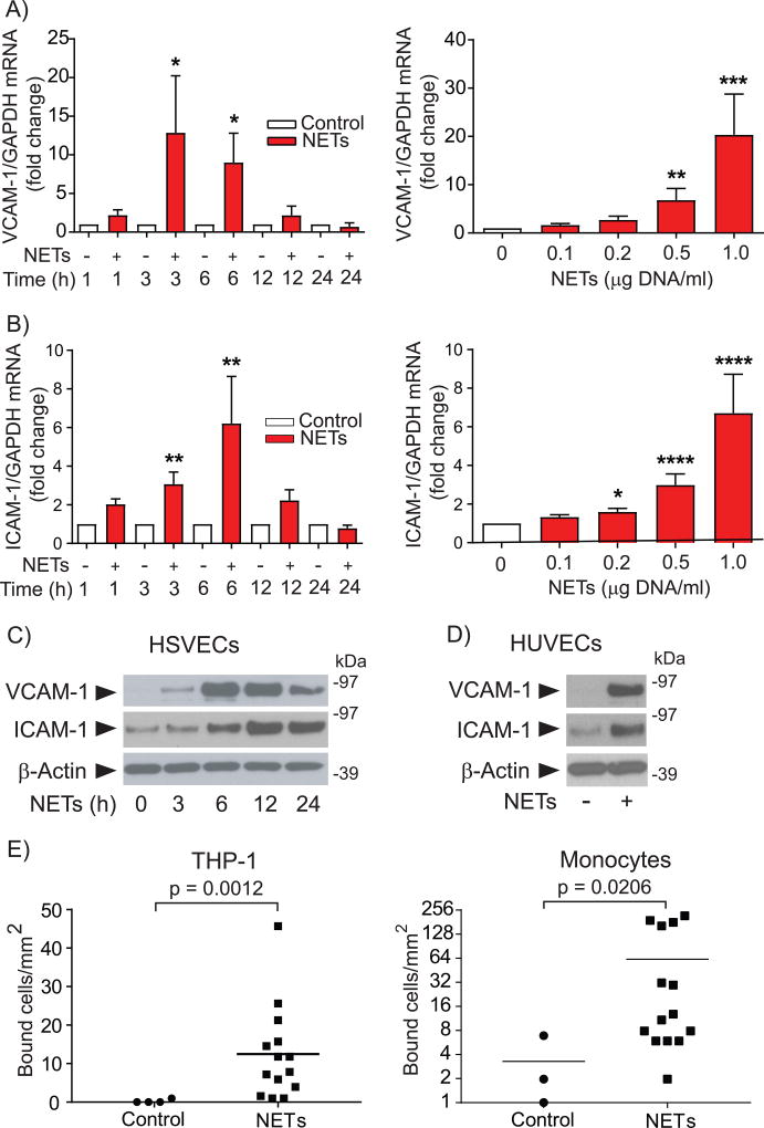
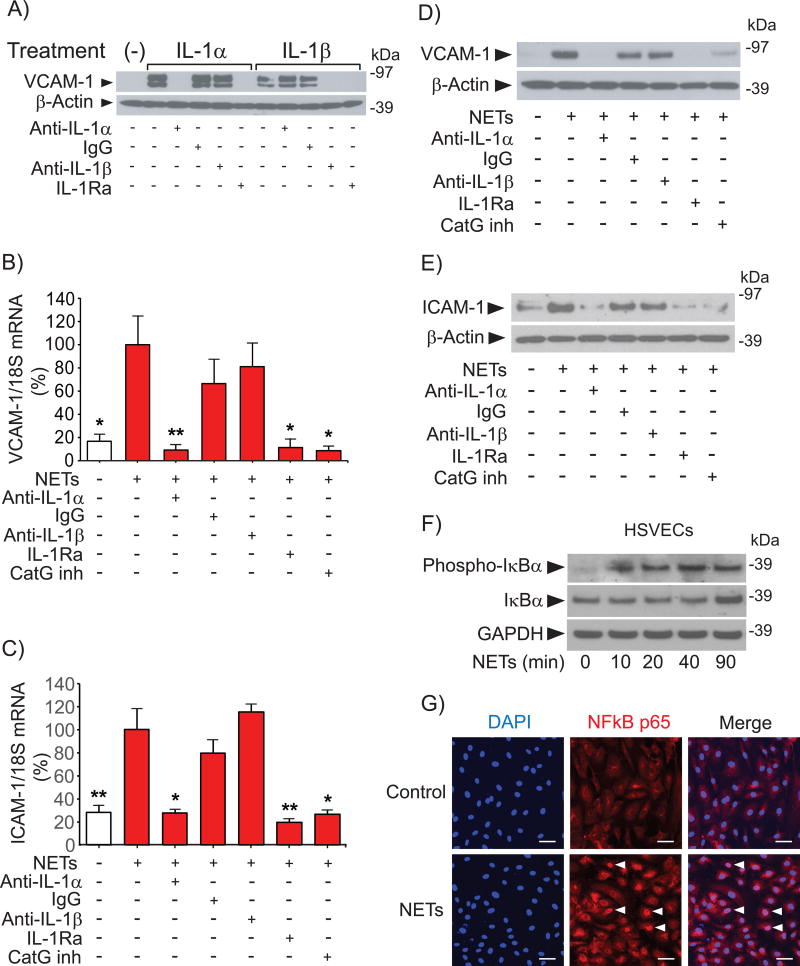
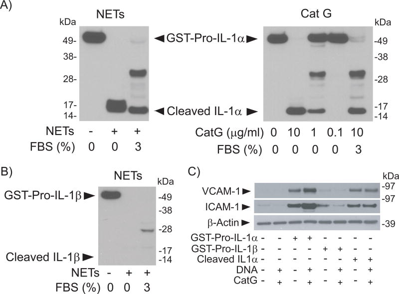
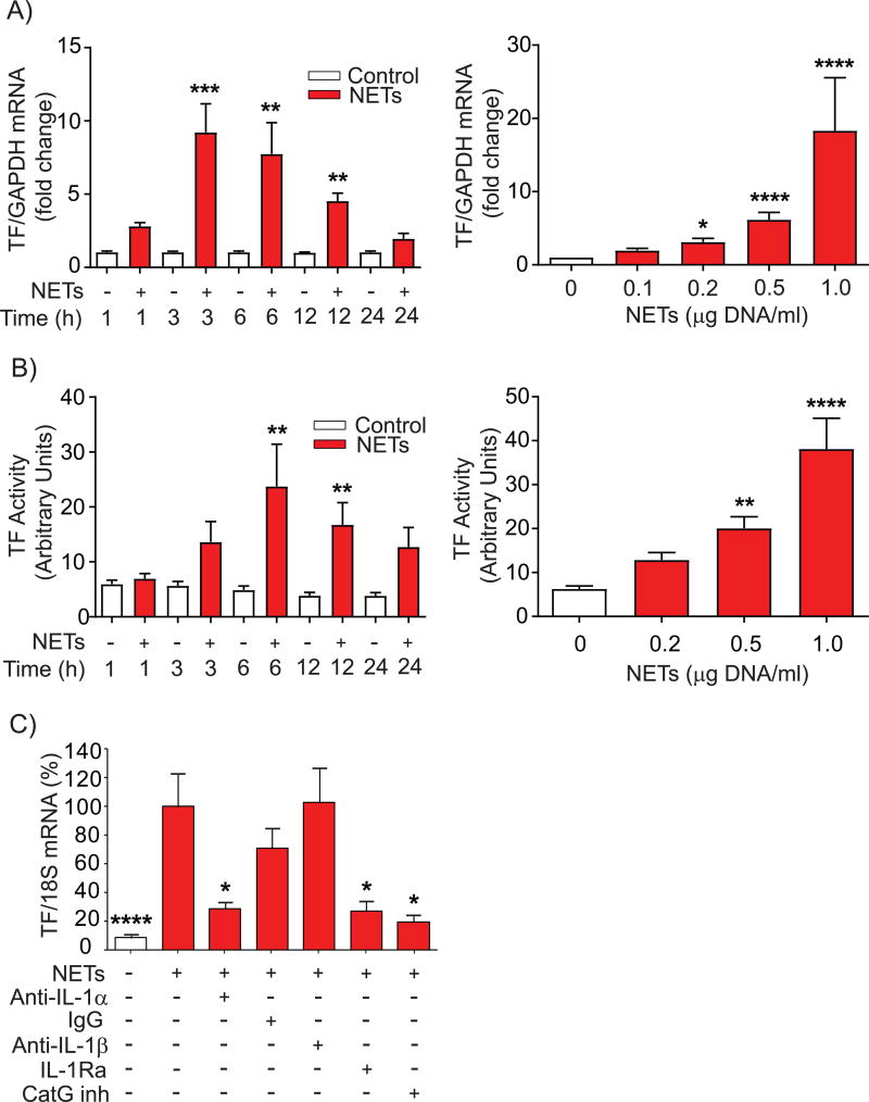
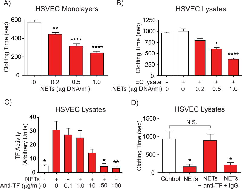
Objective- Coronary artery thrombosis can occur in the absence of plaque rupture because of superficial erosion. Erosion-prone atheromata associate with more neutrophil extracellular traps (NETs) than lesions with stable or rupture-prone characteristics. The effects of NETs on endothelial cell (EC) inflammatory and thrombogenic properties remain unknown. We hypothesized that NETs alter EC functions related to erosion-associated thrombosis. Approach and Results- Exposure of human ECs to NETs increased VCAM-1 (vascular cell adhesion molecule 1) and ICAM-1 (intercellular adhesion molecule 1) mRNA and protein expression in a time- and concentration-dependent manner. THP-1 monocytoid cells and primary human monocytes bound more avidly to NET-treated human umbilical vein ECs than to unstimulated cells under flow. Treatment of human ECs with NETs augmented the expression of TF (tissue factor) mRNA, increased EC TF activity, and hastened clotting of recalcified plasma. Anti-TF-neutralizing antibody blocked NET-induced acceleration of clotting by ECs. NETs alone did not exhibit TF activity or acceleration of clotting in cell-free assays. Pretreatment of NETs with anti-interleukin (IL)-1α-neutralizing antibody or IL-1Ra (IL-1 receptor antagonist)-but not with anti-IL-1β-neutralizing antibody or control IgG-blocked NET-induced VCAM-1, ICAM-1, and TF expression. Inhibition of cathepsin G, a serine protease abundant in NETs, also limited the effect of NETs on EC activation. Cathepsin G potentiated the effect of IL-1α on ECs by cleaving the pro-IL-1α precursor and releasing the more potent mature IL-1α form. Conclusions- NETs promote EC activation and increased thrombogenicity through concerted action of IL-1α and cathepsin G. Thus, NETs may amplify and propagate EC dysfunction related to thrombosis because of superficial erosion.

Keywords: cathepsin G; endothelial cells; extracellular traps; interleukin-1; neutrophils; vascular cell adhesion molecule 1.


Figures

Figure 1
Figure 1. NETs increase the endothelial expression of leukocyte adhesion molecules and augment leukocyte adhesion to ECs under flow
(A–B) HSVECs were incubated with NETs (0.5 µg DNA/ml) for the indicated periods of time (left panels) or for 3 hours with various concentrations of NETs (right panels), followed by RNA extraction and determination of VCAM-1 and ICAM-1 mRNA levels by RT-qPCR. Levels of GAPDH mRNA served as an internal control for adjustment between samples. Left panels, n = 7; right panels, n = 9. P values: *<0.05, **<0.01, ***<0.001, ****<0.0001 vs. the respective controls. (C) HSVECs were incubated with NETs (0.5 µg DNA/ml) for the indicated periods of time. Whole-cell extracts were fractionated by SDS-PAGE and immunoblotted with antibodies to VCAM-1, ICAM-1, or β-actin. (D) HUVECs were treated with NETs (0.5 µg DNA/ml) for 6 hours and analyzed by immunoblot as in (C). (E) HUVECs were treated with NETs (0.5 µg DNA/ml) for 6 hours, followed by analysis of the adhesion of THP-1 cells (left panel, n = 4) or primary human monocytes (right panel, n = 3) under flow as described in the Materials and Methods section.
Figure 2
Figure 2. IL-1α and CatG mediate NET-induced expression of leukocyte adhesion molecules in HSVECs
(A) Cells were incubated with 50 pg/ml IL-1α or IL-1β for 3 hours in the presence of the indicated antibodies (20 ng/ml) or IL-1Ra (1 µg/ml). Whole-cell extracts were fractionated by SDS-PAGE and immunoblotted with antibodies to VCAM-1 or β-actin. (B–C) Cells were incubated with NETs (0.5 µg DNA/ml) for 3 hours in the presence of the indicated antibodies (20 ng/ml), IL-1Ra (1 µg/ml), or CatG inhibitor I (50 µM), followed by RNA extraction and determination of VCAM-1 and ICAM-1 mRNA levels by RT-qPCR. N = 3–7. P values: *<0.05, **<0.01 vs. NETs alone, defined as 100%. (D–E) Cells were incubated as in (B-C) for 6 hours, followed by fractionation of whole-cell extracts by SDS-PAGE and immunoblotting with antibodies to VCAM-1, ICAM-1, or β-actin. (F) Cells were incubated with NETs (0.5 µg DNA/ml) for the indicated periods of time, followed by fractionation of whole-cell extracts by SDS-PAGE and immunoblotting with antibodies to phospho-IκBa, IκBα, or GAPDH. (G) Cells were incubated with NETs (0.5 µg DNA/ml) for 90 minutes, fixed, and stained with anti-NFκB p65 (1/1000 dilution) and DAPI as described in the Materials and Methods section. The arrowheads indicate NFκB localized to the nucleus. Scale bar = 50 µM. Omission of the primary antibody yielded no signal.
Figure 3
Figure 3. NETs act differentially on Pro-IL-1α or Pro-IL-1β
(A) GST-Pro-IL-1α (3.6 µg/ml) was incubated with NETs (left panel, 4 µg DNA/ml), or with various concentrations of recombinant CatG (right panel) for 1 hour at 37°C in the presence or absence of 3% FBS. Samples were fractionated by SDS-PAGE and immunoblotted with antibodies to IL-1α. (B) GST-Pro-IL-1β (3.8 µg/ml) was incubated with NETs (4 µg DNA/ml) for 1 hour at 37°C in the presence or absence of 3% FBS. Samples were fractionated by SDS-PAGE and immunoblotted with antibodies to IL-1β. (C) HSVECs were incubated for 6 hours with GST-Pro-IL-1α (20 pg/ml), GST-Pro-IL-1β (20 pg/ml), or cleaved IL-1α (100 pg/ml) in the presence or absence of DNA (0.16 µg/ml) and CatG (0.032 units/ml). Whole-cell extracts were fractionated by SDS-PAGE and immunoblotted with antibodies to VCAM-1, ICAM-1, or β-actin.
Figure 4
Figure 4. NETs induce endothelial TF expression and pro-coagulant activity
(A) HSVECs were incubated with NETs (0.5 µg DNA/ml) for the indicated periods of time (left panel, n = 6) or for 3 hours with various concentrations of NETs (right panel, n = 9), followed by RNA extraction and determination of TF mRNA levels by RT-qPCR. Levels of GAPDH mRNA served as an internal control for adjustment between samples. (B) HSVECs were incubated with NETs (0.5 µg DNA/ml) for the indicated periods of time (left panel, n = 8) or for 6 hours with various concentrations of NETs (right panel, n = 6), followed by cell lysis and determination of TF activity as described in the Materials and Methods section. (C) HSVECs were incubated with NETs (0.5 µg DNA/ml) for 3 hours in the presence of the indicated antibodies (20 ng/ml), IL-1Ra (1 µg/ml), or CatG inhibitor I (50 µM), followed by RNA extraction and determination TF mRNA levels by RT-qPCR. N = 4–9. P values: *<0.05, **<0.01, ***<0.001, ****<0.0001 vs. the respective controls (A–D) or vs. NETs alone, defined as 100% (E).
Figure 5
Figure 5. NETs accelerate EC-mediated plasma clotting in a TF-dependent manner
(A) HSVECs were incubated with various concentrations of NETs for 8 hours, followed by washing and determination of plasma clotting activity of the intact monolayers as described in Materials and Methods. N = 4. (B) HSVECs were incubated with NETs (0.5 µg DNA/ml) as in (A) for 6 hours, followed by cell lysis and determination of plasma clotting activity. N = 7. (C–D) HSVECs were incubated with NETs (0.5 µg DNA/ml) or left unstimulated for 6 hours. TF activity of cell lysates was measured in the presence of various concentrations of anti-TF neutralizing antibody (C). The plasma clotting activity of cell lysates was determined in the presence or absence of 50 µg/ml anti-TF neutralizing antibody or control IgG (D). N = 5. P values: *<0.05, **<0.01, ****<0.0001 vs. the respective controls (A, B, and D) or vs. NETs alone (C). N.S., not significant.
Figure 6
Figure 6. Interleukin-1α mediates endothelial cell activation by neutrophil extracellular traps
When neutrophils undergo NETosis, the extruded DNA strands associate with numerous proteins including the precursor forms of IL-1α and β and serine proteinases produced by the granulocytes. The results presented here show that the neutrophil-derived proteinase cathepsin G processes pro-IL-1α to the more active mature form by limited proteolysis, but degrades pro-IL-1b to inactive fragments. This pathway likely operates in vivo, as cathepsin G retains activity even in the presence of plasma proteinase inhibitors. NETs bearing mature IL-1α can activate endothelial cells to express adhesion molecules that can recruit further leukocytes, and elicit the local production of the potent procoagulant tissue factor. Thus, activation of endothelial cells by this NET-associated cytokine can amplify, sustain, and propagate local vascular inflammation and also promote thrombosis. These results have particular importance for postulated mechanisms of thrombosis due to superficial erosion. We have proposed a "multi-hit" pathway for the pathogenesis of this mode of arterial thrombosis: an initial endothelial desquamation with inadequate local repair, followed by an amplification phase. NET-associated IL-1α could participate in the amplification phase by aggravating the consequences of local endothelial erosion and promotion of the formation and persistence of arterial thrombi that lead to clinical events.

Similar articles

Cited by

References

    1. Libby P. Mechanisms of acute coronary syndromes and their implications for therapy. N Engl J Med. 2013;368:2004–2013. - PubMed
    1. Quillard T, Franck G, Mawson T, Folco E, Libby P. Mechanisms of erosion of atherosclerotic plaques. Curr Opin Lipidol. 2017 - PMC - PubMed
    1. Partida RA, Libby P, Crea F, Jang IK. Plaque erosion: a new in vivo diagnosis and a potential major shift in the management of patients with acute coronary syndromes. Eur Heart J. 2018 - PMC - PubMed
    1. Farb A, Burke AP, Tang AL, Liang TY, Mannan P, Smialek J, Virmani R. Coronary plaque erosion without rupture into a lipid core. A frequent cause of coronary thrombosis in sudden coronary death. Circulation. 1996;93:1354–1363. - PubMed
    1. Kolodgie FD, Burke AP, Farb A, Weber DK, Kutys R, Wight TN, Virmani R. Differential accumulation of proteoglycans and hyaluronan in culprit lesions: insights into plaque erosion. Arterioscler Thromb Vasc Biol. 2002;22:1642–1648. - PubMed

Publication types

MeSH terms