. 2018 Jul 31;38(4):BSR20180563.
doi: 10.1042/BSR20180563. Print 2018 Aug 31.

Induced pluripotent stem cell-derived endothelial cells promote angiogenesis and accelerate wound closure in a murine excisional wound healing model

Affiliations

Induced pluripotent stem cell-derived endothelial cells promote angiogenesis and accelerate wound closure in a murine excisional wound healing model

Zoë E Clayton et al. Biosci Rep. .

Abstract

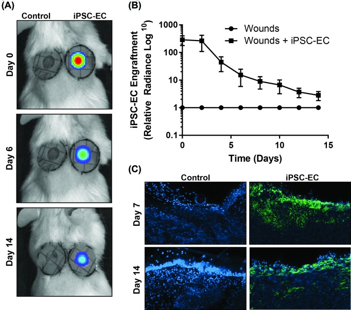
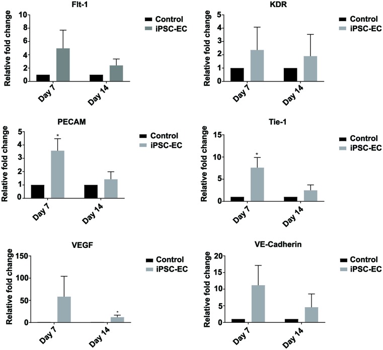
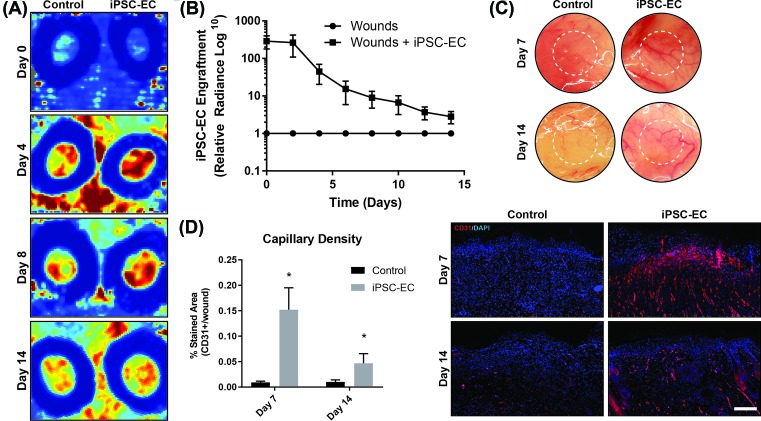
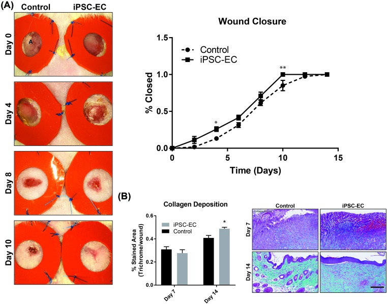
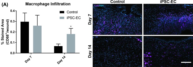
Chronic wounds are a major complication in patients with cardiovascular diseases. Cell therapies have shown potential to stimulate wound healing, but clinical trials using adult stem cells have been tempered by limited numbers of cells and invasive procurement procedures. Induced pluripotent stem cells (iPSCs) have several advantages of other cell types, for example they can be generated in abundance from patients' somatic cells (autologous) or those from a matched donor. iPSCs can be efficiently differentiated to functional endothelial cells (iPSC-ECs). Here, we used a murine excisional wound model to test the pro-angiogenic properties of iPSC-ECs in wound healing. Two full-thickness wounds were made on the dorsum of NOD-SCID mice and splinted. iPSC-ECs (5 × 10⁵) were topically applied to one wound, with the other serving as a control. Treatment with iPSC-ECs significantly increased wound perfusion and accelerated wound closure. Expression of endothelial cell (EC) surface marker, platelet endothelial cell adhesion molecule (PECAM-1) (CD31), and pro-angiogenic EC receptor, Tie1, mRNA was up-regulated in iPSC-EC treated wounds at 7 days post-wounding. Histological analysis of wound sections showed increased capillary density in iPSC-EC wounds at days 7 and 14 post-wounding, and increased collagen content at day 14. Anti-GFP fluorescence confirmed presence of iPSC-ECs in the wounds. Bioluminescent imaging (BLI) showed progressive decline of iPSC-ECs over time, suggesting that iPSC-ECs are acting primarily through short-term paracrine effects. These results highlight the pro-regenerative effects of iPSC-ECs and demonstrate that they are a promising potential therapy for intractable wounds.

Keywords: angiogenesis; endothelial cells; induced pluripotent stem cells; wound healing.


Conflict of interest statement

The authors declare that there are no competing interests associated with the manuscript.

Figures

Figure 1
Figure 1. iPSC-EC engraftment in wounds
iIPSC-ECs were transduced with a double fusion reporter construct encoding GFP for fluorescence imaging and firefly luciferase for BLI. (A) Representative IVIS images, showing bioluminescent signal present in iPSC-EC-treated wounds (right) on days 0, 6 and 14 post-wounding. (B) Declining bioluminescent signal in wounds over time, relative to control wound background signal. (C) GFP staining (green) with DAPI nuclear stain (blue) in control and iPSC-EC treated wounds at on days 7 and 14 post-wounding (n=6 per treatment group).
Figure 2
Figure 2. iPSC-EC treatment increases expression of pro-angiogenic genes
qPCR analysis of wound mRNA revealed a significant up-regulation of PECAM (CD31) and endothelial cell receptor, Tie-1, expression in iPSC-EC wounds on day 7 post-wounding. This was resolved by day 14. No significant differences in endothelial cell surface marker VE-cadherin (CD144), VEGF, VEGF receptor 1 (Flt1) or VEGF receptor 2 (KDR) expression were measured (*P<0.05 compared with controls, n=6 per treatment group).
Figure 3
Figure 3. iPSC-EC treatment increases vascular density and wound perfusion
(A) Representative laser Doppler images, showing perfusion in control and iPSC-EC treated wounds on days 0, 4, 8 and 18 post-wounding. (B) Wound perfusion in iPSC-EC treated wounds, relative to their respective control wounds. Increased perfusion in iPSC-EC treated wounds was most pronounced during the first week of healing. (C) Representative photomicrographs of wounds, showing increased neo-vessel formation in iPSC-EC wounds relative to controls. (D) Capillary density, as measured by CD31+ staining, was significantly increased in iPSC-EC wounds at both early and late time points (*P<0.05, n=6 per treatment group).
Figure 4
Figure 4. iPSC-EC treatment increases collagen deposition and accelerates wound closure
(A) Representative photomicrographs, showing progressive wound closure over a 14-day period. Rate of closure was significantly increased in iPSC-EC treated wounds compared with controls. (B) Milligan’s Trichrome staining for collagen content. No differences were observed on day 7; by day 14 iPSC-EC treated wounds had significantly higher collagen content (*P<0.05, **P<0.01, n=6 per treatment group).
Figure 5
Figure 5. iPSC-EC treatment increases macrophage infiltration
(A) Anti-CD68 staining for macrophage infiltration. No differences were observed on day 7; by day 14 iPSC-EC treated wounds had significantly macrophage infiltration (*P<0.05, n=6 per treatment group).

Similar articles

Cited by

References

    1. Leavitt T., Hu M.S., Marshall C.D., Barnes L.A., Longaker M.T. and Lorenz H.P. (2016) Stem cells and chronic wound healing: state of the art. Chronic Wound Care Management Res. 3, 7–27
    1. Hinchliffe R.J., Brownrigg J.R.W., Andros G., Apelqvist J., Boyko E.J., Fitridge R. et al. (2016) Effectiveness of revascularization of the ulcerated foot in patients with diabetes and peripheral artery disease: a systematic review. Diabetes Metab. Res. Rev. 32, 136–144 10.1002/dmrr.2705 - DOI - PubMed
    1. Dreifke M.B., Jayasuriya A.A. and Jayasuriya A.C. (2015) Current wound healing procedures and potential care. Mater. Sci. Eng. C Mater. Biol. Appl. 48, 651–662 10.1016/j.msec.2014.12.068 - DOI - PMC - PubMed
    1. Greaves N.S., Ashcroft K.J., Baguneid M. and Bayat A. (2013) Current understanding of molecular and cellular mechanisms in fibroplasia and angiogenesis during acute wound healing. J. Dermatol. Sci. 72, 206–217 10.1016/j.jdermsci.2013.07.008 - DOI - PubMed
    1. Asahara T., Masuda H., Takahashi T., Kalka C., Pastore C., Silver M. et al. (1999) Bone marrow origin of endothelial progenitor cells responsible for postnatal vasculogenesis in physiological and pathological neovascularization. Circ. Res. 85, 221–228 10.1161/01.RES.85.3.221 - DOI - PubMed

Publication types

LinkOut - more resources