Review
. 2018 Aug;10(8):e8712.
doi: 10.15252/emmm.201708712.

Reprogramming of basic metabolic pathways in microbial sepsis: therapeutic targets at last?

Affiliations
Review

Reprogramming of basic metabolic pathways in microbial sepsis: therapeutic targets at last?

Lise Van Wyngene et al. EMBO Mol Med. 2018 Aug.

Abstract

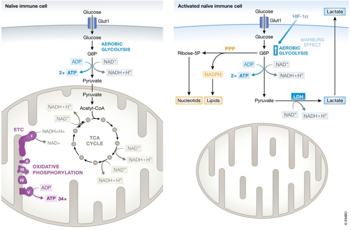
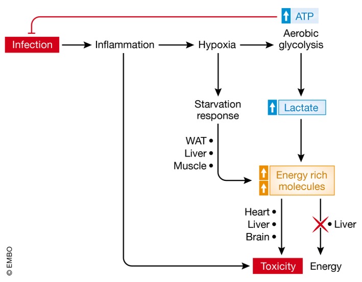
Sepsis is a highly lethal and urgent unmet medical need. It is the result of a complex interplay of several pathways, including inflammation, immune activation, hypoxia, and metabolic reprogramming. Specifically, the regulation and the impact of the latter have become better understood in which the highly catabolic status during sepsis and its similarity with starvation responses appear to be essential in the poor prognosis in sepsis. It seems logical that new interventions based on the recognition of new therapeutic targets in the key metabolic pathways should be developed and may have a good chance to penetrate to the bedside. In this review, we concentrate on the pathological changes in metabolism, observed during sepsis, and the presumed underlying mechanisms, with a focus on the level of the organism and the interplay between different organ systems.

Keywords: hypoxia; inflammation; interventions; metabolic reprogramming; sepsis.


Figures

Figure 1
Figure 1. Impact of sepsis on carbohydrate metabolism
Naïve immune cells rely on glycolysis and oxidative phosphorylation as the main metabolic pathways to generate ATP. Glycolysis is the catabolic process in which glucose is converted into pyruvate. Extracellular glucose is imported into cells by GLUT1 (SLC2A1 gene). Subsequent intracellular processing of glucose yields two pyruvate molecules and two ATP molecules by a series of enzymatic reactions. At sufficient O2 tension, pyruvate is imported in mitochondria and converted into acetyl‐coA and enters the tricarboxylic acid cycle (TCA cycle, a.k.a. Kreb's cycle), after which each pyruvate yields 17 ATP molecules by the electron transport chain (ETC) and oxidative phosphorylation. Upon activation of immune cells, the glycolysis pathway is upregulated under the control of HIF‐1α. The metabolic pathway shifts from oxidative phosphorylation to aerobic glycolysis, also referred to as the Warburg effect, to meet the increased energy demand in activated immune cells. Despite the presence of abundant O2 in the environment, glucose is then directly metabolized into lactate. Although energetically less favorable (glycolysis generates 2 molecules of ATP out of 1 molecule glucose, whereas oxidative phosphorylation provides 36 ATP molecules), aerobic glycolysis allows higher velocity of glycolysis and thus faster ATP production, as well as provides important precursors for the synthesis of lipids, amino acids, and nucleotides required for proliferation. Interfering with aerobic glycolysis in immune cells has been shown to undermine their anti‐infectious activities, highlighting the importance of this altered metabolism in activated immune cells.
Figure 2
Figure 2. The pathogenesis of sepsis is associated with a deregulation in glucose metabolism
Both high and low glucose levels correlate with sepsis severity. Initially, a hyperglycemic response is observed in both animal models and in human patients, presumably resulting from insulin resistance and altered glycogen metabolism. This allows redirection of glucose to immune cells supporting aerobic glycolysis and thus immune function, and also leads to lactate production. In later stages, hypoglycemia could be observed as a consequence of several factors. In contrast to animal models, hypoglycemia is less frequently observed in human patients.
Figure 3
Figure 3. An overview of lipolysis and lipid oxidation in healthy conditions
(A) Lipolysis is the hydrolytic conversion of triglycerides (TG) into glycerol and free fatty acids (FFAs) and is most abundant in white and brown adipose tissue. This process is tightly regulated by glucagon, (nor)epinephrine, and other hormones, but also by pro‐inflammatory cytokines. Hydrolysis of the ester bonds between long‐chain fatty acids and the glycerol backbone is executed by lipases. Up to now, three enzymes have been implicated in performing the complete hydrolysis of TG into FFAs and glycerol: adipose triglyceride lipase (ATGL), hormone sensitive lipase (HSL), and monoglyceride lipase (MGL). The FFAs are released into the blood stream and can be taken up by peripheral organs to produce energy via mitochondrial β‐oxidation. (B) Fatty acid β‐oxidation is a multistep process breaking down fatty acids in the mitochondria of the cell to produce acetyl‐CoA, which can be used by the tricarboxylic acid (TCA) cycle to produce ATP. In brief, FFAs are transported across the cell membrane by members of the FATP transporter family. Once inside the cytosol, the FFA is coupled to coenzyme A (CoA) by acyl‐CoA synthetase (ACS) and shuttled across the inner mitochondrial membrane by carnitine palmitoyltransferase II (CPT2) and the carnitine acyltransferase (CAT) after being coupled to carnitine by carnitine palmitoyltransferase I (CPT1). In the mitochondrial matrix, β‐oxidation is conducted by cleaving two carbon molecules in every oxidation cycle to form acetyl‐CoA. The cycle is repeated until the complete fatty acid has been reduced to acetyl‐CoA, which is subsequently enters the TCA cycle.
Figure 4
Figure 4. Lipolysis, fatty acid oxidation, and ketogenesis in sepsis
Sepsis is associated with the development of an anorectic response since patients are often unwilling or unable to eat. During a normal starvation response and during sepsis, lipolysis in white and brown adipose tissue is being upregulated by several pro‐lipolytic signals. The inhibitory effect on lipolysis of insulin, which is upregulated in sepsis due to high glucose levels, is however absent due to insulin resistance. Free fatty acids (FFAs) in the blood are upregulated in both conditions and can be taken up by peripheral organs to produce energy. The increased FFA levels activate and upregulate the expression of PPAR‐α, the main transcription factor responsible for the induction of genes involved in the β‐oxidation of fatty acids and the production of ketone bodies (KBs). During sepsis, PPAR‐α levels are downregulated and the breakdown of fatty acids through β‐oxidation is compromised, causing FFAs to accumulate in organs such as the liver, heart, and kidney, but also in the blood. Overall, the deficits in FFA breakdown during sepsis cause a shortage of energy and lipotoxicity and mitochondrial damage due to FFA accumulation. Green represents the normal starvation response, and red represents the response during sepsis.
Figure 5
Figure 5. Overview of protein catabolism in sepsis
In sepsis conditions, catabolism of proteins in skeletal muscle is a recurrent feature, but the main regulators are still not identified. Branched‐chain amino acids (AAs) are oxidized to branched‐chain keto acids (BCKAs), which can be used in the TCA cycle. Glutamine (Gln) and alanine (Ala) find their way to kidney and intestine and liver. In the former two, Gln is de‐aminated to glutamate (Glu) and ammonia (NH3), which is removed. Glu and pyruvate can yield Ala and α‐ketoglutaric acid (αKG), which can enter the TCA cycle. Ala is mainly used as a gluconeogenic substrate and is transformed to pyruvate, whereby the NH2 group is removed via the urea cycle. During liver failure, ammonia may leak into the blood, leading to brain damage and coma.
Figure 6
Figure 6. The toxic consequences of metabolic reprogramming in sepsis
Infection is the start of sepsis. It leads to direct tissue damage and to inflammation, which in turn leads to hypoxia, which is essential to allow white blood cells (WBCs) to produce fast ATP from glucose and act fast on the infectious agents. The hypoxic response also leads to mobilization of energy‐rich molecules such as lactate and fatty acids, which however can also lead to toxicity, when over abundant.

References

    1. Arts RJW, Gresnigt MS, Joosten LAB, Netea MG (2016) Cellular metabolism of myeloid cells in sepsis. J Leukoc Biol 100: 1–14 - PubMed
    1. Arulkumaran N, Deutschman CS, Pinsky MR, Zuckerbraun B, Schumacker PT, Gomez H, Gomez A, Murray P, Kellum JA (2016) Mitochondrial function in sepsis. Shock 45: 271–281 - PMC - PubMed
    1. Askanazi J, Carpentier Y, Elwyn D, Noenström J, Jeevanandam M, Rosenum S, Gump F, Kinney J (1980) Influence of total parenteral nutrition on fuel utilization in injury and sepsis. Ann Surg 191: 40 - PMC - PubMed
    1. Baldwin AC, Green CD, Olson KL, Moxley MA, Corbett JA (2012) A role for aberrant protein palmitoylation in FFA‐induced ER stress and β‐cell death. Am J Physiol Endocrinol Metab 302: E1390–E1398 - PMC - PubMed
    1. Balmer ML, Hess C (2017) Starving for survival—how catabolic metabolism fuels immune function. Curr Opin Immunol 46: 8–13 - PubMed

Publication types

MeSH terms

Substances