. 2018 Aug 10;361(6402):604-607.
doi: 10.1126/science.aat5671. Epub 2018 Jul 5.

A liquid phase of synapsin and lipid vesicles

Affiliations

A liquid phase of synapsin and lipid vesicles

Dragomir Milovanovic et al. Science. .

Abstract

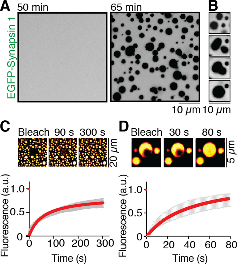
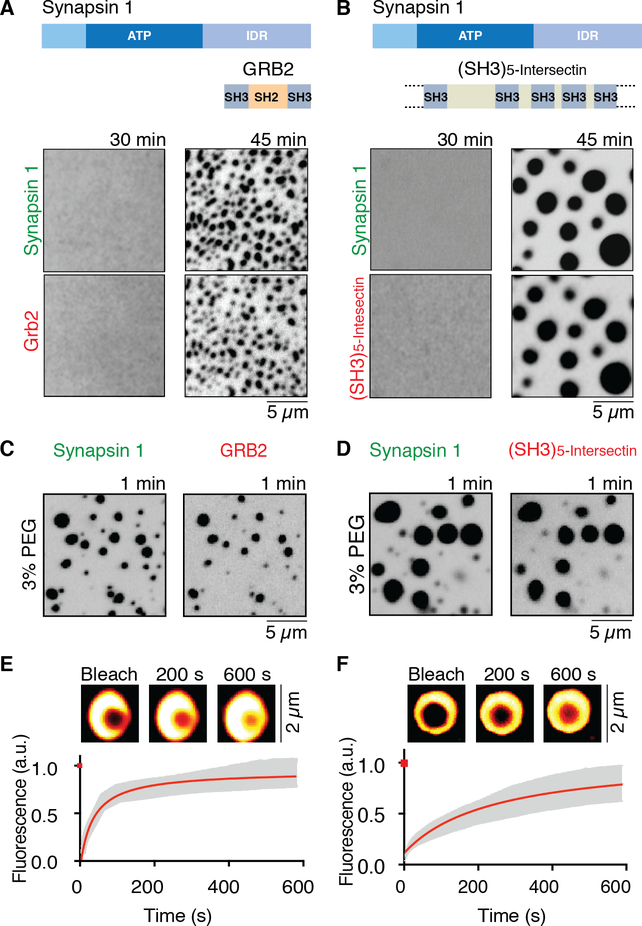
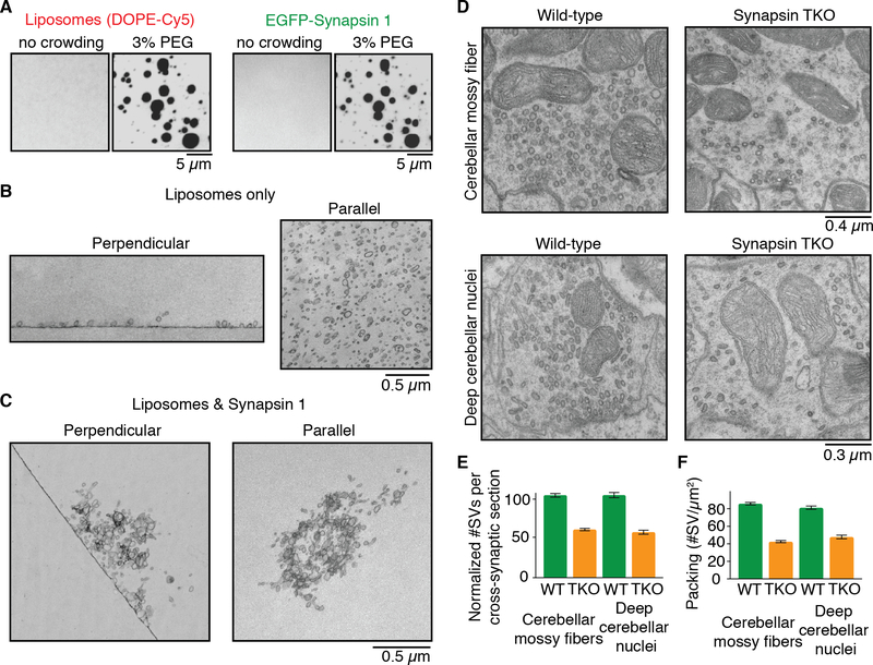
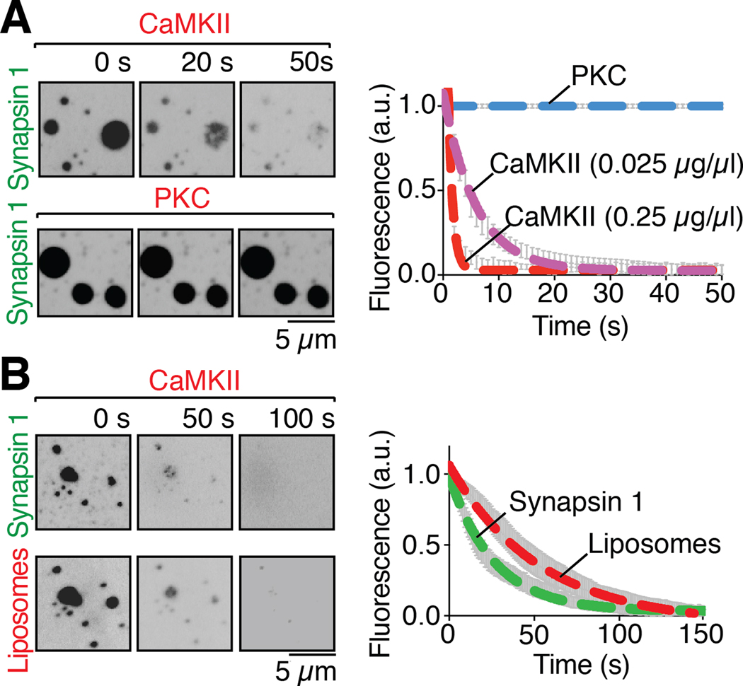
Neurotransmitter-containing synaptic vesicles (SVs) form tight clusters at synapses. These clusters act as a reservoir from which SVs are drawn for exocytosis during sustained activity. Several components associated with SVs that are likely to help form such clusters have been reported, including synapsin. Here we found that synapsin can form a distinct liquid phase in an aqueous environment. Other scaffolding proteins could coassemble into this condensate but were not necessary for its formation. Importantly, the synapsin phase could capture small lipid vesicles. The synapsin phase rapidly disassembled upon phosphorylation by calcium/calmodulin-dependent protein kinase II, mimicking the dispersion of synapsin 1 that occurs at presynaptic sites upon stimulation. Thus, principles of liquid-liquid phase separation may apply to the clustering of SVs at synapses.


Conflict of interest statement

Competing interests:

Authors declare no competing interests.

Figures

Fig. 1.
Fig. 1.. Synapsin 1 undergoes liquid-liquid phase separation.
(A) EGFP-Synapsin 1 (10 μM) forms droplets when incubated for one hour in a buffer of physiological salt concentration at RT (B) Droplets of synapsin 1 show liquid behavior by fusing with each other and relaxing into a round-shaped structure, minimizing surface tension. (C) Photobleaching of several synapsin 1-droplets with subsequent recovery of fluorescence, as shown by micrograph and by quantification of the fluorescence recovery in the bleached region. Error bars represent s.e.m., and red is the fit with hyperbolic function. (D) Fluorescence recovery of synapsin 1 after photobleaching a region within a droplet (see inset). Error bars represent s.e.m., red is the fit with a hyperbolic function.
Fig. 2.
Fig. 2.. Synapsin 1 drives phase separation of SH3 domain containing binding partners.
(A) and (B) Full-length synapsin 1 (10 μM) and either Grb2 (10 μM), or the SH3 domain containing region of intersectin (10 μM) form droplets under physiological conditions. Top: domain organization of the proteins. Bottom: fluorescence images of the protein mixtures at 30 and 45min. (C) and (D) Fluorescence images of the solution immediately (within one min) after mixing of the two proteins in the presence of the crowding reagent (3 % PEG 8,000). (E) and (F) Fluorescence recovery of synapsin 1 after photobleaching a region within a synapsin 1-Grb2 droplet, or a synapsin 1-(SH3)5-intersectin droplet (see insets). Error bars represent s.e.m., red is the fit with a hyperbolic function.
Fig. 3.
Fig. 3.. Synapsin 1 condensates are reaction centers able to sequester lipid vesicles.
(A) Fluorescence images of mixture of liposome (left, Cy5-DOPE) and synapsin 1 (right, EGFPSynapsin 1) without or with crowding agent (3% PEG). (B) EM images of liposomes incubated without synapsin 1 in the same buffer conditions used for A. Left: section perpendicular to the liposome-glass interface. Right: section comprising the layer of liposomes absorbed to the glass surface. (C) Same as in B, but showing liposomes incubated with synapsin 1. The field shown at right is from a section parallel to the glass surface but above the glass interface. (D) EM images of synapses from cerebellar mossy fibers (top) and deep cerebellar nuclei (bottom) obtained from adult wild-type (WT, left) and synapsin triple knockout (TKO, right) mice. (E) SV number in synaptic cross-sections of wild-type and synapsin TKO mice, normalized to wild-type. (F) Synaptic vesicles number per unit area of synaptic section in wild-type and synapsin TKO mice. For each condition, fifty sections from three independent animals were examined. Error bars represent s.e.m.
Fig. 4.
Fig. 4.. Phosphorylation of the intrinsically disordered region of synapsin 1 disperses condensates of either synapsin 1 alone or synapsin 1 and liposomes.
(A) Left: fluorescence images of EGFP-Synapsin 1 condensates preincubated with either CaMKII (0.025 μg/μl), calmodulin and calcium, or PKC, PS, DAG and calcium, upon addition, at 0 seconds, of ATP (200 μM), demonstrating dispersion of synapsin by CaMKII, but not by PKC. Right: time course of the effect of the kinases on the condensates, as assessed by the decrease of florescence on ROIs corresponding to randomly selected droplets (B) Left: Fluorescence images of liposome-synapsin condensates preincubated with CaMKII (0.25 μg/μl), calmodulin and calcium, upon addition, at 0 seconds, of ATP (200 μM), demonstrating dispersion of both synapsin and liposomes. Right: Time-course of the effect of CaMKII on liposome-synapsin droplets dispersion. Error bars represent s.e.m.; dashed lines represent the fit with a single exponential function. Incubations were carried out at RT in a buffer of physiological salt concentration supplemented with 3% PEG 8,000.

Comment in

References

    1. Milovanovic D, De Camilli P, Synaptic Vesicle Clusters at Synapses: A Distinct Liquid Phase? Neuron. 93, 995–1002 (2017). - PMC - PubMed
    1. Rizzoli SO, Betz WJ, The structural organization of the readily releasable pool of synaptic vesicles. Science. 303, 2037–2039 (2004). - PubMed
    1. Darcy KJ, Staras K, Collinson LM, Goda Y, Constitutive sharing of recycling synaptic vesicles between presynaptic boutons. Nat. Neurosci 9, 315–321 (2006). - PubMed
    1. Acuna C, Liu X, Südhof TC, How to Make an Active Zone: Unexpected Universal Functional Redundancy between RIMs and RIM-BPs. Neuron. 91, 792–807 (2016). - PubMed
    1. Wang SSH et al., Fusion Competent Synaptic Vesicles Persist upon Active Zone Disruption and Loss of Vesicle Docking. Neuron. 91, 777–791 (2016). - PMC - PubMed

Publication types