. 2018 Jul 5;10(7):357.
doi: 10.3390/v10070357.

Dengue Virus Induces the Release of sCD40L and Changes in Levels of Membranal CD42b and CD40L Molecules in Human Platelets

Affiliations

Dengue Virus Induces the Release of sCD40L and Changes in Levels of Membranal CD42b and CD40L Molecules in Human Platelets

Daniel Núñez-Avellaneda et al. Viruses. .

Abstract

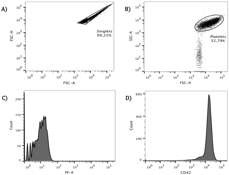
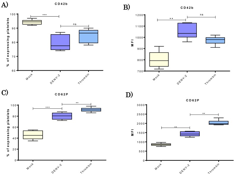
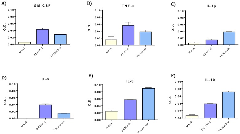
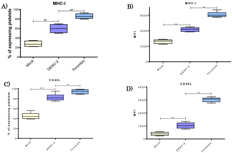
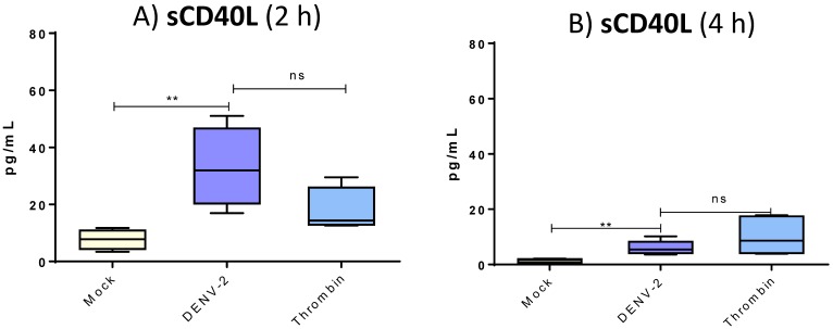
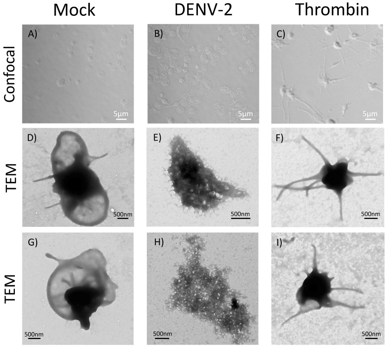
Platelets are considered as significant players in innate and adaptive immune responses. The adhesion molecules they express, including P-selectin, CD40L, and CD42b, facilitate interactions with many cellular effectors. Upon interacting with a pathogen, platelets rapidly express and enhance their adhesion molecules, and secrete cytokines and chemokines. A similar phenomenon occurs after exposure of platelets to thrombin, an agonist extensively used for in vitro activation of these cells. It was recently reported that the dengue virus not only interacts with platelets but possibly infects them, which triggers an increased expression of adhesion molecule P-selectin as well as secretion of IL-1β. In the present study, surface molecules of platelets like CD40L, CD42b, CD62P, and MHC class I were evaluated at 4 h of interaction with dengue virus serotype 2 (DENV-2), finding that DENV-2 induced a sharp rise in the membrane expression of all these molecules. At 2 and 4 h of DENV-2 stimulation of platelets, a significantly greater secretion of soluble CD40L (sCD40L) was found (versus basal levels) as well as cytokines such as GM-CSF, IL-6, IL-8, IL-10, and TNF-α. Compared to basal, DENV-2 elicited more than two-fold increase in these cytokines. Compared to the thrombin-induced response, the level generated by DENV-2 was much higher for GM-CSF, IL-6, and TNF-α. All these events induced by DENV end up in conspicuous morphological changes observed in platelets by confocal microscopy and transmission electron microscopy, very different from those elicited by thrombin in a more physiological scenery.

Keywords: CD40L; Immune response; dengue virus; platelets.


Conflict of interest statement

The authors declare no conflict of interest.

Figures

Figure 1
Figure 1
Platelet selection by cytometric analysis. Cells were obtained from wash platelet rich plasma (PRP) from healthy donors, carefully washed and resuspended in Tyrode’s solution. (A) Washed platelet suspension was acquired in the cytometer and singlets selected; these represented 98 ± 1.25%; (B) Platelet population was selected by size and granularity and separated from smaller size particles, which accounted for 52.79% in this particular analysis presented. Finally, to validate the selected population, the CD42b molecule was used as platelet marker, auto-fluorescence was measured (C); and anti-CD42-PE identified platelets, which exhibit this marker (D).
Figure 2
Figure 2
DENV-2 induced changes in CD42b molecule and activation CD62P marker on human platelets. Platelets from healthy donors (n = 10) were analyzed. Platelets were incubated with DENV2 to a MOI of 0.5 for 4 h at 37 °C. For each experiment, mock and thrombin groups were included. Percentages of platelets expressing CD42b molecule in each group are shown (A); as well as median fluorescence intensity (MFI) that indicate density of of CD42b molecule on the cell surface (B); as a measure of platelet activation, the percentage of CD62P-expressing platelets was determined (C); as well as MFI of CD62P expression (D). Unpaired t-test and Mann-Whitney U distribution were used for these experiments, where ** p < 0.01 and *** p < 0.001, ns = not significant.
Figure 3
Figure 3
Higher levels of TNF-α, IL-6, IL-8, GM-CSF, and IL-10 were released from DENV-2 exposed platelets. Cytokines and chemokines were evaluated, two pools from six supernatants for each experimental group. Original supernatants were obtained from 3 × 10⁸/mL human platelets after 4 h of stimulation with DENV-2 (MOI 0.2), mock or thrombin. Cytokines were measured by optical density GM-CSF (A); TNF-α (B); IL-1β (C); IL-6 (D); IL-8 (E) and IL-20 (F) at 450 nm. TNF-α, IL-8, GM-CSF, and IL-10 were elevated after DENV-2 incubation. IL-6 showed to be more elevated than thrombin control, and IL-1β only showed a small increment. Since each bar contains values of only two pools, only standard deviation is shown. No statistical analysis was performed on these data.
Figure 4
Figure 4
DENV-2 induced an increase of MHC-I and CD40L molecules on platelets. Platelets were obtained from healthy donors (n = 10) and divided in the three experimental groups. Platelets, DENV-2, and MOI 0.5 were incubated for 4 h at 37 °C; mock and thrombin groups were also processed. Percentage of MHC-I expressing platelets (A) and MFI of MHC-I (B) were determined. Additionally; percentage of platelets expression CD40L (C); and MFI in platelets of CD40L (D) were evaluated. Significant increases of MHC class I and CD40L were observed in the DENV-2 treated platelets for both percentages and density of molecules in the cell surface. Unpaired t test and Mann-Whitney U distribution were used, where ** p < 0.01 and *** p < 0.001).
Figure 5
Figure 5
Human platelets release sCD40L after DENV stimulus. In supernatants of 3 × 10⁸/mL treated platelets from healthy donors (n = 15), soluble CD40L was measured by ELISA test. At 2 and 4 h, platelets were DENV-2 stimulated, MOI 0.2, at 37 °C. Optical density was read at 450 nm and amount of sCD40L calculated for each experimental group. Unpaired t test was used, ** p < 0.01 and ns = not significant.
Figure 6
Figure 6
DENV-2 induced conspicuous morphological changes in human platelets. Platelets stimulated with DENV-2, MOI 0.2 (B) and MOI 0.5 (E,H) for 4 h, mock (and thrombin were incubated at 37 °C, and analyzed by TEM in triplicate. A representative image of the observed changes is presented. Conspicuous morphological changes occur in DENV-2-treated platelets (B,E,H), very different to those observed in thrombin positive control (C,F,I), meanwhile, mock control showed a normal platelet morphology (A,D,G). Images were captured at 50,000×. Duplicate assays were independently repeated three times.

Similar articles

Cited by

References

    1. Boulaftali Y., Hess P.R., Kahn M.L., Bergmeier W. Platelet immunoreceptor tyrosine-based activation motif (ITAM) signaling and vascular integrity. Circ. Res. 2014;114:1174–1184. doi: 10.1161/CIRCRESAHA.114.301611. - DOI - PMC - PubMed
    1. Lindemann S., Tolley N.D., Dixon D.A., McIntyre T.M., Prescott S.M., Zimmerman G.A., Weyrich A.S. Activated platelets mediate inflammatory signaling by regulated interleukin 1beta synthesis. J. Cell Biol. 2001;154:485–490. doi: 10.1083/jcb.200105058. - DOI - PMC - PubMed
    1. Herter J.M., Rossaint J., Zarbock A. Platelets in inflammation and immunity. J. Thromb. Haemost. 2014;12:1764–1775. doi: 10.1111/jth.12730. - DOI - PubMed
    1. Chapman L.M., Aggrey A.A., Field D.J., Srivastava K., Ture S., Yui K., Topham D.J., Baldwin W.M., 3rd, Morrell C.N. Platelets present antigen in the context of MHC class I. J. Immunol. 2012;189:916–923. doi: 10.4049/jimmunol.1200580. - DOI - PMC - PubMed
    1. Assinger A. Platelets and infection—An emerging role of platelets in viral infection. Front. Immunol. 2014;5:649. doi: 10.3389/fimmu.2014.00649. - DOI - PMC - PubMed

Publication types

MeSH terms