. 2018 Jul 5;8(1):10166.
doi: 10.1038/s41598-018-28257-5.

Zika Virus Replication in Dorsal Root Ganglia Explants from Interferon Receptor1 Knockout Mice Causes Myelin Degeneration

Affiliations

Zika Virus Replication in Dorsal Root Ganglia Explants from Interferon Receptor1 Knockout Mice Causes Myelin Degeneration

Vera Giulia Volpi et al. Sci Rep. .

Abstract

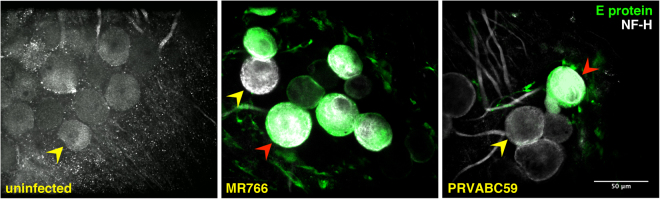
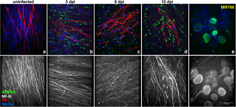
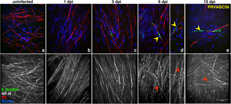
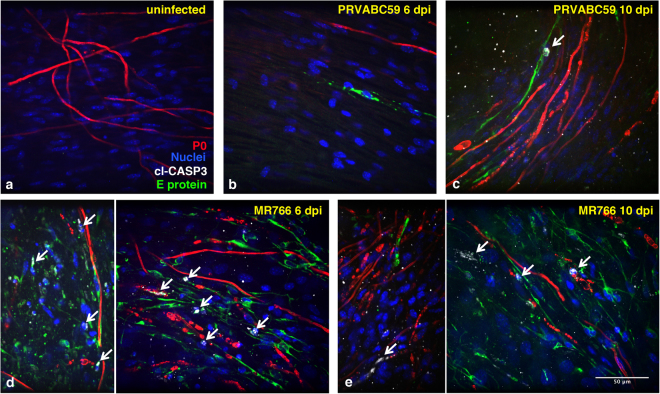
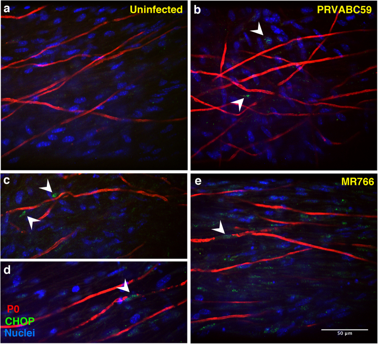
Zika virus (ZIKV) is a neurotropic agent that targets the developing fetal brain in women infected during pregnancy. In addition to the developing central nervous system, ZIKV has been recently shown to infect cells of the peripheral nervous system (PNS), highlighting its potential to cause acute peripheral neuropathies in adults, such as Guillain-Barré Syndrome (GBS). Here we show that myelinating dorsal root ganglia (DRG) explants obtained from interferon-alpha/beta receptor knock-out mice are productively infected by ZIKV. Virus replication is cytopathic in both peripheral neurons and myelinating Schwann cells leading to myelin disruption. These results confirm and extend previous observations suggesting that the PNS is indeed a potential site of ZIKV infection, replication and cytopathicity.


Conflict of interest statement

The authors declare no competing interests.

Figures

Figure 1
Figure 1
ZIKV infects peripheral DRG neurons in myelinating DRG explants cultures. Immunofluorescence staining on myelinating Ifnar1-KO DRG explants infected with MR766 or PRVABC59 strains after 6 dpi. In green, the anti-E protein mAb marks the viral particles detected in the cell body of the infected neurons and undetectable in uninfected DRG explants; in gray, anti-neurofilament-H (NF-H) Ab stains both neuronal bodies and axons. Red and yellow arrowheads indicate examples of infected and uninfected neurons, respectively. Scale bar, 50 μm.
Figure 2
Figure 2
Kinetics of MR766 replication in myelinating DRG explants. Myelinating Ifnar1-KO DRG explants were infected with MR766 ZIKV strain after 2 weeks of myelination induction. Staining against viral E protein (green), NF-H (white) and P0 (red) was performed 1 (b), 3 (c), 6 (d) and 10 (e) days post-infection (dpi) and compared to uninfected control (a). Hoechst dye was used to stain the nuclei (blue). White and blue arrowheads indicate single uninfected SC with intact myelin and the associated axon, respectively (panels a and d). Yellow and red arrowheads point at infected myelinating SC and their corresponding axons. Some SC display ongoing demyelination with or without axonal degeneration. At 10 dpi, diffuse demyelination, axonal degeneration and abundant cytolysis are detectable. Scale bar, 50 μm.
Figure 3
Figure 3
Active replication of MR766 ZIKV strain in myelinating DRG explants. Immunofluorescence staining of myelinating Ifnar1-KO DRG explants in presence or absence of ZIKV infection. Viral dsRNA staining (in green) is undetectable in uninfected DRG explants (a) and positive in infected myelinating co-cultures after 3 (b), 6 (c) and 10 (d) days post-infection (dpi). Panel e illustrates active viral replication in infected neuronal bodies. Scale bar, 50 μm.
Figure 4
Figure 4
Kinetics of PRVABC59 replication in myelinating DRG explants. Myelinating Ifnar1-KO DRG explants were infected with PRVABC59 strain after 2 weeks of myelination induction. Staining with anti-E viral protein (green), anti-NF-H (white) and anti-P0 (red) Abs was performed 1 (b), 3 (c), 6 (d) and 10 (e) days post-infection (dpi) and compared to uninfected control (a). Hoechst dye was used to stain the nuclei (blue). Yellow and red arrowheads point at infected myelinating SC and at the corresponding axons, respectively. Some infected myelinating SC display demyelination with or without associated axonal degeneration. Scale bar, 50 μm.
Figure 5
Figure 5
ZIKV infection activates caspase 3 in myelinating DRG explants. Myelinating Ifnar1-KO DRG explants were infected with either PRVABC59 (b,c) or MR766 (d,e) strains after 2 weeks of myelination induction. In panels (d,e), two representative images of MR766 infection at 6 and 10 days post-infection (dpi), respectively, are shown. Staining with anti-E viral protein (green), anti-cl-CASP3 (white) and anti-P0 (red) Abs was performed and compared to uninfected control (a). Hoechst dye was used to stain the nuclei (blue). White arrows point at cells in which activated cl-CASP3 signal, marker of apoptosis induction, is detected. Scale bar, 50 μm.
Figure 6
Figure 6
Kinetics of ZIKV productive infection and virus-induced cell death in DRG explants. Co-cultures were infected as described above. Culture supernatants were harvested at regular intervals after infection up to 10 days post-infection (dpi) and viral titers were determined by plaque assay on Vero cells (a). Evaluation of necrotic cell death was determined by the levels of AK activity released in the culture supernatants and expressed as relative luminescence units (RLU) (b). Means ± SD of 7 independent wells are reported. P values were determined using one-way ANOVA with Bonferronis multiple comparison test of day 1 post-infection vs. each following day (*p < 0.05; **p < 0.01; ****p < 0.0001).
Figure 7
Figure 7
ZIKV infection activates the ER stress-mediated expression of CHOP in myelinating DRG co-cultures. Myelinating Ifnar1-KO DRG explants were infected with either PRVABC59 (b) or MR766 (c,d,e) strains after 2 weeks of myelination induction and compared to uninfected control (a). Staining with anti-CHOP (green) and anti-P0 (red) Abs was performed and Hoechst dye was used to stain the nuclei (blue). White arrowheads point at CHOP-positive nuclei detected at 3 dpi, index of activated ER stress response following ZIKV infection. Scale bar, 50 µm.

References

    1. Marchette NJ, Garcia R, Rudnick A. Isolation of Zika virus from Aedes aegypti mosquitoes in Malaysia. Am J Trop Med Hyg. 1969;18:411–415. doi: 10.4269/ajtmh.1969.18.411. - DOI - PubMed
    1. Dick GW. Zika virus. II. Pathogenicity and physical properties. Trans R Soc Trop Med Hyg. 1952;46:521–534. doi: 10.1016/0035-9203(52)90043-6. - DOI - PubMed
    1. Dick GW. Epidemiological notes on some viruses isolated in Uganda; Yellow fever, Rift Valley fever, Bwamba fever, West Nile, Mengo, Semliki forest, Bunyamwera, Ntaya, Uganda S and Zika viruses. Trans R Soc Trop Med Hyg. 1953;47:13–48. doi: 10.1016/0035-9203(53)90021-2. - DOI - PubMed
    1. Cao-Lormeau VM, et al. Guillain-Barre Syndrome outbreak associated with Zika virus infection in French Polynesia: a case-control study. Lancet. 2016;387:1531–1539. doi: 10.1016/S0140-6736(16)00562-6. - DOI - PMC - PubMed
    1. Faria NR, et al. Establishment and cryptic transmission of Zika virus in Brazil and the Americas. Nature. 2017;546:406–410. doi: 10.1038/nature22401. - DOI - PMC - PubMed

Publication types

MeSH terms