. 2018 Jul 5;8(1):10145.
doi: 10.1038/s41598-018-28544-1.

Exposure to podophyllotoxin inhibits oocyte meiosis by disturbing meiotic spindle formation

Affiliations

Exposure to podophyllotoxin inhibits oocyte meiosis by disturbing meiotic spindle formation

Lin-Lin Hu et al. Sci Rep. .

Abstract

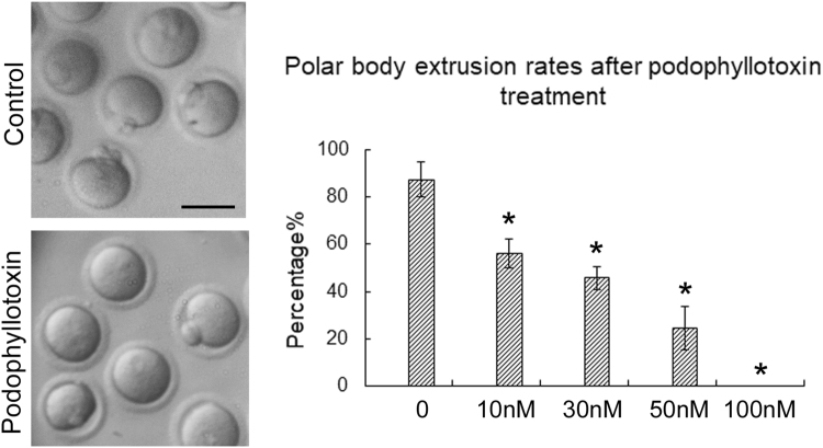
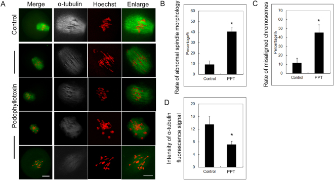
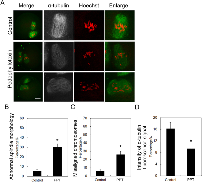
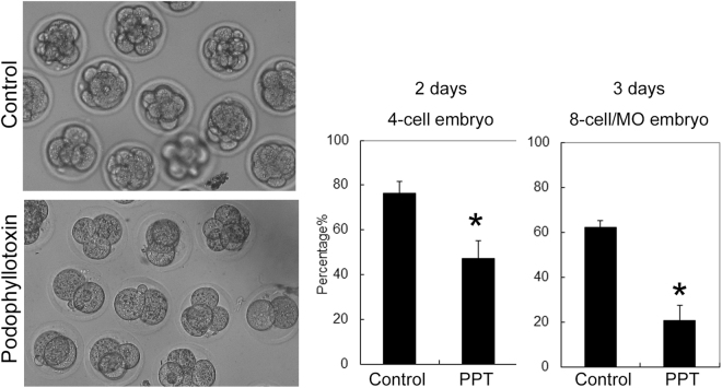
Podophyllotoxin is used as medical cream which is widely applied to genital warts and molluscum contagiosum. Although previous study showed that podophyllotoxin had minimal toxicity, it was forbidden to use during pregnancy since it might be toxic to the embryos. In present study we used mouse as the model and tried to examine whether podophyllotoxin exposure was toxic to oocyte maturation, which further affected embryo development. Our results showed that podophyllotoxin exposure inhibited mouse oocyte maturation, showing with the failure of polar body extrusion, and the inhibitory effects of podophyllotoxin on oocytes was dose-depended. Further studies showed that the meiotic spindle formation was disturbed, the chromosomes were misaligned and the fluorescence signal of microtubule was decreased, indicating that podophyllotoxin may affect microtubule dynamics for spindle organization. Moreover, the oocytes which reached metaphase II under podophyllotoxin exposure also showed aberrant spindle morphology and chromosome misalignment, and the embryos generated from these oocytes showed low developmental competence. We also found that the localization of p44/42 MAPK and gamma-tubulin was disrupted, which further confirmed the effects of podophyllotoxin on meiotic spindle formation. In all, our results indicated that podophyllotoxin exposure could affect mouse oocyte maturation by disturbing microtubule dynamics and meiotic spindle formation.


Conflict of interest statement

The authors declare no competing interests.

Figures

Figure 1
Figure 1
Podophyllotoxin exposure affected mouse oocyte maturation. The oocytes were cultured with 10 nM, 30 nM, 50 nM and 100 nM podophyllotoxin for 12 h. Most oocytes extruded the first polar body in the control group, however, a big proportion of oocytes failed to extrude the polar body in the podophyllotoxin treatment group. The polar body extrusion rate was significantly decreased with podophyllotoxin treatment in a dose dependent manner. *The statistical test was performed (p < 0.05).
Figure 2
Figure 2
Podophyllotoxin exposure affected spindle formation and chromosome alignment in the meiosis I of oocytes. (A) Fluorescence staining for the spindle and chromosomes after podophyllotoxin exposure. In the control group, the spindle showed well barrel shape morphology, and the chromosomes aligned well. However, after podophyllotoxin exposure the spindle morphology was abnormal, the chromosomes were mis-aligned. Moreover, the fluorescence signals of microtubule were much weaker compared with the control group. Green, alpha-tubulin; red, chromosomes. Bar = 20 μm. (B) Rates of abnormal spindle morphology in the podophyllotoxin exposure oocyte group. *The statistical test was performed (p < 0.05). (C) Intensity of alpha-tubulin fluorescence signals in the podophyllotoxin exposure oocyte group. *The statistical test was performed (p < 0.05). (D) Rates of misaligned chromosomes in the podophyllotoxin exposure oocyte group. *The statistical test was performed (p < 0.05).
Figure 3
Figure 3
Podophyllotoxin exposure affected spindle formation and chromosome alignment in meiosis II of oocytes. (A) Fluorescence staining for the spindle and chromosomes after podophyllotoxin exposure. In the control group, the oocytes extruded the first polar body, the spindle showed well barrel shape morphology, and the chromosomes aligned well. However, after podophyllotoxin exposure the spindle morphology was disrupted, the chromosomes were mis-aligned. Similar with meiosis I, the fluorescence signals of microtubule were much weaker compared with the control group. Green, alpha-tubulin; red, chromosomes. Bar = 20 μm. (B) Rates of abnormal spindle morphology in the podophyllotoxin exposure oocyte group. *The statistical test was performed (p < 0.05). (C) Intensity of alpha-tubulin fluorescence signals in the podophyllotoxin exposure oocyte group. *The statistical test was performed (p < 0.05). (D) Rates of misaligned chromosomes in the podophyllotoxin exposure oocyte group. *The statistical test was performed (p < 0.05).
Figure 4
Figure 4
Podophyllotoxin exposure affected the localization pattern of p44/42 MAPK and gamma-tubulin in mouse oocytes. (A) In the control oocytes, p44/42 MAPK localized at the poles of spindle, while in the podophyllotoxin treatment oocytes, the signals of p44/42 MAPK scattered around the spindle. Green, alpha-tubulin; red, p44/42 MAPK; blue, chromosomes. Bar = 20 μm. (B) In the control oocytes, gamma-tubulin localized at the poles of spindle, while in the podophyllotoxin treatment oocytes, the signals of gamma-tubulin dispersed from the spindle. Green, alpha-tubulin; red, gamma-tubulin; blue, chromosomes. Bar = 5 μm.
Figure 5
Figure 5
Podophyllotoxin exposure affected embryo development competence. The oocytes were cultured 30 nM podophyllotoxin for 12 h. The oocytes which extruded the first polar body were collected. And the embryos generated from the parthenogenetic activation oocytes with podophyllotoxin treatment group showed lower developmental rates after 2 days and 3 days culture. *The statistical test was performed (p < 0.05).

Similar articles

Cited by

References

    1. Sun SC, Kim NH. Molecular mechanisms of asymmetric division in oocytes. Microscopy and microanalysis: the official journal of Microscopy Society of America, Microbeam Analysis Society, Microscopical Society of Canada. 2013;19:883–897. doi: 10.1017/S1431927613001566. - DOI - PubMed
    1. Sun SC, Kim NH. Spindle assembly checkpoint and its regulators in meiosis. Human reproduction update. 2012;18:60–72. doi: 10.1093/humupd/dmr044. - DOI - PubMed
    1. Sun SC, Xiong B, Lu SS, Sun QY. MEK1/2 is a critical regulator of microtubule assembly and spindle organization during rat oocyte meiotic maturation. Molecular reproduction and development. 2008;75:1542–1548. doi: 10.1002/mrd.20891. - DOI - PubMed
    1. Sun SC, et al. Perturbation of survivin expression affects chromosome alignment and spindle checkpoint in mouse oocyte meiotic maturation. Cell cycle. 2009;8:3365–3372. doi: 10.4161/cc.8.20.9855. - DOI - PubMed
    1. Sun SC, et al. Arp2/3 complex regulates asymmetric division and cytokinesis in mouse oocytes. PloS one. 2011;6:e18392. doi: 10.1371/journal.pone.0018392. - DOI - PMC - PubMed

Publication types

LinkOut - more resources