. 2018 Jul 5;8(1):10215.
doi: 10.1038/s41598-018-28282-4.

A novel endoscopic classification for craniopharyngioma based on its origin

Affiliations

A novel endoscopic classification for craniopharyngioma based on its origin

Bin Tang et al. Sci Rep. .

Abstract

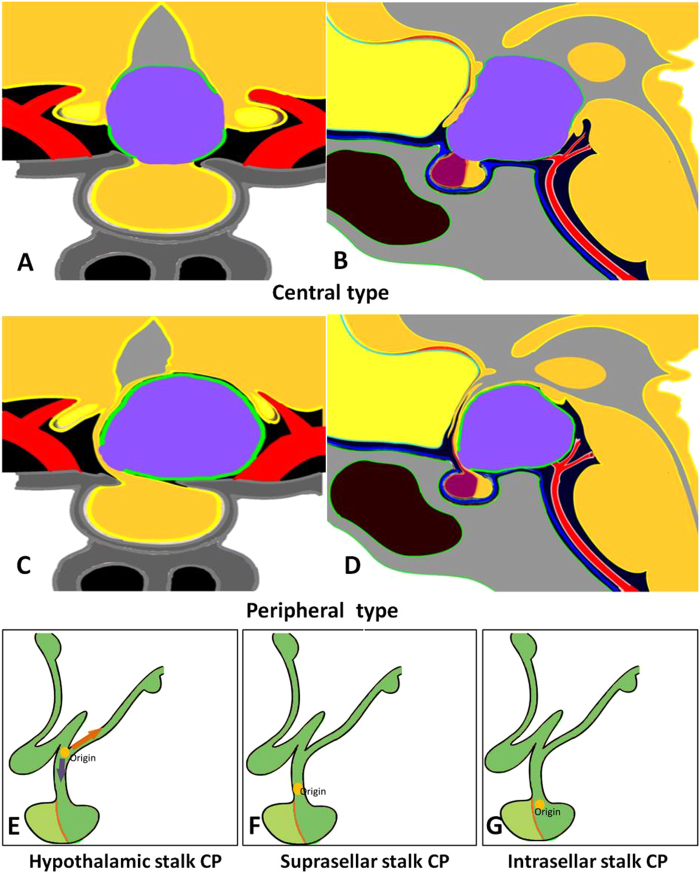
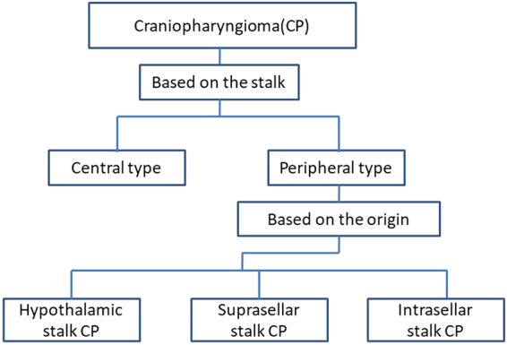
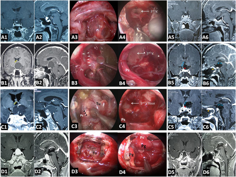
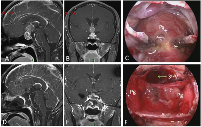
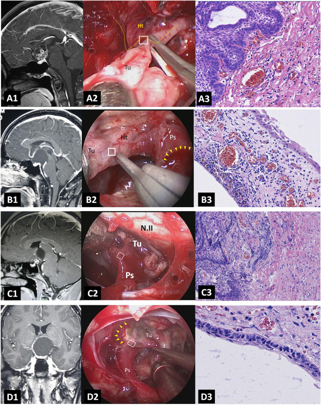
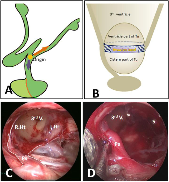
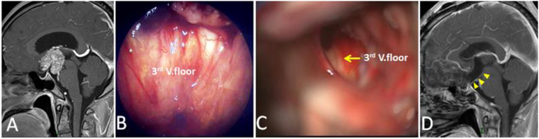
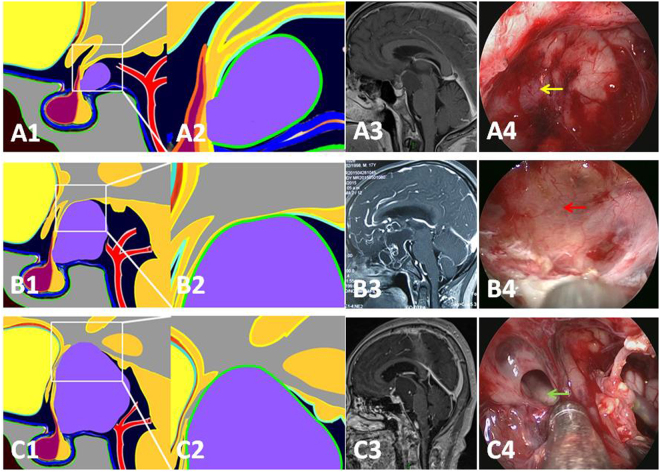
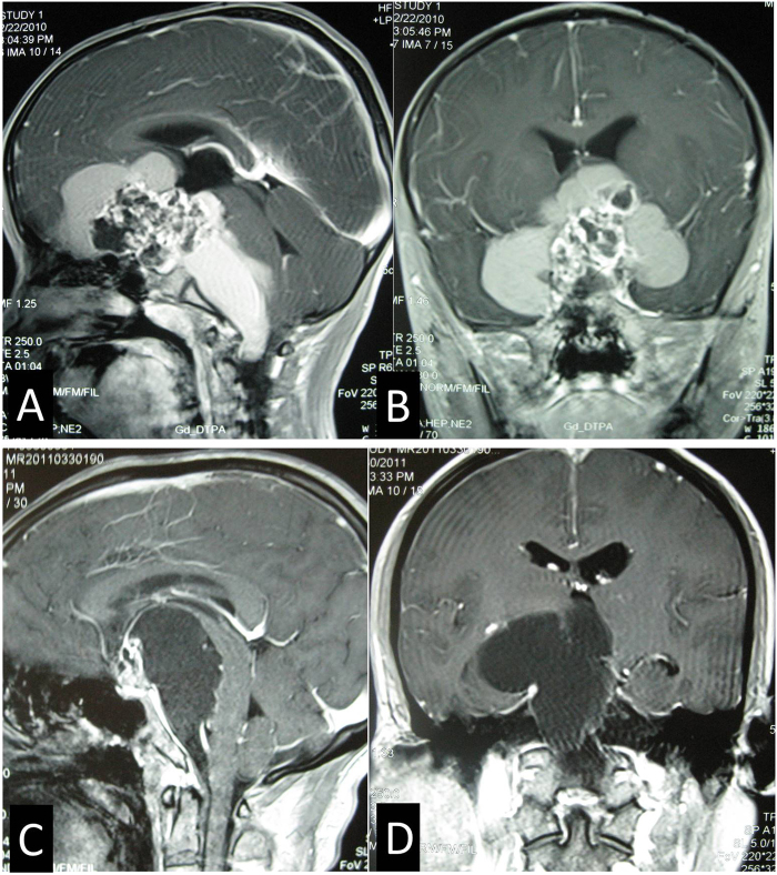
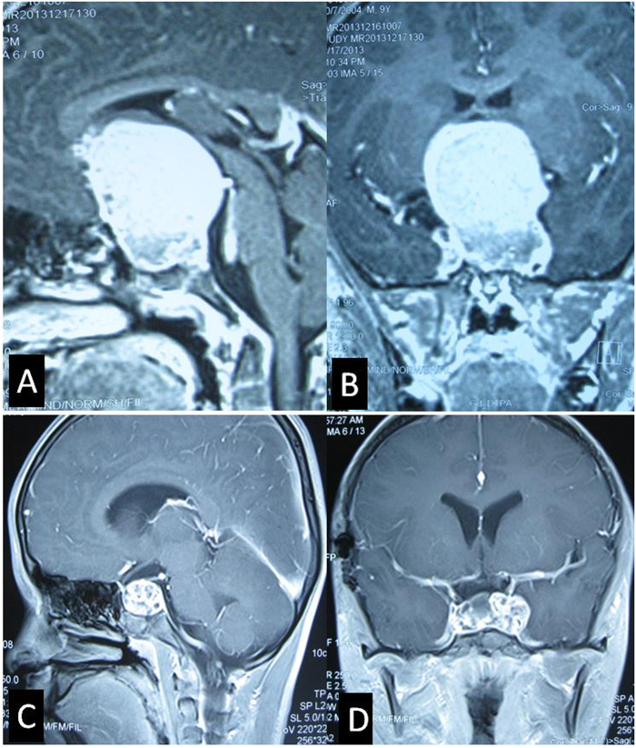
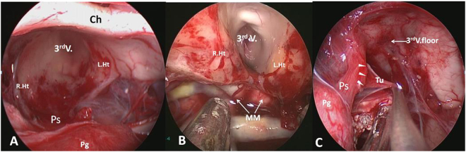
Endoscopic endonasal approach for craniopharyngioma (CP) resection provides a wide view and direct observation of hypothalamus and origin of tumor. Under endoscopy, 92 CPs were classified into 2 types: Peripheral and Central, according to its relation to pituitary stalk. Peripheral type was further divided into 3 subtypes: Hypothalamic stalk, Suprasellar stalk and Intrasellar stalk CP, according to the different origin site along hypothalamus-pituitary axis. Peripheral type arisen from the stalk but expanded and grown laterally in an exophytic pattern, accounting for 71.7% of all CPs, preservation rate of stalk was higher (76.0%). Central type grew within and along pituitary stalk and located strictly in the midline. The pituitary stalk was hardly preserved (only15.4%). Hypothalamic stalk CPs (n = 36, 54.6%) developed from the junction of hypothalamus and stalk, hypothalamus damage was found in all of this subtype after surgery. Suprasellar stalk CPs (n = 14, 21.2%) originated from the lower portion of stalk and displaced hypothalamus upward rather than infiltrated it. Intrasellar stalk CPs (n = 16, 24.2%) arose from the subdiaphragma portion of the stalk, with less hypothalamus damage. Recoginzing the origin of CP is helpful to understand its growth pattern and relation to hypothalamus, which is critical in planning the most appropriate surgical approach and degree of excision.


Conflict of interest statement

The authors declare no competing interests.

Figures

Figure 1
Figure 1
Schematic diagrams of the endoscopic subclassification of CPs. (A,B) Central type CP grows within and along the stalk and no pedicle or definite origin site can be identified, tumor is always located strictly in the midline. (C,D) Peripheral type CP arises from the stalk but expands and grows laterally in an exophytic pattern, the residual stalk is usually displaced to circumferential surface of the tumor. (E,F and G) Different origin sites of 3 subtypes of Peripheral type CPs along the pituitary stalk. (E) Hypothalamic stalk CP develops at the junction of the hypothalamus and the stalk, which usually extends up to the hypothalamus (brown arrow) and/or down to the up portion of stalk (black arrow), and usually invaded into the third ventricle. (F) Suprasellar stalk CP derives from suprasellar segment, usually low portion, of the stalk, and commonly locates extraventriclely. (G) Intrasellar stalk CP originates from the part of stalk under diaphragma, which is also known as intrasellar CP. Yellow discs in schematic diagrams indicate the origin site of the tumor.
Figure 2
Figure 2
Scheme of the endoscopic classification of CPs based on the relation with stalk and the origin of tumor.
Figure 3
Figure 3
Pre- and post-operative images and intraoperative findings of each subtype of CPs via endoscopic endonasal approach. (A1-6) A case of Central type CP. (A1-2) Coronal and sagittal postcontrast T1-weighted MR images showing a Central type CP which is located in the midline. (A3-4) Intraoperative photographs showing that tumor grew within and along the stalk, and the characteristic structure of stalk could be clearly identified on the surface of tumor (A3). Bilateral hypothalamus damage limited to tuber cinereum area could be seen after tumor removal (A4). (A5-6) Coronal and sagittal postcontrast T1-weighted MR images obtained after GTR. The pituitary gland was preserved and intact, but the stalk was not preserved. (B1-6) A case of Hypothalamic stalk CP. (B1-2) Coronal and sagittal postcontrast T1-weighted MR images showing a suprasellar CP located slightly toward to the left side and grows into the third ventricle, with the anterior third ventricle shifted to the right side (yellow arrowheads in B1). (B3-4) Intraoperative photographs showing that tumor originated from the junction of the hypothalamus and the stalk (white arrowheads in B3), the stalk was pushed to the right side. After tumor removal, a defect at the left side of the third ventricle floor could be found, and the remnant of stalk was pushed to the right side and connected to the right side of the third ventricle floor, with a relative normal hypothalamus at the right side (B4). (B5-6) Coronal and sagittal postcontrast T1-weighted MR images showing total tumor removal achieved. The stalk was preserved and pushed to the right side (red arrowhead in B5), the right hypothalamus was intact (blue arrowheads in B5-6). Note the vascularized nasoseptal flap at the posterior aspect of the sphenoid sinus on the sagittal image (orange arrowhead in B6). (C1-6) A case of Suprasellar stalk CP. (C1-2) Coronal and sagittal postcontrast T1-weighted MR images showing a suprasellar CP located toward to the left side, with the anterior third ventricle shifted to the right side (yellow arrowheads in C1). (C3-4) Intraoperative photographs showing that tumor derived from suprasellar segment, low portion of the stalk (black arrowheads in C3). After tumor removal, the third ventricle floor was intact, with no hypothalamus damage, and the remanent of stalk was pushed to the right side (C4). (C5-6) Coronal and sagittal postcontrast T1-weighted MR images showing total tumor removal achieved. The stalk was preserved and pushed to the right side (red arrowhead in C5), and the hypothalamus was intact (blue arrowheads in C5-6). Note the vascularized nasoseptal flap at the posterior aspect of the sphenoid sinus on the sagittal image (orange arrowhead in C6). (D1-6) A case of Intrasellar stalk CP. (D1-2) Coronal and sagittal postcontrast T1-weighted MR images showing a typical intrasellar CP. (D3-4) Intraoperative photographs showing that the solid tumor located in the sellar region. Residual pituitary gland and stalk can be seen after tumor removal (D4). (D5-6) Coronal and sagittal postcontrast T1-weighted MR images showing total tumor removal achieved. Ht, hypothalamus; Tu, tumor; Pg, pituitary gland; Ps, pituitary stalk. 3rd V., the third ventricle.
Figure 4
Figure 4
Example of a Central type Cp grows longer in vertical direction. (A and B) Sagittal (A) and Coronal (B) postcontrast T1-weighted MR images showing that tumor grows up to the third ventricle and down to the intrasellar region through pitiutary stalk. (C and F) Intraoperative photographs showing that tumor grew within and along the stalk and down to the intrasellar region (C). The third ventricle floor was destroyed, residual pituitary gland pushed and compressed by the tumor could be seen after tumor removal (F). (D and E) Sagittal (D) and Coronal (E) postcontrast T1-weighted MR images obtained after GTR demonstrating extensive, safe resection of the tumor. 3rd V., the third ventricle; Pg, pituitary gland.
Figure 5
Figure 5
Histopathological examination of the origin and non-origin capsule of CPs. (A1-3) Relationship between origin site of Hypothalamic stalk CP and hypothalamus. (A1) Sagittal postcontrast T1-weighted MR image showing a typical Hypothalamic stalk CP which grows up to the third ventricle. (A2) Intraoperative photograph showing that the specimen was obtained from the junction of hypothalamus and stalk (white box), yellow dashed line indicates the board of hypothalamus. (A3) Histopathological examination (×200) of tissue in the white box (B) showing that tumor invades the hypothalamus with interdigitation or finger-like infiltration. (B1-3) Relationship between non-origin capsule of Suprasellar stalk CP and hypothalamus. (B1) Sagittal postcontrast T1-weighted MR image showing a typical Suprasellar stalk CP which is located extraventriclely and pushes hypothalamus upward. (B2) Intraoperative photograph showing that the specimen was obtained from the non-origin capsule of tumor attached to hypothalamus (white box). Yellow arrowheads indict the origin site of tumor which located in the lower part of stalk. (B3) Histopathological examination (×200) of tissue in the white box (B) showing that the non-origin capsule of tumor is full of tumor cells, without any hypothalamus tissues. (C1-3) Relationship between origin site of Hypothalamic stalk CP and pituitary stalk. (C1) Sagittal postcontrast T1-weighted MR image showing a Hypothalamic stalk CP which grows up to the third ventricle. (C2) Intraoperative photograph showing that the specimen was obtained from the inferior part of the tumor base attached to the top of stalk (white box). (C3) Histopathological examination (×200) of tissue in the white box (B) showing the invasion of the tumor tissue into pituitary stalk. (D1-3) Relationship between non-origin capsule of Hypothalamic stalk CP and pituitary stalk. (D1) Coronal postcontrast T1-weighted MR image showing a Hypothalamic stalk CP which pushes the anterior third ventricle to the right side. (D2) Intraoperative photograph showing that the specimen was obtained from the non-origin capsule of tumor closed to the stalk (white box). yellow arrowheads indicate the origin site of tumor. (D3) Histopathological examination (×200) of tissue in the white box (B) showing the clear boundary between tumor and pituitary stalk which is without any tumor cells invasion. N.II, optic nerve; Ht, hypothalamus; Tu, tumor. Ps, pituitary stalk.
Figure 6
Figure 6
Illustration of the steps to diagnose the subtype of CP preoperatively and its corresponding degree of hypothalamus and stalk injury.
Figure 7
Figure 7
(A and B) Schematic drawing of Hypothalamic stalk CPs, which originate from the junction of hypothalamus and stalk, and can grow along the hypothalamus upward, or along the stalk downward, or both directions. The most common growth pattern is on both directions, and between the two parts, there is a circumferential band surrounded by tight adherence of the hypothalamic remnants (B). (C and D) Intraoperative photographs showing that a large defect at the left side of the third ventricle floor was observed after completely resection of a Hypothalamic stalk CP. The stalk was pushed to contralateral side and connected to the remnants of contralateral hypothalamus. Ps, pituitary stalk; 3rd V., the third ventricle, L.Ht, left hypothalamus, R.Ht, right hypothalamus.
Figure 8
Figure 8
Example of a purely intraventricle Cp. (A) Sagittal postcontrast T1-weighted MR image showing a solid tumor only grows into the third ventricle. (B and C) Intraoperative photographs showing that the third ventricle floor was intact after tumor removal via the callosal-interforniceal approach. (D) Sagittal postcontrast T1-weighted MR images obtained after GTR. Yellow arrowheads indicate the intact the third ventricle floor. 3rd V., the third ventricle.
Figure 9
Figure 9
Example of a Hypothalamic stalk Cp (pathological confirmed as adamantinomatous Cp) grows in vertical direction. (A and B) Sagittal (A) and Coronal (B) postcontrast T1-weighted MR images showing that the upward growth tumo towards to the third ventricle is cystic, while the downward growth tumor towards to suprasellar cistern is solid. (C) The CT scan showing that plaque calcification can be found in the cisternal part (red arrow).
Figure 10
Figure 10
Three stages in the Suprasellar stalk CPs according to the relationship between tumor and hypothalamus. (A1 and A2) Schematic drawing of a Suprasellar stalk CP in Grade 1: tumor contacts the hypothalamus but there exists a subaracnoid and pial membrane between them. (A3) The T1-GDPA image of a typical case in the sagittal plane of MR image. (A4) Intraoperative photographs showing that the third ventricle floor was thick and intact after tumor resection (yellow arrow). (B1 and B2) Schematic drawing of a Suprasellar stalk CP in Grade 2: tumor obviously pushes the hypothalamus upward with no membrane between them. (B3) The T1-GDPA image of a typical case in the sagittal plane of MR image. (B4) Intraoperative photographs showing that the third ventricle floor was thin and intact after tumor resection (red arrow). (C1 and C2) Schematic drawing of a Suprasellar stalk CP in Grade 3: tumor severely pushes the hypothalamus upward and breaks it into the third ventricle with tight adherence to the remnants of hypothalamus. (C3) The T1-GDPA image of a typical case in the sagittal plane of MR image. (C4) Intraoperative photographs showing that a defect of the third ventricle floor could be seen after tumor resection (blue arrow).
Figure 11
Figure 11
Examples of giant Suprasellar stalk CPs grow in roughly horizontal direction. (A and B) Sagittal (A) and Coronal (B) postcontrast T1-weighted MR images showing a giant Suprasellar stalk CP extends to the anterior fossa, sylvian fissure, the prepontine and posterior fossa. (C and D) Sagittal (C) and Coronal (D) postcontrast T1-weighted MR images showing a giant Suprasellar stalk CP extends to the prepontine and posterior fossa.
Figure 12
Figure 12
Growth pattern of Intrasellar stalk CPs. (A and B) Sagittal (A) and Coronal (B) postcontrast T1-weighted MR images showing a giant solid Intrasellar stalk CP grows in vertical direction and pushes the hypothalamus upward, at the same time, sella turcica is obviously much enlarged by the tumor. (C and D) Sagittal (C) and Coronal (D) postcontrast T1-weighted MR images showing a Intrasellar stalk CP grows laterally into the left cavernous sinus.
Figure 13
Figure 13
Different subtype of CPs presents different relation pattern to hypothalamus. (A) Ipsilateral hypothalamus injury (left): after removal of a Hypothalamic stalk CP, a defect at the left side of the third ventricle floor could be seen, the remnant of stalk was pushed to the right side and connected to the right side of the third ventricle floor, with no hypothalamus damage at the right side. (B) Bilateral hypothalamus injury: after removal of a Central type CP, a wide defect of bilateral floor of the third ventricle was observed, making the third ventricle totally opened with no or a small area of remnant third ventricle floor and stalk. (C) Removal of a Suprasellar stalk CP which originated from the lower-middle portion of stalk (white arrowheads), pituitary stalk was pushed to the right side, and the third ventricle floor was intact, with no hypothalamus damage. Tu, tumor; Pg, pituitary gland; Ps, pituitary stalk; 3rd V., the third ventricle, L.Ht, left hypothalamus, R.Ht, right hypothalamus; MM, mammillary body.

Similar articles

Cited by

References

    1. Fuller, G. N. & Scheithauer, B. W. The 2007 Revised World Health Organization (WHO) Classification of Tumours of the Central Nervous System: newly codified entities. Brain Pathol 17, 304-307 (2007). - PMC - PubMed
    1. Lewin R, Ruffolo E, Saraceno C. Craniopharyngioma arising in the pharyngeal hypophysis. South Med J. 1984;77:1519–1523. doi: 10.1097/00007611-198412000-00011. - DOI - PubMed
    1. Karavitaki N, Cudlip S, Adams CB, Wass JA. Craniopharyngiomas. Endocr Rev. 2006;27:371–397. doi: 10.1210/er.2006-0002. - DOI - PubMed
    1. Jane JA, Laws ER. Craniopharyngioma. Pituitary. 2006;9:323–326. doi: 10.1007/s11102-006-0413-8. - DOI - PubMed
    1. Wang KC, Hong SH, Kim SK, Cho BK. Origin of craniopharyngiomas: implication on the growth pattern. Childs Nerv Syst. 2005;21:628–634. doi: 10.1007/s00381-005-1203-8. - DOI - PubMed