. 2018 Jul 5;8(1):10164.
doi: 10.1038/s41598-018-28453-3.

The multidomain flavodiiron protein from Clostridium difficile 630 is an NADH:oxygen oxidoreductase

Affiliations

The multidomain flavodiiron protein from Clostridium difficile 630 is an NADH:oxygen oxidoreductase

Filipe Folgosa et al. Sci Rep. .

Abstract

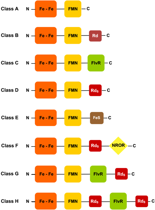
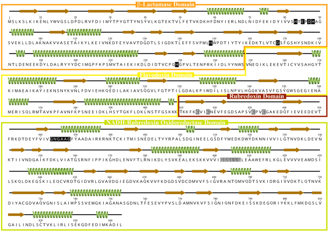
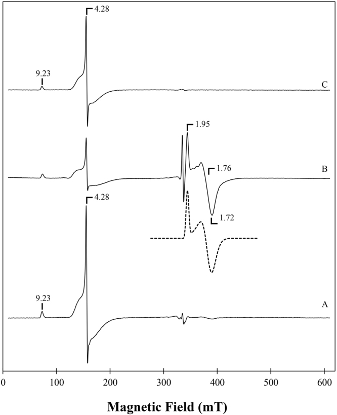
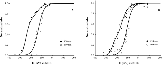
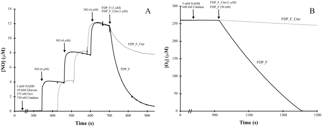
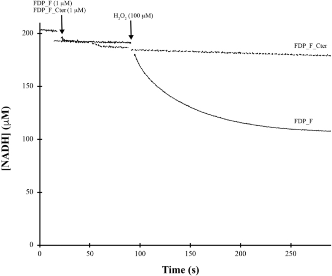
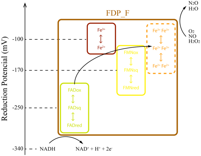
Flavodiiron proteins (FDPs) are enzymes with a minimal core of two domains: a metallo-β-lactamase-like, harbouring a diiron center, and a flavodoxin, FMN containing, domains. FDPs are O2 or NO reducing enzymes; for many pathogens, they help mitigate the NO produced by the immune system of the host, and aid survival during fluctuating concentrations concentrations of oxygen. FDPs have a mosaic structure, being predicted to contain multiple extra domains. Clostridium difficile, a threatening human pathogen, encodes two FDPs: one with the two canonical domains, and another with a larger polypeptide chain of 843 amino acids, CD1623, with two extra domains, predicted to be a short-rubredoxin-like and an NAD(P)H:rubredoxin oxidoreductase. This multi-domain protein is the most complex FDP characterized thus far. Each of the predicted domains was characterized and the presence of the predicted cofactors confirmed by biochemical and spectroscopic analysis. Results show that this protein operates as a standalone FDP, receiving electrons directly from NADH, and reducing oxygen to water, precluding the need for extra partners. CD1623 displayed negligible NO reductase activity, and is thus considered an oxygen selective FDP, that may contribute to the survival of C. difficile in the human gut and in the environment.


Conflict of interest statement

The authors declare no competing interests.

Figures

Figure 1
Figure 1
Schematic representation of the several Classes of FDPs. Fe-Fe – metallo-β-lactamase domain; FMN – flavodoxin domain; Rd - canonical rubredoxin domain; RdS - short rubredoxin domain; NROR – NADH:rubredoxin oxidoreductase domain; FlvR – flavin reductase-like domain.
Figure 2
Figure 2
Schematic representation of FDP_F protein. Amino acid sequence of FDP_F, with predicted alfa helixes and beta strands represented as green coils and as brown arrows, respectively. Secondary structure prediction was performed with Phyre2. The four domains predicted by amino acid sequence analysis are highlighted in the coloured boxes; metallo-β-lactamase domain in orange, Flavodoxin domain in yellow, Rubredoxin domain in dark red and the NADH:Rubredoxin oxidoreductase domain in green. The ligands for the di-iron and iron centers in the β-Lactamase and Rubredoxin domains are marked with black and grey boxes, respectively; the two glycine motifs of the NADH:rubredoxin oxidoreductase domain are highlighted with black (FAD binding) and grey (NADH binding) boxes.
Figure 3
Figure 3
SDP-PAGE gel from FDP_F and FDP_F_Cter. Lane 1 contains FDP_F_Cter and lane 2 contains FDP_F. Low-range molecular mass markers from Bio-Rad were used as standards.
Figure 4
Figure 4
UV-Visible spectra of FDP_F and FDP_F_Cter. Panel A- Spectra of FDP_F_Cter. The black line represents the full spectrum of the as isolated (oxidized) protein, while the dashed and the dashed-dot lines correspond to the FAD and rubredoxin components. The FAD component was obtained after substoichiometric reduction with sodium dithionite, and this spectrum was used to obtain the rubredoxin component after subtraction from the spectrum of the fully oxidized protein. The insert shows the spectra after complete reduction by NADH (black line) and dithionite (dashed line). Panel B- spectra of the full FDP_F. The black line represents the full spectrum of the protein as isolated (oxidized), while the dashed and the dashed-dot lines represent the FMN, FAD and rubredoxin components. FMN component was calculated by subtraction of the full spectra minus the spectra of the rubredoxin (x1) and FAD (x0.8) components from FDP_F_Cter. The insert shows the spectra after complete reduction of the protein with NADH (black line) and dithionite (dashed line). In both spectra protein concentration was 20 µM in 50 mM Tris–HCl, pH 7.5 containing 18% glycerol.
Figure 5
Figure 5
EPR spectra of FDP_F and FDP_F_Cter. Spectrum A- EPR spectrum of FDP_F (300 μM as isolated (oxidized), Spectrum B- EPR spectrum of FDP_F (sample A) after anaerobic incubation with substoichiometric menadiol. The dashed line below Spectrum B is the simulation spectrum obtained for the diiron component using g values of 1.96, 1.76 and 1.72. Spectrum C – EPR spectrum of FDP_F_Cter as isolated (200 μM). Temperature, 7 K; Microwave frequency, 9.39 GHz; Modulation amplitude, 1.0 mT; Microwave frequency, 2 mW.
Figure 6
Figure 6
Anaerobic redox titration curves of FDP_F and FDP_F_Cter. Both panels correspond to the normalized intensities measured at the indicated wavelengths. Panel A represents the titration curve for FDP_F_Cter and Panel B for FDP_F. In both titrations protein concentration was 25 µM and the experiments were performed in 50 mM Tris–HCl, pH 7.5 containing 18% glycerol. The solid lines correspond to Nernst equations adjusted as described in the text, with the following reduction potentials: FDP_F_Cter, −130 mV (Rd), −250 mV, −250 mV (FAD); FDP_F, −110 mV (Rd), −170 mV, −170 mV (FMN), −250 mV, −250 mV (FAD).
Figure 7
Figure 7
NO and O2 reductase activities of FDP_F and FDP_F_Cter. Panel A represents the NO reductase activity of both enzymes. Assays were performed anaerobically in a modified Clark type electrode. In both assays protein concentration was 1 µM in 50 mM Tris–HCl, pH 7.5 containing 18% glycerol. Panel B represents the O2 reductase activity of both enzymes. Assays were performed in a modified Clark type electrode. Protein concentrations were 50 nM and 1 µM for FDP_F and FDP_F_Cter, respectively, in 50 mM Tris–HCl, pH 7.5 containing 18% glycerol. NADH (5 mM) was used as reductant.
Figure 8
Figure 8
H2O2 reductase activity of FDP_F and FDP_F_Cter. H2O2 reductase activity of both enzymes. Reaction was performed anaerobically in a glove box, and monitored at 340 nm, following NADH (0.2 mM) consumption. In all assays protein concentrations were 1 µM in 50 mM Tris–HCl, pH 7.5 containing 18% glycerol.
Figure 9
Figure 9
Scheme of the intramolecular electron transfer chain within C. difficile FDP_F. The relative position of each domain as well as an electron flow from NADH to the substrate are represented, considering the determined reduction potentials. Although not determined, the reduction potentials for the diiron center are expected to be in the range indicated, as explained in the text. Each domain is encircled with boxes of different colors: metallo-β-lactamase domain in orange, Flavodoxin domain in yellow, Rubredoxin domain in dark red and the NADH:Rubredoxin oxidoreductase domain in green. The whole protein is encircled in brown.

References

    1. Romao CV, Vicente JB, Borges PT, Frazao C, Teixeira M. The dual function of flavodiiron proteins: oxygen and/or nitric oxide reductases. J Biol Inorg Chem. 2016;21:39–52. doi: 10.1007/s00775-015-1329-4. - DOI - PubMed
    1. Frederick RE, et al. Dioxygen and nitric oxide scavenging by Treponema denticola flavodiiron protein: a mechanistic paradigm for catalysis. Journal of Biological Inorganic Chemistry. 2015;20:603–613. doi: 10.1007/s00775-015-1248-4. - DOI - PMC - PubMed
    1. Ermakova M, et al. Heterocyst-specific flavodiiron protein Flv3B enables oxic diazotrophic growth of the filamentous cyanobacterium Anabaena sp. PCC 7120. Proc Natl Acad Sci USA. 2014;111:11205–11210. doi: 10.1073/pnas.1407327111. - DOI - PMC - PubMed
    1. Vicente JB, Tran V, Pinto L, Teixeira M, Singh U. A detoxifying oxygen reductase in the anaerobic protozoan Entamoeba histolytica. Eukaryot Cell. 2012;11:1112–1118. doi: 10.1128/EC.00149-12. - DOI - PMC - PubMed
    1. Kawasaki S, et al. Adaptive responses to oxygen stress in obligatory anaerobes Clostridium acetobutylicum and Clostridium aminovalericum. Appl Environ Microbiol. 2005;71:8442–8450. doi: 10.1128/AEM.71.12.8442-8450.2005. - DOI - PMC - PubMed

Publication types